Abstract
Background: Diaphyseal nonunion following failed intramedullary nailing is a challenging orthopedic condition. Secondary dynamization using an Ilizarov external ring fixator while retaining the intramedullary nail may improve fracture healing and functional recovery. Objective: To evaluate the clinical and radiological outcomes of secondary dynamization using an Ilizarov external ring fixator after failure of diaphyseal nonunion with intramedullary nailing while keeping the nail in situ. Methods: This prospective study was conducted at the Department of Orthopaedic Surgery, NITOR, Dhaka, Bangladesh, from June 2024 to May 2026. Thirty patients with diaphyseal nonunion following failed intramedullary nailing were treated with secondary dynamization using an Ilizarov external ring fixator while retaining the intramedullary nail. Clinical, radiological, and functional outcomes were analyzed using SPSS version 22.0. Results: The mean age of the participants was 35.7 ± 9.2 years, and 80.0% were male. Fracture union was achieved in 93.3% of patients, with a mean union time of 18.8 ± 3.4 weeks. At final follow-up, 86.7% achieved full weight bearing, 80.0% regained full range of motion, and the mean VAS pain score improved from 7.4 ± 1.2 to 1.9 ± 0.8. Pin tract infection was the most common complication (23.3%), and infection was significantly associated with persistent nonunion (p = 0.014). Conclusion: Secondary dynamization with an Ilizarov external ring fixator while retaining the intramedullary nail is an effective and reliable treatment for diaphyseal nonunion, providing a high union rate and good functional outcome.
Keywords
Diaphyseal Nonunion, Ilizarov External Fixator, Intramedullary Nail, Secondary Dynamization, Fracture Union
1. Introduction
Diaphyseal nonunion remains one of the most challenging complications in orthopedic trauma surgery, resulting in prolonged disability, repeated surgical interventions, increased healthcare costs, and reduced quality of life. Although modern fracture fixation techniques have significantly improved union rates, nonunion continues to occur in approximately 1-10% of long bone fractures depending on the fracture pattern, anatomical location, degree of soft tissue injury, and patient-related risk factors such as smoking, diabetes mellitus, infection, and osteoporosis
| [1] | Mohamed A, Francis D, Fuad U, Elmaleh N, Nagi A. Nonunion in long bone fractures: a comprehensive review of current treatment strategies. Cureus. 2025; 17 (11): e97599.
https://doi.org/10.7759/cureus.97599 |
| [2] | Rupp M, Biehl C, Budak M, Thormann U, Heiss C, Alt V. Diaphyseal long bone nonunions: types, aetiology, economics, and treatment recommendations. Int Orthop. 2018; 42(2): 247-258. https://doi.org/10.1007/s00264-017-3734-5 |
[1, 2]
. Tibial and femoral shaft fractures are particularly prone to delayed union and nonunion because of their limited blood supply, extensive soft tissue damage in high-energy trauma, and the mechanical demands placed upon these weight-bearing bones
.
Intramedullary nailing (IMN) is considered the gold standard for the management of most diaphyseal fractures of the femur and tibia because it provides stable fixation while preserving the periosteal blood supply and allowing early mobilization
. Despite these advantages, failure of fracture healing following IMN remains a recognized complication. Nonunion after intramedullary nailing may result from inadequate mechanical stability, impaired biological healing potential, infection, bone loss, or a combination of these factors
| [5] | Krappinger D, Wolf B, Dammerer D, Thaler M, Schwendinger P, Lindtner RA. Risk factors for nonunion after intramedullary nailing of subtrochanteric femoral fractures. Arch Orthop Trauma Surg. 2019; 139(6): 769-777.
https://doi.org/10.1007/s00402-019-03132-0 |
[5]
. Traditionally, management options include exchange reamed nailing, augmentative plating with or without bone grafting, nail dynamization, bone grafting, and external fixation. However, no single treatment modality is universally effective for all types of nonunion, particularly in cases with multiple previous surgeries or compromised biology
.
The concept of dynamization has been widely adopted to stimulate fracture healing by converting a statically locked intramedullary nail into a load-sharing construct through the removal of one or more locking screws. Controlled axial micromotion enhances callus formation by increasing physiological compression across the fracture site while maintaining alignment
| [7] | Yu P, Huang L, Wang L, Cai L, Tu D. Nail dynamization for delayed union and nonunion in femur and tibia fractures following intramedullary nailing: a systematic review and meta-analysis. Injury. 2025; 56(11): 112748.
https://doi.org/10.1016/j.injury.2025.112748 |
[7]
. Although primary nail dynamization is effective in selected delayed unions, its success decreases substantially in established nonunion, especially when significant instability, deformity, or biological deficiency exists
| [8] | Vaughn JE, Shah RV, Samman T, Stirton J, Liu J, Ebraheim NA. Systematic review of dynamization vs exchange nailing for delayed/non-union femoral fractures. World J Orthop. 2018; 9(7): 92-99. https://doi.org/10.5312/wjo.v9.i7.92 |
[8]
. Furthermore, removal of locking screws alone may not provide sufficient compression in long-standing nonunion or in fractures with bone defects.
The Ilizarov external ring fixator has revolutionized the treatment of complex nonunions through the principles of tension-stress, stable circular fixation, and controlled compression-distraction osteogenesis. Since its introduction by Gavriil Ilizarov, the technique has demonstrated high union rates in aseptic and infected nonunion, bone defects, deformity correction, and limb length discrepancy
| [9] | Gubin A, Borzunov D, Malkova T. Ilizarov method for bone lengthening and defect management: review of contemporary literature. Bull Hosp Jt Dis (2013). 2016; 74(2): 145-154. |
[9]
. Circular external fixation offers multiplanar stability while permitting progressive compression, correction of deformity, early weight bearing, and preservation of local blood supply. Importantly, it facilitates biological stimulation of osteogenesis without extensive surgical exposure, thereby minimizing additional soft tissue trauma
.
Secondary dynamization using an Ilizarov external ring fixator while keeping the intramedullary nail in situ has emerged as an attractive limb-salvage strategy in selected patients with failed diaphyseal nonunion after IMN. This combined technique integrates the internal stability of the retained nail with the adjustable compression provided by the circular fixator. The intramedullary nail maintains alignment and rotational stability, whereas the Ilizarov frame allows gradual axial compression and controlled dynamization across the nonunion site. Consequently, extensive implant removal, exchange nailing, or repeated open surgery may be avoided, reducing operative time, blood loss, and soft tissue disruption
| [11] | Gulabi D, Erdem M, Cecen GS, Avci CC, Saglam N, Saglam F. Ilizarov fixator combined with an intramedullary nail for tibial nonunions with bone loss: is it effective? Clin Orthop Relat Res. 2014; 472(12): 3892-3901.
https://doi.org/10.1007/s11999-014-3932-2 |
[11]
.
Several clinical studies have reported encouraging outcomes using combined nail-Ilizarov constructs, demonstrating union rates exceeding 90% in difficult tibial and femoral nonunions, including cases complicated by failed previous fixation, deformity, infection, and segmental bone loss
| [12] | Xie L, Huang Y, Zhang L, Si S, Yu Y. Ilizarov method and its combined methods in the treatment of long bone defects of the lower extremity: a systematic review and meta-analysis. BMC Musculoskelet Disord. 2023; 24(1): 891.
https://doi.org/10.1186/s12891-023-07001-9 |
| [13] | Mohd-Yusof N, Kow RY, Sulong AF, Nallayan RM, Zainal-Abidin MA. Clinical and functional outcomes of Ilizarov bone transport in traumatic tibial bone loss. Malays Orthop J. 2025; 19(2): 91-99. |
[12, 13]
. Retaining the intramedullary nail also decreases the duration of external fixation compared with Ilizarov treatment alone because the nail provides internal support after frame removal. This approach improves patient comfort, facilitates rehabilitation, and reduces complications associated with prolonged external fixation such as pin tract infection and joint stiffness
| [14] | Jeremic D, Grubor N, Bascarevic Z, Slavkovic N, Krivokapic B, Vukomanovic B, et al. Comparative analysis of complication rates in tibial shaft fractures: intramedullary nail versus Ilizarov external fixation method. J Clin Med. 2024; 13(7): 2034. https://doi.org/10.3390/jcm13072034 |
[14]
.
Despite these promising results, evidence regarding secondary dynamization with an Ilizarov external ring fixator after failed diaphyseal nonunion while retaining the original intramedullary nail remains limited. Most published studies consist of retrospective case series with heterogeneous patient populations and varying surgical protocols. Therefore, further evaluation is necessary to determine union rates, functional outcomes, complications, time to union, and factors influencing treatment success. Such evidence may help establish this combined technique as a reliable alternative to exchange nailing or extensive revision surgery in carefully selected patients with complex diaphyseal nonunion
| [15] | Yin P, Ji Q, Li T, Li J, Li Z, Liu J, et al. A systematic review and meta-analysis of Ilizarov methods in the treatment of infected nonunion of tibia and femur. PLoS One. 2015; 10(11): e0141973. https://doi.org/10.1371/journal.pone.0141973 |
[15]
.
The present study was undertaken to evaluate the effectiveness of secondary dynamization by Ilizarov external ring fixator after failure of diaphyseal nonunion with intramedullary nailing while keeping the nail in situ. The findings are expected to provide further evidence regarding the role of this minimally invasive limb-salvage technique and its potential as an alternative to exchange nailing or extensive reconstructive procedures in the management of challenging diaphyseal nonunions.
2. Objectives
The main objective was to evaluate the outcome of secondary dynamization using an Ilizarov external ring fixator with the intramedullary nail retained in situ for the treatment of diaphyseal nonunion after failed intramedullary nailing.
3. Methodology & Materials
This prospective study was conducted in the Department of Orthopaedic Surgery, National Institute of Traumatology and Orthopaedic Rehabilitation (NITOR), Dhaka, Bangladesh, over a period from June 2024 to May 2026 years. The study included patients with diaphyseal nonunion following failed intramedullary nailing who were managed by secondary dynamization using an Ilizarov external ring fixator while keeping the intramedullary nail in situ. Eligible patients were enrolled consecutively after obtaining informed written consent. A total of 30 patients were included in the study through consecutive sampling.
3.1. Selection Criteria
3.1.1. Inclusion Criteria
1) Patients aged 18 years or above with established diaphyseal nonunion following intramedullary nailing.
2) Patients treated with secondary dynamization using an Ilizarov external ring fixator while retaining the intramedullary nail in situ.
3) Patients willing to participate and provide written informed consent.
3.1.2. Exclusion Criteria
1) Patients with pathological fractures.
2) Patients with severe neurovascular injury requiring amputation.
3) Patients with incomplete clinical or radiological records.
4) Patients unwilling to participate or lost to follow-up before assessment of fracture union.
A structured case record form was used for data collection. Baseline demographic variables included age, sex, smoking status, and associated comorbidities. Injury-related variables included mechanism of injury, involved bone (femur or tibia), side of injury, AO fracture classification, duration of nonunion, and number of previous surgical procedures. Operative variables included the type of nonunion (hypertrophic, oligotrophic, or atrophic), presence of infection, duration of surgery, duration of external fixation, and time to full weight bearing. All patients underwent secondary dynamization using an Ilizarov external ring fixator while the previously inserted intramedullary nail was retained in situ. Standard postoperative rehabilitation protocols were followed. Clinical evaluation was performed during each follow-up visit and included assessment of pain using the Visual Analogue Scale (VAS), weight-bearing status, range of motion of adjacent joints, limb alignment, limb length discrepancy, and overall functional recovery. Functional outcome at the final follow-up was graded as excellent, good, fair, or poor according to the predetermined functional assessment criteria used in the study. Radiological assessment was carried out using standard anteroposterior and lateral radiographs obtained at regular follow-up intervals. Fracture union was considered achieved when bridging callus was observed across at least three cortices, accompanied by painless full weight bearing and the absence of abnormal movement at the nonunion site. Time to union and duration of external fixation were also recorded. The primary outcome measure was fracture union. Secondary outcome measures included time to fracture union, duration of external fixation, pain improvement, functional outcome, weight-bearing ability, range of motion, limb alignment, limb length discrepancy, and postoperative complications. Complications evaluated included pin tract infection, wire loosening, implant failure, delayed union, persistent nonunion, malalignment, refracture, and joint stiffness.
3.2. Statistical Analysis
All collected data were checked for completeness and consistency before analysis. Data were entered into a computer and analyzed using Statistical Package for the Social Sciences (SPSS) version 22.0. Continuous variables were expressed as mean ± standard deviation (SD), while categorical variables were presented as frequency and percentage. Descriptive statistics were used to summarize demographic characteristics and treatment outcomes. Where appropriate, paired or independent t-tests and Chi-square or Fisher's exact tests were used for comparison. A p-value <0.05 was considered statistically significant. The findings were presented using tables, charts, and graphical illustrations.
3.3. Quality Assurance
A standardized data collection form was used for data collection. Clinical and radiological assessments were performed using uniform criteria. Data were checked for completeness and accuracy before analysis, and statistical analysis was conducted under the supervision of the principal investigator.
3.4. Ethical Considerations
Ethical approval was obtained from the Institutional Review Board (IRB)/Ethical Review Committee of NITOR. Written informed consent was obtained from all participants. Confidentiality was maintained throughout the study, and participation was voluntary. All procedures were conducted in accordance with the Declaration of Helsinki.
4. Result
Table 1 shows the demographic characteristics of the study participants. The mean age was 35.7 ± 9.2 years, with the highest proportion (40.0%) aged 30-39 years. Most participants were male (80.0%). Half of the patients were smokers (50.0%). Regarding comorbidities, 66.7% had no associated comorbidity, while diabetes mellitus, hypertension, and both diabetes with hypertension were present in 16.7%, 10.0%, and 6.6% of patients, respectively.
Table 1. Demographic characteristics of the study participants (n=30).
Variable | Frequency (n) | Percentage (%) |
Age (years) |
18-29 | 8 | 26.7% |
30-39 | 12 | 40% |
40-49 | 7 | 23.3% |
≥50 | 3 | 10% |
Mean ± SD | 35.7 ± 9.2 |
Sex |
Male | 24 | 80% |
Female | 6 | 20% |
Smoking status |
Smoker | 15 | 50% |
Non-smoker | 15 | 50% |
Comorbidity |
None | 20 | 66.7% |
Diabetes mellitus | 5 | 16.7% |
Hypertension | 3 | 10% |
Diabetes + Hypertension | 2 | 6.6% |
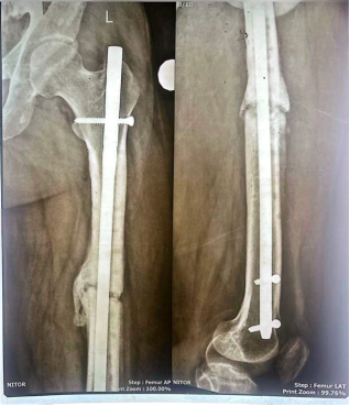
Figure 1. Preoperative anteroposterior (AP) and lateral radiographs of a 26-year-old male patient showing diaphyseal nonunion of the femur following failed intramedullary nailing. Preoperative anteroposterior (AP) and lateral radiographs of a 26-year-old male patient showing diaphyseal nonunion of the femur following failed intramedullary nailing.
Table 2. Injury and fracture characteristics of the study participants (n=30).
Variable | Frequency (n) | Percentage (%) |
Mechanism of injury |
Road traffic accident | 18 | 60% |
Fall from height | 9 | 30% |
Assault | 2 | 6.7% |
Sports injury | 1 | 3.3% |
Bone involved |
Tibia | 19 | 63.3% |
Femur | 11 | 36.7% |
Side involved |
Right | 17 | 56.7% |
Left | 13 | 43.3% |
AO Classification |
Type A | 11 | 36.7% |
Type B | 12 | 40% |
Type C | 7 | 23.3% |
Duration of nonunion |
6-9 months | 10 | 33.3% |
10-12 months | 12 | 40% |
>12 months | 8 | 26.7% |
Mean ± SD | 10.4 ± 2.8 months | |
Previous surgeries |
One | 18 | 60% |
Two | 9 | 30% |
≥3 | 3 | 10% |
Table 2 presents the injury and fracture characteristics of the study participants. Road traffic accident was the most common mechanism of injury (60.0%), followed by fall from height (30.0%). The tibia was involved in 63.3% of cases, and the right side was affected in 56.7%. According to the AO classification, Type B fractures (40.0%) were the most frequent. The mean duration of nonunion was 10.4 ± 2.8 months, and 60.0% of patients had undergone one previous surgical procedure.
Table 3. Operative characteristics (n=30).
Variable | Frequency (n) | Percentage (%) |
Type of nonunion |
Hypertrophic | 18 | 60% |
Oligotrophic | 8 | 26.7% |
Atrophic | 4 | 13.3% |
Infection |
Present | 4 | 13.3% |
Absent | 26 | 86.7% |
Operative variables |
Operative time (minutes) | 94.8 ± 15.6 |
External fixation duration (weeks) | 18.9 ± 3.8 |
Time to full weight bearing (weeks) | 7.1 ± 1.8 |
Table 3 shows the operative characteristics of the study participants. Hypertrophic nonunion was the most common type (60.0%), while infection was present in 13.3% of patients. The mean operative time was 94.8 ± 15.6 minutes, the mean duration of external fixation was 18.9 ± 3.8 weeks, and patients achieved full weight bearing after a mean of 7.1 ± 1.8 weeks.
Figure 2. Intraoperative AP and lateral radiographs showing secondary dynamization with an Ilizarov external ring fixator while retaining the intramedullary nail in situ. Intraoperative AP and lateral radiographs showing secondary dynamization with an Ilizarov external ring fixator while retaining the intramedullary nail in situ.
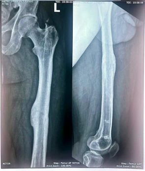
Figure 3. Postoperative AP and lateral radiographs demonstrating successful fracture union at final follow-up. Postoperative AP and lateral radiographs demonstrating successful fracture union at final follow-up.
Table 4. Clinical and radiological outcome (Primary outcome).
Variable | Frequency (n) | Percentage (%) |
Fracture union achieved | 28 | 93.3% |
Persistent nonunion | 2 | 6.7% |
Radiological union | 28 | 93.3% |
External fixator removed successfully | 28 | 93.3% |
Table 4 presents the primary clinical and radiological outcomes of the study. Fracture union was successfully achieved in 93.3% of patients, while persistent nonunion occurred in 6.7%. Radiological union was observed in 93.3% of cases, and the external fixator was successfully removed in the same proportion of patients. The mean time to fracture union was 18.8 ± 3.4 weeks.
Table 5. Time to Fracture Union among Patients with Successful Union.
Time | Frequency | Percentage (%) |
≤16 weeks | 8 | 28.6% |
17-20 weeks | 12 | 42.8% |
>20 weeks | 8 | 28.6% |
Table 5 shows the distribution of fracture union time among patients who achieved union. The largest proportion (42.8%) achieved union between 17 and 20 weeks, whereas 28.6% achieved union within 16 weeks and another 28.6% required more than 20 weeks. The mean union time was 18.8 ± 3.4 weeks.
Table 6. Functional outcome after treatment.
Variable | Frequency (n) | Percentage (%) |
Weight bearing | Full | 26 | 86.7% |
Partial | 3 | 10% |
Unable | 1 | 3.3% |
Range of motion | Full | 24 | 80% |
Mild restriction | 5 | 16.7% |
Moderate restriction | 1 | 3.3% |
Limb alignment | Normal | 27 | 90% |
Mild malalignment | 3 | 10% |
Limb-length discrepancy | None | 22 | 73.3% |
<1 cm | 6 | 20% |
≥1 cm | 2 | 6.7% |
Pain score |
Preoperative VAS | 7.4 ± 1.2 |
Final follow-up VAS | 1.9 ± 0.8 |
Table 6 presents the functional outcomes following treatment. At final follow-up, 86.7% of patients achieved full weight bearing and 80.0% regained full range of motion. Normal limb alignment was observed in 90.0% of patients, while 73.3% had no limb-length discrepancy. The mean pain score (VAS) improved markedly from 7.4 ± 1.2 preoperatively to 1.9 ± 0.8 at final follow-up.
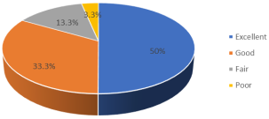
Figure 4. Functional grading of the study participants. Functional grading of the study participants.
Figure 4 illustrates the functional grading of the study participants. Most patients had excellent (50.0%) or good (33.3%) functional outcomes, while 13.3% had fair and 3.3% had poor outcomes after treatment.
Table 7. Complications following treatment (n=30).
Complication | Frequency (n) | Percentage (%) |
None | 16 | 53.3% |
Pin tract infection | 7 | 23.3% |
Wire loosening | 2 | 6.7% |
Joint stiffness | 2 | 6.7% |
Delayed union | 1 | 3.3% |
Implant failure | 1 | 3.3% |
Persistent nonunion | 1 | 3.3% |
Table 7 shows the postoperative complications observed among the study participants. More than half of the patients (53.3%) experienced no complications. The most common complication was pin tract infection (23.3%), followed by wire loosening and joint stiffness (6.7% each). Delayed union, implant failure, and persistent nonunion were each observed in 3.3% of patients.
Table 8. Association between type of nonunion and fracture union.
Type of nonunion | Union | Nonunion | Total | P-value |
Hypertrophic | 18 | 0 | 18 | 0.143 |
Oligotrophic | 7 | 1 | 8 |
Atrophic | 3 | 1 | 4 |
Total | 28 | 2 | 30 |
Table 8 presents the association between the type of nonunion and fracture union. Fracture union was achieved in all patients with hypertrophic nonunion, whereas nonunion occurred in one patient each with oligotrophic and atrophic nonunion. However, the association between the type of nonunion and fracture union was not statistically significant (χ
2 = 4.82, p = 0.143).
Table 9. Association between infection and fracture union.
Infection | Union | Nonunion | Total | P-value |
Present | 2 | 2 | 4 | 0.014 |
Absent | 26 | 0 | 26 |
Total | 28 | 2 | 30 |
Table 9 shows the association between infection and fracture union. Persistent nonunion occurred only among patients with infection, whereas all patients without infection achieved successful fracture union. This association was statistically significant (Fisher's Exact Test, p = 0.014), indicating that infection was significantly associated with poorer fracture healing.
5. Discussion
The present prospective study evaluated the effectiveness of secondary dynamization using an Ilizarov external ring fixator while retaining the intramedullary nail in situ for the management of diaphyseal nonunion following failed intramedullary nailing. A total of 30 patients were included. The study assessed demographic characteristics, injury profile, operative variables, fracture union, functional outcome, and postoperative complications. The mean age of the study participants was 35.7 ± 9.2 years, with the majority of patients (40.0%) belonging to the 30-39 years age group. This finding indicates that diaphyseal nonunion predominantly affects young and middle-aged adults, who represent the economically productive population and are more frequently exposed to high-energy trauma. Similar findings were reported by Menon et al. (2002), who investigated augmentative Ilizarov external fixation after failed intramedullary nailing and found that most patients were young adults with a mean age of approximately 31 years
| [16] | Menon DK, Dougall TW, Pool RD, Simonis RB. Augmentative Ilizarov external fixation after failure of diaphyseal union with intramedullary nailing. J Orthop Trauma. 2002; 16(7): 491-497. |
[16]
. Their study also demonstrated that this treatment modality was effective in restoring union in active individuals with resistant diaphyseal nonunion. A comparable age distribution was also described by Rozbruch and colleagues and by subsequent studies evaluating Ilizarov techniques for complex long-bone nonunion, which reported that these injuries are primarily encountered in adults between the third and fifth decades of life because of their greater involvement in road traffic accidents and occupational injuries
. The present study demonstrated a marked male predominance (80.0%), whereas females accounted for only 20.0% of the study population. This observation is consistent with the epidemiological pattern of long-bone fractures worldwide, where males sustain significantly more high-energy trauma than females. Menon et al. likewise reported that most patients requiring augmentative Ilizarov fixation after failed intramedullary nailing were male
| [16] | Menon DK, Dougall TW, Pool RD, Simonis RB. Augmentative Ilizarov external fixation after failure of diaphyseal union with intramedullary nailing. J Orthop Trauma. 2002; 16(7): 491-497. |
[16]
. Similar male predominance has also been reported in studies evaluating infected tibial nonunion treated by the Ilizarov method
| [18] | Megas P, Saridis A, Kouzelis A, Kallivokas A, Mylonas S, Tyllianakis M. The treatment of infected nonunion of the tibia following intramedullary nailing by the Ilizarov method. Injury. 2010; 41(3): 294-299.
https://doi.org/10.1016/j.injury.2009.09.006 |
[18]
. Smoking is a well-recognized biological risk factor for delayed fracture healing because nicotine causes peripheral vasoconstriction, impairs osteoblast activity, and reduces callus formation. In the present study, 50.0% of the patients were smokers. Although the current study did not specifically evaluate the independent effect of smoking on fracture union, previous orthopedic literature has consistently demonstrated that smoking adversely affects fracture healing and increases the risk of delayed union and nonunion. Therefore, smoking cessation should be emphasized before and after reconstructive procedures for long-bone nonunion.
Regarding associated medical conditions, 66.7% of patients had no significant comorbidity, while diabetes mellitus and hypertension were present in a smaller proportion of participants. Diabetes is recognized as an important factor contributing to impaired fracture healing through reduced angiogenesis, delayed osteogenesis, and an increased susceptibility to infection. Consequently, careful optimization of glycaemic control before definitive reconstructive surgery is recommended to improve the likelihood of successful fracture union. Similar observations have been reported by Maini et al., who found that systemic comorbidities, particularly diabetes and infection, adversely affected the management of long-bone nonunion treated with the Ilizarov technique
| [19] | Maini L, Chadha M, Vishwanath J, Kapoor S, Mehtani A, Dhaon BK. The Ilizarov method in infected nonunion of fractures. Injury. 2000; 31(7): 509-517. |
[19]
. Likewise, Ding et al. demonstrated in a systematic review and meta-analysis that patients with diabetes had more than twice the risk of impaired fracture healing, including delayed union and nonunion, compared with non-diabetic patients
| [20] | Ding ZC, Zeng WN, Rong X, Liang ZM, Zhou ZK. Do patients with diabetes have an increased risk of impaired fracture healing? A systematic review and meta-analysis. ANZ J Surg. 2020; 90(7-8): 1259-1264. https://doi.org/10.1111/ans.15944 |
[20]
. Overall, the demographic profile of the present study closely resembles that reported in previous investigations of Ilizarov-assisted treatment of diaphyseal nonunion, suggesting that young adult males with high-energy traumatic injuries remain the population most frequently requiring secondary reconstructive procedures after failure of intramedullary nailing. In the present study, road traffic accident (RTA) was the predominant mechanism of injury, accounting for 60.0% of all cases, followed by falls from height (30.0%). This finding is consistent with the epidemiology of diaphyseal fractures reported worldwide, where high-energy trauma remains the leading cause of long-bone fractures and subsequent nonunion. Menon et al. reported that most patients requiring augmentative Ilizarov external fixation after failed intramedullary nailing sustained injuries following high-energy trauma, particularly road traffic accidents
| [16] | Menon DK, Dougall TW, Pool RD, Simonis RB. Augmentative Ilizarov external fixation after failure of diaphyseal union with intramedullary nailing. J Orthop Trauma. 2002; 16(7): 491-497. |
[16]
. The authors emphasized that severe soft tissue injury and extensive bone damage associated with these injuries increase the risk of delayed union and nonunion. The tibia was the most commonly affected bone in the present series (63.3%), whereas femoral involvement constituted 36.7%. This observation agrees with Gulabi et al., who reported that tibial shaft nonunion is encountered more frequently because of the subcutaneous location of the tibia, its relatively poor soft-tissue envelope, and its vulnerability to open injuries and infection
| [11] | Gulabi D, Erdem M, Cecen GS, Avci CC, Saglam N, Saglam F. Ilizarov fixator combined with an intramedullary nail for tibial nonunions with bone loss: is it effective? Clin Orthop Relat Res. 2014; 472(12): 3892-3901.
https://doi.org/10.1007/s11999-014-3932-2 |
[11]
. Their study demonstrated favorable outcomes using an Ilizarov external fixator combined with an intramedullary nail for tibial nonunion with bone loss.
The right lower limb was involved in 56.7% of patients, which is comparable to previous orthopedic series where no significant difference between right- and left-sided injuries has been demonstrated. The predominance of right-sided injuries may simply reflect the dominance of the right lower extremity during daily activities and driving, although side involvement has not been shown to influence fracture healing significantly. According to the AO fracture classification, Type B fractures (40.0%) were the most frequent, followed by Type A (36.7%) and Type C (23.3%). Increasing fracture complexity is known to influence the biological and mechanical environment required for fracture healing. More complex fracture configurations generally require prolonged healing time and are associated with a higher incidence of delayed union and nonunion because of greater periosteal disruption, comminution, and reduced cortical contact. Recent studies have demonstrated that fracture comminution, higher AO/OTA fracture types, residual fracture gap, and inadequate cortical contact are significant predictors of nonunion following intramedullary nailing
| [21] | Kraus KR, Flores JW, Slaven JE, Sharma I, Arnold PK, Mullis BH, et al. A scoring system for predicting nonunion after intramedullary nailing of femoral shaft fractures. JAAOS Glob Res Rev. 2024; 8(9): e24.
https://doi.org/10.5435/JAAOSGlobal-D-24-00000 |
[21]
. The mean duration of nonunion before secondary dynamization in the present study was 10.4 ± 2.8 months, indicating that most patients had established nonunion before undergoing definitive reconstruction. Menon et al. described patients referred after multiple failed procedures with longstanding diaphyseal nonunion requiring salvage using the Ilizarov external fixator while retaining the intramedullary nail. Patients had undergone an average of 2.4 previous operations before referral, emphasizing the complexity of this patient population. Similarly, 60.0% of patients in the present study had undergone only one previous operation before Ilizarov dynamization, while 30.0% had undergone two procedures. Brinker and O'Connor reported that patients with femoral nonunion treated by Ilizarov compression over an intramedullary nail had undergone multiple previous exchange nailings before successful union was achieved, highlighting the role of retained intramedullary fixation combined with external compression in resistant nonunion
| [22] | Brinker MR, O'Connor DP. Ilizarov compression over a nail for aseptic femoral nonunions that have failed exchange nailing: a report of five cases. J Orthop Trauma. 2003; 17(10): 668-676. |
[22]
.
Hypertrophic nonunion represented 60.0% of all cases in the present study, whereas oligotrophic and atrophic nonunion accounted for 26.7% and 13.3%, respectively. Hypertrophic nonunion is generally characterized by adequate biological activity but insufficient mechanical stability. Therefore, restoring compression and stability through secondary dynamization using an Ilizarov external ring fixator while retaining the intramedullary nail is biologically rational. Similar observations were reported by Rouse et al. (2024), who concluded that hypertrophic nonunion is predominantly a mechanical problem with preserved biological healing potential and responds favorably to compression-based stabilization techniques such as circular external fixation and distraction osteogenesis
| [23] | Rouse BJ, Sheridan GA, Page BJ, Fragomen AT, Rozbruch SR. Hypertrophic nonunion management with distraction osteogenesis: a scoping review of the literature. OTA Int. 2024; 7(4): e342. https://doi.org/10.1097/OI9.0000000000000342 |
[23]
. Only 13.3% of patients had evidence of infection before definitive treatment. Infection remains one of the most important adverse prognostic factors affecting fracture healing because it compromises both biological activity and implant stability. Previous reports have consistently shown lower union rates and prolonged treatment duration in infected nonunion compared with aseptic nonunion
| [19] | Maini L, Chadha M, Vishwanath J, Kapoor S, Mehtani A, Dhaon BK. The Ilizarov method in infected nonunion of fractures. Injury. 2000; 31(7): 509-517. |
[19]
. Nevertheless, the Ilizarov method remains one of the most effective treatment strategies because it provides stable fixation while permitting simultaneous management of infection and mechanical instability. The mean operative time in the present study was 94.8 ± 15.6 minutes, the mean duration of external fixation was 18.9 ± 3.8 weeks, and patients achieved full weight bearing after a mean of 7.1 ± 1.8 weeks. These findings indicate that retention of the intramedullary nail provides additional stability, allowing earlier mobilization and reducing the duration of external fixation compared with conventional Ilizarov fixation alone. Similar advantages have been reported by Gulabi et al., who observed shortened external fixation time and excellent functional recovery when combining an intramedullary nail with an Ilizarov frame
| [11] | Gulabi D, Erdem M, Cecen GS, Avci CC, Saglam N, Saglam F. Ilizarov fixator combined with an intramedullary nail for tibial nonunions with bone loss: is it effective? Clin Orthop Relat Res. 2014; 472(12): 3892-3901.
https://doi.org/10.1007/s11999-014-3932-2 |
[11]
. The primary objective of the present study was to evaluate fracture union following secondary dynamization using an Ilizarov external ring fixator while retaining the intramedullary nail. Fracture union was achieved in 28 of 30 patients (93.3%), while persistent nonunion occurred in only 6.7%. These findings demonstrate a high success rate for this technique and support its use as an effective salvage procedure following failed intramedullary nailing. The union rate observed in the present study is comparable with the findings of Menon et al., who reported successful bone union in all patients treated with augmentative Ilizarov external fixation while retaining the intramedullary nail
| [16] | Menon DK, Dougall TW, Pool RD, Simonis RB. Augmentative Ilizarov external fixation after failure of diaphyseal union with intramedullary nailing. J Orthop Trauma. 2002; 16(7): 491-497. |
[16]
. They concluded that the technique provides excellent stability, allows progressive compression at the nonunion site, relieves pain, and restores function without removal of the existing implant. Likewise, Brinker and O'Connor demonstrated complete fracture union in patients with resistant femoral nonunion treated using Ilizarov compression over a retained intramedullary nail after failed exchange nailing
| [22] | Brinker MR, O'Connor DP. Ilizarov compression over a nail for aseptic femoral nonunions that have failed exchange nailing: a report of five cases. J Orthop Trauma. 2003; 17(10): 668-676. |
[22]
. They reported substantial pain reduction, improved ambulatory function, and successful healing without additional major reconstructive procedures. The mean time to fracture union in the present study was 18.8 ± 3.4 weeks, with most patients achieving union between 17 and 20 weeks. This duration is comparable to previously published series evaluating combined internal and external fixation techniques for resistant diaphyseal nonunion. Retention of the intramedullary nail reduces rotational instability while the Ilizarov frame permits controlled compression and early functional loading, thereby accelerating biological healing and shortening rehabilitation. Similarly, Menon et al. reported successful union in all patients treated with augmentative Ilizarov external fixation while retaining the intramedullary nail, emphasizing the synergistic effect of internal stability and external compression
| [16] | Menon DK, Dougall TW, Pool RD, Simonis RB. Augmentative Ilizarov external fixation after failure of diaphyseal union with intramedullary nailing. J Orthop Trauma. 2002; 16(7): 491-497. |
[16]
. Functional recovery is an important indicator of successful treatment in patients with diaphyseal nonunion. In the present study, 86.7% of patients achieved full weight bearing, 80.0% regained full range of motion, and 90.0% demonstrated normal limb alignment at the final follow-up. Furthermore, 73.3% of patients had no measurable limb-length discrepancy, indicating satisfactory restoration of limb function following secondary dynamization with an Ilizarov external ring fixator while retaining the intramedullary nail. The mean Visual Analogue Scale (VAS) pain score improved remarkably from 7.4 ± 1.2 before surgery to 1.9 ± 0.8 at the final follow-up, demonstrating substantial pain relief after treatment. This finding is comparable with the study by Brinker and O'Connor, who treated aseptic femoral nonunion using Ilizarov compression over a retained intramedullary nail after failed exchange nailing
| [22] | Brinker MR, O'Connor DP. Ilizarov compression over a nail for aseptic femoral nonunions that have failed exchange nailing: a report of five cases. J Orthop Trauma. 2003; 17(10): 668-676. |
[22]
. They reported that all patients became full weight bearing, ambulatory function improved considerably, and the average pain score decreased from 8/10 preoperatively to 1/10 postoperatively, closely resembling the pain improvement observed in the present study. The favorable functional recovery observed in the present study is also consistent with the findings of Menon et al., who reported significant pain reduction, restoration of limb function, and high patient satisfaction after augmentative Ilizarov external fixation while retaining the intramedullary nail
| [16] | Menon DK, Dougall TW, Pool RD, Simonis RB. Augmentative Ilizarov external fixation after failure of diaphyseal union with intramedullary nailing. J Orthop Trauma. 2002; 16(7): 491-497. |
[16]
. Their study demonstrated that preservation of the existing intramedullary nail provides additional rotational stability, allowing early mobilization and progressive weight bearing without compromising fracture healing. Similarly, Gulabi et al. evaluated Ilizarov fixation combined with intramedullary nailing for tibial nonunion with bone loss and reported excellent functional outcomes according to the Paley functional criteria in most patients
| [11] | Gulabi D, Erdem M, Cecen GS, Avci CC, Saglam N, Saglam F. Ilizarov fixator combined with an intramedullary nail for tibial nonunions with bone loss: is it effective? Clin Orthop Relat Res. 2014; 472(12): 3892-3901.
https://doi.org/10.1007/s11999-014-3932-2 |
[11]
. They concluded that combining internal and external fixation reduced external fixation time while permitting early rehabilitation and satisfactory restoration of lower-limb function. Complications remain an important concern following reconstruction of resistant long-bone nonunion. In the present study, 53.3% of patients experienced no postoperative complications. The most common complication was pin tract infection (23.3%), followed by wire loosening (6.7%), joint stiffness (6.7%), implant failure (3.3%), delayed union (3.3%), and persistent nonunion (3.3%).
Pin tract infection was the most frequent complication in the present series. This finding is consistent with previous studies by Maini et al., Yin et al. (systematic review), which reported superficial pin-site infection as the most common complication associated with Ilizarov external fixation. In these studies, most pin tract infections were successfully managed with local pin-site care and oral antibiotics without removal of the external fixator or interruption of treatment
| [15] | Yin P, Ji Q, Li T, Li J, Li Z, Liu J, et al. A systematic review and meta-analysis of Ilizarov methods in the treatment of infected nonunion of tibia and femur. PLoS One. 2015; 10(11): e0141973. https://doi.org/10.1371/journal.pone.0141973 |
| [19] | Maini L, Chadha M, Vishwanath J, Kapoor S, Mehtani A, Dhaon BK. The Ilizarov method in infected nonunion of fractures. Injury. 2000; 31(7): 509-517. |
[15, 19]
. Gulabi et al. similarly reported pin-site infection as the most frequent complication during treatment with combined Ilizarov fixation and intramedullary nailing, although all infections resolved with conservative management
| [11] | Gulabi D, Erdem M, Cecen GS, Avci CC, Saglam N, Saglam F. Ilizarov fixator combined with an intramedullary nail for tibial nonunions with bone loss: is it effective? Clin Orthop Relat Res. 2014; 472(12): 3892-3901.
https://doi.org/10.1007/s11999-014-3932-2 |
[11]
. Brinker and O'Connor also reported that minor pin-site irritation occurred in all patients treated with Ilizarov compression over an intramedullary nail; however, no patient developed deep infection or required further reconstructive surgery
| [22] | Brinker MR, O'Connor DP. Ilizarov compression over a nail for aseptic femoral nonunions that have failed exchange nailing: a report of five cases. J Orthop Trauma. 2003; 17(10): 668-676. |
[22]
. Their findings support the safety of retaining the intramedullary nail when meticulous pin placement and postoperative care are maintained. The relatively low incidence of implant failure and persistent nonunion observed in the present study suggests that the combination of retained intramedullary fixation with external compression provides adequate mechanical stability throughout the healing process. This observation agrees with Menon et al., who achieved successful fracture union without implant-related failure in their series and concluded that augmentative Ilizarov fixation is an effective salvage procedure for resistant diaphyseal nonunion after failed intramedullary nailing
| [16] | Menon DK, Dougall TW, Pool RD, Simonis RB. Augmentative Ilizarov external fixation after failure of diaphyseal union with intramedullary nailing. J Orthop Trauma. 2002; 16(7): 491-497. |
[16]
.
The present study demonstrated that all patients with hypertrophic nonunion achieved successful fracture union, whereas persistent nonunion occurred only among patients with oligotrophic and atrophic nonunion. Although this trend suggests better healing in hypertrophic nonunion, the association was not statistically significant (p = 0.143).
Hypertrophic nonunion is characterized by abundant biological activity with inadequate mechanical stability. Consequently, compression-based techniques such as secondary dynamization with an Ilizarov frame are particularly effective because they restore stability without compromising the biological healing potential. Similarly, Gómez-Barrena et al. emphasized that hypertrophic nonunion is primarily a mechanical problem and that successful treatment requires restoration of stability, whereas atrophic nonunion often requires biological augmentation in addition to stable fixation
. Infection significantly influenced fracture healing in the present study. Persistent nonunion occurred only among patients with infection, whereas all patients without infection achieved successful fracture union. This association was statistically significant (Fisher's Exact Test, p = 0.014), indicating that infection remains an important predictor of poor fracture healing. This finding is supported by numerous studies demonstrating that infection adversely affects osteogenesis through persistent inflammation, impaired vascularity, bone destruction, and instability. Brinker and O'Connor reported that eradication of infection is essential before definitive reconstruction because persistent infection substantially reduces the likelihood of successful fracture union
| [22] | Brinker MR, O'Connor DP. Ilizarov compression over a nail for aseptic femoral nonunions that have failed exchange nailing: a report of five cases. J Orthop Trauma. 2003; 17(10): 668-676. |
[22]
. Likewise, Gulabi et al. emphasized that careful debridement, stable fixation, and biological reconstruction are essential for achieving satisfactory union in infected or complex tibial nonunion
| [11] | Gulabi D, Erdem M, Cecen GS, Avci CC, Saglam N, Saglam F. Ilizarov fixator combined with an intramedullary nail for tibial nonunions with bone loss: is it effective? Clin Orthop Relat Res. 2014; 472(12): 3892-3901.
https://doi.org/10.1007/s11999-014-3932-2 |
[11]
. Their study demonstrated that combining an intramedullary nail with an Ilizarov frame provides sufficient stability to facilitate fracture healing while minimizing prolonged external fixation.
Overall, the findings of the present study are consistent with the available literature and suggest that secondary dynamization using an Ilizarov external ring fixator while retaining the intramedullary nail is an effective salvage technique for resistant diaphyseal nonunion. The procedure achieved a high fracture union rate, satisfactory functional recovery, acceptable complication rates, and significant pain reduction, particularly among patients without infection and those with biologically active hypertrophic nonunion.
6. Limitations of the Study
The present study has several limitations. First, the sample size was relatively small (n=30), which may limit the generalizability of the findings. Second, this was a single-center study conducted at a tertiary referral hospital; therefore, the results may not be representative of the broader population. Third, the absence of a control or comparison group limited the ability to compare the effectiveness of secondary dynamization using the Ilizarov external ring fixator with other treatment modalities. Finally, the follow-up period was limited to the assessment of fracture union and early functional outcomes, and long-term outcomes could not be evaluated.
7. Conclusion
The findings of this study suggest that secondary dynamization using an Ilizarov external ring fixator while retaining the intramedullary nail in situ is an effective treatment option for diaphyseal nonunion following failed intramedullary nailing. The procedure achieved a high fracture union rate, satisfactory functional recovery, early restoration of weight-bearing ability, and significant pain reduction, with an acceptable rate of postoperative complications. Patients with hypertrophic nonunion demonstrated favorable healing outcomes, whereas the presence of infection was significantly associated with persistent nonunion. The retained intramedullary nail provided additional mechanical stability, while the Ilizarov external ring fixator allowed controlled compression and biological stimulation of fracture healing. Overall, this technique appears to be a reliable limb-salvage procedure for appropriately selected patients with diaphyseal nonunion after failed intramedullary nailing. Further multicenter studies with larger sample sizes and longer follow-up are recommended to validate these findings and establish the long-term effectiveness of this treatment approach.
Abbreviations
AO | Association for the Study of Internal Fixation |
AP | Anteroposterior |
CI | Confidence Interval |
DM | Diabetes Mellitus |
ERC | Ethical Review Committee |
IRB | Institutional Review Board |
IM | Intramedullary |
ION | Infected Osteosynthesis Nonunion (if Used) |
NITOR | National Institute of Traumatology and Orthopaedic Rehabilitation |
OR | Odds Ratio |
RTA | Road Traffic Accident |
ROM | Range of Motion |
SD | Standard Deviation |
SPSS | Statistical Package for the Social Sciences |
VAS | Visual Analogue Scale |
WHO | World Health Organization |
Acknowledgments
We thank all participants, staff, and authors for their support and contributions to this study.
Author Contributions
Hamidul Islam: Conceptualization, Methodology, Supervision, Validation, Writing – review & editing
Muhammad Anwar Hossen: Methodology, Investigation, Supervision, Writing – review & editing
Akman Hossain: Investigation, Data curation, Writing – review & editing
Shorfuddin: Investigation, Data curation, Validation, Writing – review & editing
Sazzad Hossain: Data curation, Formal Analysis, Validation, Visualization
Asif Rahman: Formal Analysis, Writing – original draft, Writing – review & editing
Nasir Uddin: Data curation, Validation, Formal Analysis, Writing – review & editing
Faisal Haque: Writing – original draft, Writing – review & editing, Project administration
Conflicts of Interest
The authors declare no conflicts of interest.
References
| [1] |
Mohamed A, Francis D, Fuad U, Elmaleh N, Nagi A. Nonunion in long bone fractures: a comprehensive review of current treatment strategies. Cureus. 2025; 17 (11): e97599.
https://doi.org/10.7759/cureus.97599
|
| [2] |
Rupp M, Biehl C, Budak M, Thormann U, Heiss C, Alt V. Diaphyseal long bone nonunions: types, aetiology, economics, and treatment recommendations. Int Orthop. 2018; 42(2): 247-258.
https://doi.org/10.1007/s00264-017-3734-5
|
| [3] |
Bowers KM, Anderson DE. Delayed union and nonunion: current concepts, prevention, and correction: a review. Bioengineering (Basel). 2024; 11(6): 525.
https://doi.org/10.3390/bioengineering11060525
|
| [4] |
Pihlajamäki HK, Salminen ST, Böstman OM. The treatment of nonunions following intramedullary nailing of femoral shaft fractures. J Orthop Trauma. 2002; 16(6): 394-402.
https://doi.org/10.1097/00005131-200207000-00004
|
| [5] |
Krappinger D, Wolf B, Dammerer D, Thaler M, Schwendinger P, Lindtner RA. Risk factors for nonunion after intramedullary nailing of subtrochanteric femoral fractures. Arch Orthop Trauma Surg. 2019; 139(6): 769-777.
https://doi.org/10.1007/s00402-019-03132-0
|
| [6] |
Calori GM, Phillips M, Jeetle S, Tagliabue L, Giannoudis PV. Classification of non-union: need for a new scoring system? Injury. 2008; 39 Suppl 2: S59-S63.
https://doi.org/10.1016/S0020-1383(08)70015-0
|
| [7] |
Yu P, Huang L, Wang L, Cai L, Tu D. Nail dynamization for delayed union and nonunion in femur and tibia fractures following intramedullary nailing: a systematic review and meta-analysis. Injury. 2025; 56(11): 112748.
https://doi.org/10.1016/j.injury.2025.112748
|
| [8] |
Vaughn JE, Shah RV, Samman T, Stirton J, Liu J, Ebraheim NA. Systematic review of dynamization vs exchange nailing for delayed/non-union femoral fractures. World J Orthop. 2018; 9(7): 92-99.
https://doi.org/10.5312/wjo.v9.i7.92
|
| [9] |
Gubin A, Borzunov D, Malkova T. Ilizarov method for bone lengthening and defect management: review of contemporary literature. Bull Hosp Jt Dis (2013). 2016; 74(2): 145-154.
|
| [10] |
Hadeed A, Werntz RL, Varacallo M. External fixation principles and overview. In: StatPearls [Internet]. Treasure Island (FL): StatPearls Publishing; 2026. Available from:
https://www.ncbi.nlm.nih.gov/books/NBK547694/
|
| [11] |
Gulabi D, Erdem M, Cecen GS, Avci CC, Saglam N, Saglam F. Ilizarov fixator combined with an intramedullary nail for tibial nonunions with bone loss: is it effective? Clin Orthop Relat Res. 2014; 472(12): 3892-3901.
https://doi.org/10.1007/s11999-014-3932-2
|
| [12] |
Xie L, Huang Y, Zhang L, Si S, Yu Y. Ilizarov method and its combined methods in the treatment of long bone defects of the lower extremity: a systematic review and meta-analysis. BMC Musculoskelet Disord. 2023; 24(1): 891.
https://doi.org/10.1186/s12891-023-07001-9
|
| [13] |
Mohd-Yusof N, Kow RY, Sulong AF, Nallayan RM, Zainal-Abidin MA. Clinical and functional outcomes of Ilizarov bone transport in traumatic tibial bone loss. Malays Orthop J. 2025; 19(2): 91-99.
|
| [14] |
Jeremic D, Grubor N, Bascarevic Z, Slavkovic N, Krivokapic B, Vukomanovic B, et al. Comparative analysis of complication rates in tibial shaft fractures: intramedullary nail versus Ilizarov external fixation method. J Clin Med. 2024; 13(7): 2034.
https://doi.org/10.3390/jcm13072034
|
| [15] |
Yin P, Ji Q, Li T, Li J, Li Z, Liu J, et al. A systematic review and meta-analysis of Ilizarov methods in the treatment of infected nonunion of tibia and femur. PLoS One. 2015; 10(11): e0141973.
https://doi.org/10.1371/journal.pone.0141973
|
| [16] |
Menon DK, Dougall TW, Pool RD, Simonis RB. Augmentative Ilizarov external fixation after failure of diaphyseal union with intramedullary nailing. J Orthop Trauma. 2002; 16(7): 491-497.
|
| [17] |
Rozbruch SR, Pugsley JS, Blyakher A, Ilizarov S. Treatment of tibial nonunions and bone defects with the Ilizarov/Taylor Spatial Frame [poster]. New York (NY): Hospital for Special Surgery; [cited 2026 Jul 11]. Available from:
https://www.hss.edu/globalassets/files/ll/ll-treatment-tibial-nonunions-poster.pdf
|
| [18] |
Megas P, Saridis A, Kouzelis A, Kallivokas A, Mylonas S, Tyllianakis M. The treatment of infected nonunion of the tibia following intramedullary nailing by the Ilizarov method. Injury. 2010; 41(3): 294-299.
https://doi.org/10.1016/j.injury.2009.09.006
|
| [19] |
Maini L, Chadha M, Vishwanath J, Kapoor S, Mehtani A, Dhaon BK. The Ilizarov method in infected nonunion of fractures. Injury. 2000; 31(7): 509-517.
|
| [20] |
Ding ZC, Zeng WN, Rong X, Liang ZM, Zhou ZK. Do patients with diabetes have an increased risk of impaired fracture healing? A systematic review and meta-analysis. ANZ J Surg. 2020; 90(7-8): 1259-1264.
https://doi.org/10.1111/ans.15944
|
| [21] |
Kraus KR, Flores JW, Slaven JE, Sharma I, Arnold PK, Mullis BH, et al. A scoring system for predicting nonunion after intramedullary nailing of femoral shaft fractures. JAAOS Glob Res Rev. 2024; 8(9): e24.
https://doi.org/10.5435/JAAOSGlobal-D-24-00000
|
| [22] |
Brinker MR, O'Connor DP. Ilizarov compression over a nail for aseptic femoral nonunions that have failed exchange nailing: a report of five cases. J Orthop Trauma. 2003; 17(10): 668-676.
|
| [23] |
Rouse BJ, Sheridan GA, Page BJ, Fragomen AT, Rozbruch SR. Hypertrophic nonunion management with distraction osteogenesis: a scoping review of the literature. OTA Int. 2024; 7(4): e342.
https://doi.org/10.1097/OI9.0000000000000342
|
| [24] |
Gómez-Barrena E, Padilla-Eguiluz NG, Rosset P. Frontiers in non-union research. EFORT Open Rev. 2020; 5(10): 574-583.
https://doi.org/10.1302/2058-5241.5.190037
|
Cite This Article
-
APA Style
Islam, H., Hossen, M. A., Hossain, A., Shorfuddin, Hossain, S., et al. (2026). Secondary Dynamization by Ilizarov External Ring Fixator After Failure of Diaphyseal Nonunion with Intramedullary Nailing Keeping Nail in Situ. American Journal of Orthopaedics and Traumatology, 1(2), 46-57. https://doi.org/10.11648/j.ajot.20260102.12
 Copy
|
Copy
|
 Download
ACS Style
Islam, H.; Hossen, M. A.; Hossain, A.; Shorfuddin; Hossain, S., et al. Secondary Dynamization by Ilizarov External Ring Fixator After Failure of Diaphyseal Nonunion with Intramedullary Nailing Keeping Nail in Situ. Am. J. Orthop. Traumatol. 2026, 1(2), 46-57. doi: 10.11648/j.ajot.20260102.12
Copy
|
Download
AMA Style
Islam H, Hossen MA, Hossain A, Shorfuddin, Hossain S, et al. Secondary Dynamization by Ilizarov External Ring Fixator After Failure of Diaphyseal Nonunion with Intramedullary Nailing Keeping Nail in Situ. Am J Orthop Traumatol. 2026;1(2):46-57. doi: 10.11648/j.ajot.20260102.12
Copy
|
Download
-
@article{10.11648/j.ajot.20260102.12,
  author = {Hamidul Islam and Muhammad Anwar Hossen and Akman Hossain and Shorfuddin and Sazzad Hossain and Asif Rahman and Nasir Uddin and Faisal Haque},
  title = {Secondary Dynamization by Ilizarov External Ring Fixator After Failure of Diaphyseal Nonunion with Intramedullary Nailing Keeping Nail in Situ},
  journal = {American Journal of Orthopaedics and Traumatology},
  volume = {1},
  number = {2},
  pages = {46-57},
  doi = {10.11648/j.ajot.20260102.12},
  url = {https://doi.org/10.11648/j.ajot.20260102.12},
  eprint = {https://article.sciencepublishinggroup.com/pdf/10.11648.j.ajot.20260102.12},
  abstract = {Background: Diaphyseal nonunion following failed intramedullary nailing is a challenging orthopedic condition. Secondary dynamization using an Ilizarov external ring fixator while retaining the intramedullary nail may improve fracture healing and functional recovery. Objective: To evaluate the clinical and radiological outcomes of secondary dynamization using an Ilizarov external ring fixator after failure of diaphyseal nonunion with intramedullary nailing while keeping the nail in situ. Methods: This prospective study was conducted at the Department of Orthopaedic Surgery, NITOR, Dhaka, Bangladesh, from June 2024 to May 2026. Thirty patients with diaphyseal nonunion following failed intramedullary nailing were treated with secondary dynamization using an Ilizarov external ring fixator while retaining the intramedullary nail. Clinical, radiological, and functional outcomes were analyzed using SPSS version 22.0. Results: The mean age of the participants was 35.7 ± 9.2 years, and 80.0% were male. Fracture union was achieved in 93.3% of patients, with a mean union time of 18.8 ± 3.4 weeks. At final follow-up, 86.7% achieved full weight bearing, 80.0% regained full range of motion, and the mean VAS pain score improved from 7.4 ± 1.2 to 1.9 ± 0.8. Pin tract infection was the most common complication (23.3%), and infection was significantly associated with persistent nonunion (p = 0.014). Conclusion: Secondary dynamization with an Ilizarov external ring fixator while retaining the intramedullary nail is an effective and reliable treatment for diaphyseal nonunion, providing a high union rate and good functional outcome.},
 year = {2026}
}
Copy
|
Download
-
TY  - JOUR
T1  - Secondary Dynamization by Ilizarov External Ring Fixator After Failure of Diaphyseal Nonunion with Intramedullary Nailing Keeping Nail in Situ
AU  - Hamidul Islam
AU  - Muhammad Anwar Hossen
AU  - Akman Hossain
AU  - Shorfuddin
AU  - Sazzad Hossain
AU  - Asif Rahman
AU  - Nasir Uddin
AU  - Faisal Haque
Y1  - 2026/08/22
PY  - 2026
N1  - https://doi.org/10.11648/j.ajot.20260102.12
DO  - 10.11648/j.ajot.20260102.12
T2  - American Journal of Orthopaedics and Traumatology
JF  - American Journal of Orthopaedics and Traumatology
JO  - American Journal of Orthopaedics and Traumatology
SP  - 46
EP  - 57
PB  - Science Publishing Group
SN  - 3143-7192
UR  - https://doi.org/10.11648/j.ajot.20260102.12
AB  - Background: Diaphyseal nonunion following failed intramedullary nailing is a challenging orthopedic condition. Secondary dynamization using an Ilizarov external ring fixator while retaining the intramedullary nail may improve fracture healing and functional recovery. Objective: To evaluate the clinical and radiological outcomes of secondary dynamization using an Ilizarov external ring fixator after failure of diaphyseal nonunion with intramedullary nailing while keeping the nail in situ. Methods: This prospective study was conducted at the Department of Orthopaedic Surgery, NITOR, Dhaka, Bangladesh, from June 2024 to May 2026. Thirty patients with diaphyseal nonunion following failed intramedullary nailing were treated with secondary dynamization using an Ilizarov external ring fixator while retaining the intramedullary nail. Clinical, radiological, and functional outcomes were analyzed using SPSS version 22.0. Results: The mean age of the participants was 35.7 ± 9.2 years, and 80.0% were male. Fracture union was achieved in 93.3% of patients, with a mean union time of 18.8 ± 3.4 weeks. At final follow-up, 86.7% achieved full weight bearing, 80.0% regained full range of motion, and the mean VAS pain score improved from 7.4 ± 1.2 to 1.9 ± 0.8. Pin tract infection was the most common complication (23.3%), and infection was significantly associated with persistent nonunion (p = 0.014). Conclusion: Secondary dynamization with an Ilizarov external ring fixator while retaining the intramedullary nail is an effective and reliable treatment for diaphyseal nonunion, providing a high union rate and good functional outcome.
VL  - 1
IS  - 2
ER  -
Copy
|
Download

 ,
Muhammad Anwar Hossen
,
Muhammad Anwar Hossen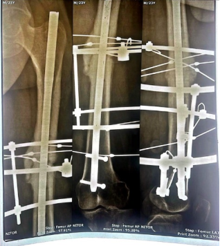



 Share This Article
 Twitter
 Linked In
 Facebook