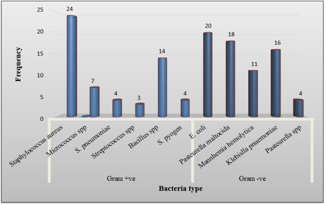
A crosswise analysis was carried out between December 2013 and March 2014 across two seasons to assess selected camel herds. The aim was to investigate how season affects disease occurrences through field surveys and collection of serum samples. A sum of 384 nasopharyngeal aspiration samples was gathered from camels. Primary biochemical tests were utilized to classify the isolates to the genus level. A multiple variable logistic regression analysis was employed to consider the relations among potential threats and immune response. The results showed a strong association between respiratory symptoms and the season, while no significant correlation was found with the districts, sex, or age (p>0.05). The most frequently identified Gram-positive bacteria were Staphylococcus aureus, Bacillus species, and Micrococcus species at rates of 19.0%, 11.1%, and 5.5%, respectively. The leading Gram-negative isolates included E. coli (15.9%), Pasteurella multocida (14.3%), and Klebsiella pneumoniae (12.7%). The study also identified a strong association between respiratory symptoms and the isolated bacteria, Mannhemia hemolytica. The study's findings suggest that various factors, such as the animals' nutritional and immune status, as well as environmental conditions, can transform the microbiota in the olfactory organ of healthy camel animals into pathogenic ones, leading to endogenous infections. The results of this study can inform the development of effective strategies for the prevention and control of camel respiratory diseases, ultimately improving their health and productivity highlighting the need for ongoing monitoring and management strategies for camel health.
| Published in | Animal and Veterinary Sciences (Volume 14, Issue 2) |
| DOI | 10.11648/j.avs.20261402.12 |
| Page(s) | 36-45 |
| Creative Commons |
This is an Open Access article, distributed under the terms of the Creative Commons Attribution 4.0 International License (http://creativecommons.org/licenses/by/4.0/), which permits unrestricted use, distribution and reproduction in any medium or format, provided the original work is properly cited. |
| Copyright |
Copyright © The Author(s), 2026. Published by Science Publishing Group |
Bacteria, Camels, Respiratory Diseases, Nasal Swabs
Variable | Category | n=384 | +ve (%) | OR | 95% CI | p value |
|---|---|---|---|---|---|---|
Districts | Moyale | 92 | 27 (29.3) | Ref | Ref | Ref |
Elewoye | 77 | 27 (35.0) | 0.76 | 0.40 - 1.47 | 0.428 | |
Gomole | 86 | 35 (40.7) | 0.59 | 0.31 - 1.10 | 0.101 | |
Yabelo | 66 | 18 (27.3) | 1.10 | 0.54 - 2.23 | 0.776 | |
Miyo | 63 | 19 (30.2) | 0.96 | 0.47 - 1.93 | 0.914 | |
Age | Adult | 210 | 74 (35.2) | Ref | Ref | Ref |
Young | 174 | 52 (30.2) | 1.28 | 0.83 - 1.97 | 0.253 | |
Sex | Female | 310 | 100 (32.2) | Ref | Ref | Ref |
Male | 74 | 26 (35.1) | 0.88 | 0.51 - 1.5 | 0.649 | |
Season | Rainy | 197 | 74 (37.5) | Ref | Ref | Ref |
Dry | 187 | 52 (27.8) | 1.50 | 1.00 - 2.38 | 0.046 | |
Clinical status | Six | 50 | 35 (70.0) | Ref | Ref | Ref |
Healthy | 334 | 91 (27.2) | 6.20 | 3.23 - 11.89 | 0.000 |
Variable | Category | n | +ve (%) | OR | 95% CI | p value |
|---|---|---|---|---|---|---|
Clinical status | Sick | 50 | 35 (70.0) | Ref | Ref | Ref |
Healthy | 334 | 91 (27.2) | 6.20 | 3.23 - 11.89 | 0.000 | |
Seasons | Rainy | 197 | 74 (37.5) | Ref | Ref | Ref |
Dry | 187 | 52 (27.8) | 1.6 | 1.07 – 2.67 | 0.024 |
BA | RS | I (%) | OR | 95% CI | p value |
|---|---|---|---|---|---|
Staphylococcus aureus | Sick | 12 (24) | Ref | Ref | Ref |
Healthy | 12 (3.5) | 8.47 | 3.55 - 20.18 | 0.000 | |
E. coli | Healthy | 15 (4.5) | Ref | Ref | Ref |
Sick | 5 (10) | 0.42 | 0.14 - 1.21 | 0.110 | |
Pasteurella maltocida | Healthy | 13 (3.8) | Ref | Ref | Ref |
Sick | 5 (10) | 0.50 | 0.15 - 1.58 | 0.241 | |
Micrococcus spp | Healthy | 7 (2.0) | Ref | Ref | Ref |
Sick | 0 (0) | 1 | |||
Mannhemia hemolytica | Sick | 6 (12) | Ref | Ref | Ref |
Healthy | 5 (1.5) | 8.97 | 2.62- 30.63 | 0.000 | |
S. pneumonia | Healthy | 3 (0.9) | Ref | Ref | Ref |
Sick | 1 (2) | 0.44 | 0.04 - 4.34 | 0.484 | |
Klebislla pnuemoniae | Healthy | 15 (4.5) | Ref | ||
Sick | 1 (2) | 2.29 | 0.29 - 17.77 | 0.426 | |
Streptococcus spp | Healthy | 3 (0.9) | Ref | Ref | Ref |
Sick | 0 (0) | 1 | |||
Pasteurella spp | Healthy | 3 (0.9) | Ref | Ref | Ref |
Sick | 1 (2) | 0.44 | 0.04 - 4.34 | 0.484 | |
Bacillus spp | Healthy | 10 (2.9) | Ref | Ref | Ref |
Sick | 4 (8) | 0.35 | 0.10 - 1.17 | 0.090 | |
S. pyogen | Healthy | 4 (1.1) | Ref | Ref | Ref |
Sick | 0 (0) | 1 |
BA | Bacterial Agents |
BHI | Brain Heart Infusion |
CI | Confident Interval |
OR | Odds Ratio |
RS | Respiratory Symptom |
YRVL | Yabelo Regional Veterinary Laboratory |
| [1] | Abbas, B.; Agab, H. A review of camel brucellosis. Preventive Veterinary Medicine. 2002, 55(1), 47-56. |
| [2] | Adney, D. R.; Clancy, C. S.; Bowen, R. A.; Munster, V. J. Camelid inoculation with Middle East respiratory syndrome coronavirus: experimental models of reservoir host infection. Viruses. 2020, 12(12), 1370. |
| [3] | Aebe, W. Traditional husbandry practices and major health problems of camels in the Ogaden. In: H. O. Anders (ed.), Nomadic People. 1991, 29, 21-30. |
| [4] | Al-Kharusi, A.; Elshafie, E. I.; Baqir, S.; Faraz, A.; Al-Ansari, A.; Burger, P.; Mahgoub, O.; Al-Kharousi, K.; Al-Duhli, H.; Al-Sinani, M.; Al-Hatali, R.; Roberts, D. Detection of Trypanosoma Infection in Dromedary Camels by Using Different Diagnostic Techniques in Northern Oman. Animals. 2022, 12(11), 1348. |
| [5] | Asante, J.; Noreddin, A.; El Zowalaty, M. E. Systematic review of important bacterial zoonoses in Africa in the last decade in light of the ‘One Health’concept. Pathogens. 2019, 8(2), 50. |
| [6] | Azizollah, E.; Bentol-hoda M.; Razieh, K. The aerobic bacterial population of the respiratory passageways of healthy dromedaries in Najaf-abbad abattoir, central Iran. Journal of Camelid Science. 2009, 2, 26-29. |
| [7] | Bekele, M. An epidemiological study of major camel diseases in the Borana lowland, Southern Ethiopia. Drylands Coordination Group (DCG) Report. 2010, 58: 1-4. |
| [8] | Bekele, T. Studies on the Respiratory Disease ‘Sonbobe’ in Camels in the Eastern Lowlands of Ethiopia. Tropical Animal Health and Production. 1999, 31, 333-345. |
| [9] | Bornstein, S.; Younan, M. Significant veterinary research on the dromedary camels of Kenya: Past and Present. Invited Review Bornstein Journal of Camelid Science. 2013, 6: 1-48. |
| [10] | Brown, JWL.; Coles, A.; Horakova, D. Association of Initial Disease-Modifying Therapy with Later Conversion to Secondary Progressive Multiple Sclerosis. JAMA. 2019, 321(2): 175-187. |
| [11] | Burger, P. A.; Ciani, E.; Faye, B. Old World camels in a modern world: a balancing act between conservation and genetic improvement. Animal Genetics. 2019, 50, 598-612 |
| [12] | Burger, PA. The history of Old World camelids in the light of molecular genetics. Tropical animal health and production. 2016, 48(5): 905-13. |
| [13] | Burt, J.; Paparella, F.; Al-Mansoori, N.; Al-Mansoori, A.; Al-Jailani, H. Causes and consequences of the 2017 coral bleaching event in the southern Persian/Arabian Gulf. Coral Reefs. 2019, 38, 567-589. |
| [14] | Cao, X.; Gu, L.; Gao, Z.; Fan, W.; Zhang, Q.; Sheng, J.; Zhang, Y.; Sun, Y. Pathogenicity and Genomic Characteristics Analysis of Pasteurella multocida Serotype A Isolated from Argali Hybrid Sheep. Microorganisms. 2024. 12(6), 1072. |
| [15] | Cawthorn, D. M., Hoffman, L. C. The role of traditional and non-traditional meat animals in feeding a growing and evolving world. Animal Frontiers, 2014, 4(4), 6-12. |
| [16] | Chauhan, R. S.; Kaushik, R. K.; Gupta, S. C.; Satija, K. C.; Kulsershtha, Prevalence of Different Diseases of Camels (Camelus dromedaries) in India. Camel News. 1986, 3: 10-14. |
| [17] | Coppock, D. L. The Borana Plateau of Southern Ethiopia: Synthesis of Pastoral Research, Development and Change 1980-91. Systems Study No. 5. International Livestock Centre for Africa, Addis Ababa. 1994, 374 pp. |
| [18] | De La Rocque, S.; Rioux, J. A.; Slingenbergh, J. Climate change: effects on animal disease systems and implications for surveillance and control. Rev Sci Tech. 2008, 27(2), 339-54. |
| [19] | Demeke, G. Prevalence of camel trypanosomes and factors associated with the disease occurrence in Liben district, Borena zone of Oromia region, Ethiopia. MSc Thesis. Free University of Berlin, FVM, Debre Zeit, Addis Ababa University, 1980. |
| [20] |
FAOSTAT. Structural data from agricultural censuses. Rome, Italy: FAO.
https://www.fao.org/faostat/en/#data/WCAD (Accessed 25 July 2022). |
| [21] | Feng, H.; Wu, K.; Yuan, Y.; Fang, M.; Wang, J.; Li, R.; Zhang, R.; Wang, X.; Ye, D.; Yang, Z.; Genomic analysis of Clostridium perfringens type D isolates from goat farms. Veterinary Microbiology. 2024, 294, 110105. |
| [22] | Feyissa, N.; Beyi, A. F.; Mamo, G.; Dawo, F.; Zewdu, E. Isolation and Identification of Aerobic Bacteria and Influenza Virus 3 from Nasal Passageway of Camels and Outbreak Investigation in Borana, Southern Ethiopia. J Vet Med Surg. 2020, 4, 24. |
| [23] | Fulton, R. W.; Cook, B. J.; Step, D. L.; Confer, A. W.; Saliki, J. T.; Payton, M. E.; Burge, L. J.; Welsh, R. D.; Blood, K. S. Evaluation of health status of calves and the impact on feedlot performance: assessment of a retained ownership program for postweaning calves. Canadian Journal of Veterinary Research. 2002. 66(3), 173. |
| [24] | Gardner, E. G.; Kelton, D.; Poljak, Z.; Van Kerkhove, M.; von Dobschuetz, S.; Greer, A. L. A case-crossover analysis of the impact of weather on primary cases of Middle East respiratory syndrome. BMC infectious diseases. 2019, 19, 1-10. |
| [25] | Gebru, M.; Tefera, G.; Dawo, F.; & Tessema, T. S. Aerobic bacteriological studies on the respiratory tracts of apparently healthy and pneumonic camels (Camelus dromedaries) in selected districts of Afar Region, Ethiopia. Tropical animal health and production. 2018, 50(3), 603-611. |
| [26] | Giro, A.; Jilo, K. Prevalence of camel trypanosomosis and associated risk factors in Arero district, Borena Zone, Southern Ethiopia. International Journal of Veterinary Science and Research. 2020, 6(1), 014-022. |
| [27] | Haile, A., Breeding strategy to improve Ethiopian Boran cattle for meat and milk production. 2011, V, 26. ILRI (aka ILCA and ILRAD). |
| [28] | Hughes, E. C.; Anderson, N. E. Zoonotic pathogens of dromedary camels in Kenya: a systematised review. Veterinary sciences. 2020, 7(3), 103. |
| [29] | Kamara, A.; Kirk, M.; Swallow, B. Property rights and land use change: implications for sustainable resource management in Borana, Southern Ethiopia. Journal of Sustainable Agriculture. 2005, 25(2), 45-61. |
| [30] | Kane, Y.; Wong, G.; Gao, G. F. Animal models, zoonotic reservoirs, and cross-species transmission of emerging human-infecting coronaviruses. Annual review of animal biosciences. 2023, 11(1), 1-31. |
| [31] | Kanchora, G.; Abebe, R. Prevalence of camel trypanosomosis and herders’ knowledge, attitude, and practices towards the disease in the pastoral area of southern Ethiopia. BMC Veterinary Research, 2024. 20(1), 400. |
| [32] | Kandeel, M. and Al-Mubarak, A. I. Camel viral diseases: current diagnostic, therapeutic, and preventive strategies. Frontiers in Veterinary Science. 2022. 9, 915475. |
| [33] | Kebede, F.; Gelaye, E. Studies on major respiratory diseases of Camel (Camelus dromedarius) in Northeastern Ethiopia. African Journal of Microbiology Research. 2010, 4(14), 1560-1564. |
| [34] | Kena, D. Review on camel production and marketing status in Ethiopia. Pastoralism. 2022. 12(1), 38. |
| [35] | Khan, N. A.; El-Menyar, A.; Al-Thani, H. The nature and consequences of camel-related injuries: a scoping review with special reference to Arab Middle Eastern countries. Injur. 2022, 53(4), 1319-1328. |
| [36] | Manzano, P.; Burgas, D.; Cadahía, L.; Eronen, J. T.; Fernández-Llamazares, Á.; Bencherif, S.; Holand, O.; Seitsonen, O.; Byambaa, B.; Fortelius, M.; Fernández-Giménez, M. E. Toward a holistic understanding of pastoralism. One Earth, 2021, 4(5), 651-665. |
| [37] | McCarthy, N.; Kamara, A.; Kirk, M. The effect of environmental variability on livestock and land-use management: The Borana plateau, southern Ethiopia. 2002. |
| [38] | Megersa, B.; Regassa, A.; Kumsa, B.; Abuna. F. Performance of camels (Camelus dromedarius) kept by pastoralists with different degrees of experience in camel keeping in Borana, Southern Ethiopia. Animal Science Journal. 2008. 79: 534-541. |
| [39] | Milkesa, A., Rufael, T., Kinfe, G., Belaineh, R., Bulbula, A., Cho, D., Naimuddin, M., Sori, T. and Dinka, H. A sero-epidemiological analysis of Coxiella burnetii infection and its risk factors in livestock from Addis Ababa, Adama, and Modjo abattoirs and pastoral areas of Oromia, Ethiopia. PLoS neglected tropical diseases. 2024, 18(7), p. e0012287. |
| [40] | Mirkena, T.; Walelign, E.; Tewolde, N.; Gari, G.; Abebe, G.; Newman, S. Camel production systems in Ethiopia: a review of literature with notes on MERS-CoV risk factors. Pastoralism. 2018, 8(1), 30. |
| [41] | Mohammed, T.; Woyessa, M; Brihanu, T.; Hiko, A. Cross sectional study of camel trypanosomosis in Babile District, Eastern Hararghe Zone, Oromia Regional State, Eastern Ethiopia. Academic Journal of Animal Diseases. 2015. 4(2): 112-117. |
| [42] | Moustafa, A. H. Study of some aerobic bacterial causes of respiratory affection in slaughtered camels in Dakahlia Governorate. Assiut Veterinary Medical Journal. 2004, 50(102), 95-105. |
| [43] | Nardone, A.; Ronchi, B.; Lacetera, N.; Ranieri, M. S.; Bernabucci, U., Effects of climate changes on animal production and sustainability of livestock systems. Livestock Science. 2010, 130(1-3), 57-69. |
| [44] | OIE. Manual of Diagnostic Tests and Vaccines for Terrestrial Animals. 5th Edition, Office International des Epizootics. Paris, 2004, pp. 409- 438. |
| [45] | Othieno, J.; Njagi, O.; Masika, S.; Apamaku, M.; Tenge, E.; Mwasa, B.; Kimondo, P.; Gardner, E.; Von Dobschuetz, S.; Muriira, J.; Adul, B. Knowledge, attitudes, and practices on camel respiratory diseases and conditions in Garissa and Isiolo, Kenya. Frontiers in Veterinary Science. 2022, 9, 1022146. |
| [46] | Owen, J. R.; Noyes, N.; Young, A. E.; Prince, D. J.; Blanchard, P. C.; Lehenbauer, T. W.; Aly, S. S.; Davis, J. H.; O’Rourke, S. M.; Abdo, Z.; Belk, K. Whole-genome sequencing and concordance between antimicrobial susceptibility genotypes and phenotypes of bacterial isolates associated with bovine respiratory disease. G3: Genes, Genomes, Genetics, 2017, 7(9), 3059-3071. |
| [47] | Palmer, D. The Marshall Illustrated Encyclopedia of Dinosaurs and Prehistoric Animals. illustrated edition, Marshall Editions, London, UK, 1999, pp. 274-277. |
| [48] | Parsons, L. M.; Somoskövi, Á.; Gutierrez, C.; Lee, E.; Paramasivan, C. N.; Abimiku, A. L.; Spector, S.; Roscigno, G.; Nkengasong, J. Laboratory diagnosis of tuberculosis in resource-poor countries: challenges and opportunities. Clinical microbiology reviews, 2011, 24(2), 314-350. |
| [49] | Quinn P. J.; Carter, M. E. Markey, B. K.; Carter, G. R. Clinical Veterinary Microbiology. Harcourt Publishers Limited, Edinburgh, London, UK, 1999, 641-648. |
| [50] | Rafu, O.; Tulu, D.; Negera, C. Camel Trypanosomosis in Yabelo and Gomole districts in Ethiopia: prevalence and associated risk factors based on parasitological examinations. Veterinary Medicine: Research and Reports. 2021, 87-94. |
| [51] | Refai, M. K. Brucellosis in animals and man in Egypt. Egypt J Vet Sci, 2003, 37, 1-31. |
| [52] | Sahoo, A. Camel: a fast-declining animal species but can strive with its unique climate resilience and ‘desert to medicine’application”. EC Veterinary science. 2020. 5, 43-57. |
| [53] | Sazmand, A.; Joachim, A.; Otranto, D. Zoonotic parasites of dromedary camels: so important, so ignored. Parasites & vectors. 2019. 12, 1-10. |
| [54] | Smith, J. A.; Doe, R. B. "The Impact of Climate Change on Agriculture. International Journal of Agricultural Research. 2020, 35(2), 123-135. |
| [55] | Swelum, A. A.; El-Saadony, M. T.; Abdo, M.; Ombarak, R. A.; Hussein, E. O.; Suliman, G.; Alhimaidi, A. R.; Ammari, A. A.; Ba-Awadh, H.; Taha, A. E.; El-Tarabily, K. A. Nutritional, antimicrobial and medicinal properties of Camel’s milk: A review. Saudi Journal of Biological Sciences, 2021 28(5): 3126-3136. |
| [56] | Tefera, M. Observations on the clinical examination of the camel (Camelus dromedarius) in the field. Tropical animal health and production, 2004, 36, 435-449. |
| [57] | Tesfaye, A.; Amaha, N. A review on cross-border livestock trade across dry land borders of Ethiopia: The trends and implications. Journal of Scientific and Innovative Research, 2018. 7(2), 36-42. |
| [58] | Teshome, D.; Tessema, T.; Kumsa, S.; Muluneh, B.; Sacchini, F.; Kumbe, A. Sero-prevalence of contagious bovine pleuropneumonia in dryland of Borana, southern Oromia, Ethiopia. Research in Veterinary Science. 2024, 166, p. 105100. |
| [59] | Thrusfield, M. Veterinary Epidemiology. 2nd Edition. Blackwell Science Ltd., London, 2005, pp. 178-198. |
| [60] | Trofin, A. E.; Ungureanu, E.; Trincă, L. C.; Fortună, M. E.; Eperjessy, D. B. Potential valorisation of Protobind 1000 as adsorbent for Pb2+ and Zn2+. Journal of Applied Life Sciences and Environment. 2022. Vol. 55, Issue 1(189): 31-44. |
| [61] | Volpato, G.; Howard, P. The material and cultural recovery of camels and camel husbandry among Sahrawi refugees of Western Sahara. Pastoralism, 2014, 4, 1-23. |
| [62] | Waktole, H.; Aden, M.; Ashenafi, H. Seroepidemiology of camel brucellosis in and around Dire Dawa, Eastern Ethiopia. Veterinary Medicine International, 2022, 1, 6624293. |
| [63] | Woolums, A. R.; Karisch, B. B.; Frye, J. G.; Epperson, W.; Smith, D. R.; Blanton Jr, J.; Austin, F.; Kaplan, R.; Hiott, L.; Woodley, T.; Gupta, S. K. Multidrug resistant Mannheimia haemolytica isolated from high-risk beef stocker cattle after antimicrobial metaphylaxis and treatment for bovine respiratory disease. Veterinary microbiology. 2018, 221, 143-152. |
| [64] | Woldearegay, Y. H.; Berhanu, M.; Mebratu, A. T. Study on management practices and production constraints of camel in Raya-Azebo district, Tigray, Northern Ethiopia. European Journal of Biological Sciences. 2015, 7(1), 01-06. |
| [65] | Wu, H., Guang, X., Al-Fageeh, M. B., Cao, J., Pan, S., Zhou, H., Zhang, L., Abutarboush, M. H., Xing, Y., Xie, Z. and Alshanqeeti, A. S., 2014. Camelid genomes reveal evolution and adaptation to desert environments. Nature communications, 5(1), p. 5188. |
| [66] | Yadamsuren, A; Daria, O.; Liu, S. The seasonal distribution of wild camels (Camelus ferus) in relation to changes of the environmental conditions in Mongolia. Open Journal of Ecology. 2019, 9(08) 293. |
| [67] | Zeleke, B. Status and growth trend of draught animals’ population in Ethiopia. Journal of Dairy, Veterinary & Animal Research, 2017, 6(1), 238-241. |
| [68] | Zhu, S.; Zimmerman, D.; Deem, S. L. A review of zoonotic pathogens of dromedary camels. EcoHealth, 2019. 16, 356-377. |
| [69] | Zorde, M. D. Bioprospecting in the Nepeta, Salvia, and Clinopodium Genera Under Controlled Growing Environments for Application in Natural Products and Cimex lectularius L. Repellency (Doctoral dissertation, Rutgers The State University of New Jersey, School of Graduate Studies) 2024. |
APA Style
Muluneh, B., Teshome, D., Tessema, T., Kumbe, A., Kumsa, S., et al. (2026). Epidemiological and Seasonal Dynamicity Studies on Major Respiratory Diseases of Camel in Selected Districts of Borana Zone, Southern Oromia, Ethiopia. Animal and Veterinary Sciences, 14(2), 36-45. https://doi.org/10.11648/j.avs.20261402.12
ACS Style
Muluneh, B.; Teshome, D.; Tessema, T.; Kumbe, A.; Kumsa, S., et al. Epidemiological and Seasonal Dynamicity Studies on Major Respiratory Diseases of Camel in Selected Districts of Borana Zone, Southern Oromia, Ethiopia. Anim. Vet. Sci. 2026, 14(2), 36-45. doi: 10.11648/j.avs.20261402.12
@article{10.11648/j.avs.20261402.12,
  author = {Bantayehu Muluneh and Dereje Teshome and Tamirat Tessema and Adem Kumbe and Sisay Kumsa and Gayo Ginbe},
  title = {Epidemiological and Seasonal Dynamicity Studies on Major Respiratory Diseases of Camel in Selected Districts of Borana Zone, Southern Oromia, Ethiopia},
  journal = {Animal and Veterinary Sciences},
  volume = {14},
  number = {2},
  pages = {36-45},
  doi = {10.11648/j.avs.20261402.12},
  url = {https://doi.org/10.11648/j.avs.20261402.12},
  eprint = {https://article.sciencepublishinggroup.com/pdf/10.11648.j.avs.20261402.12},
  abstract = {A crosswise analysis was carried out between December 2013 and March 2014 across two seasons to assess selected camel herds. The aim was to investigate how season affects disease occurrences through field surveys and collection of serum samples. A sum of 384 nasopharyngeal aspiration samples was gathered from camels. Primary biochemical tests were utilized to classify the isolates to the genus level. A multiple variable logistic regression analysis was employed to consider the relations among potential threats and immune response. The results showed a strong association between respiratory symptoms and the season, while no significant correlation was found with the districts, sex, or age (p>0.05). The most frequently identified Gram-positive bacteria were Staphylococcus aureus, Bacillus species, and Micrococcus species at rates of 19.0%, 11.1%, and 5.5%, respectively. The leading Gram-negative isolates included E. coli (15.9%), Pasteurella multocida (14.3%), and Klebsiella pneumoniae (12.7%). The study also identified a strong association between respiratory symptoms and the isolated bacteria, Mannhemia hemolytica. The study's findings suggest that various factors, such as the animals' nutritional and immune status, as well as environmental conditions, can transform the microbiota in the olfactory organ of healthy camel animals into pathogenic ones, leading to endogenous infections. The results of this study can inform the development of effective strategies for the prevention and control of camel respiratory diseases, ultimately improving their health and productivity highlighting the need for ongoing monitoring and management strategies for camel health.},
 year = {2026}
}
TY  - JOUR
T1  - Epidemiological and Seasonal Dynamicity Studies on Major Respiratory Diseases of Camel in Selected Districts of Borana Zone, Southern Oromia, Ethiopia
AU  - Bantayehu Muluneh
AU  - Dereje Teshome
AU  - Tamirat Tessema
AU  - Adem Kumbe
AU  - Sisay Kumsa
AU  - Gayo Ginbe
Y1  - 2026/05/13
PY  - 2026
N1  - https://doi.org/10.11648/j.avs.20261402.12
DO  - 10.11648/j.avs.20261402.12
T2  - Animal and Veterinary Sciences
JF  - Animal and Veterinary Sciences
JO  - Animal and Veterinary Sciences
SP  - 36
EP  - 45
PB  - Science Publishing Group
SN  - 2328-5850
UR  - https://doi.org/10.11648/j.avs.20261402.12
AB  - A crosswise analysis was carried out between December 2013 and March 2014 across two seasons to assess selected camel herds. The aim was to investigate how season affects disease occurrences through field surveys and collection of serum samples. A sum of 384 nasopharyngeal aspiration samples was gathered from camels. Primary biochemical tests were utilized to classify the isolates to the genus level. A multiple variable logistic regression analysis was employed to consider the relations among potential threats and immune response. The results showed a strong association between respiratory symptoms and the season, while no significant correlation was found with the districts, sex, or age (p>0.05). The most frequently identified Gram-positive bacteria were Staphylococcus aureus, Bacillus species, and Micrococcus species at rates of 19.0%, 11.1%, and 5.5%, respectively. The leading Gram-negative isolates included E. coli (15.9%), Pasteurella multocida (14.3%), and Klebsiella pneumoniae (12.7%). The study also identified a strong association between respiratory symptoms and the isolated bacteria, Mannhemia hemolytica. The study's findings suggest that various factors, such as the animals' nutritional and immune status, as well as environmental conditions, can transform the microbiota in the olfactory organ of healthy camel animals into pathogenic ones, leading to endogenous infections. The results of this study can inform the development of effective strategies for the prevention and control of camel respiratory diseases, ultimately improving their health and productivity highlighting the need for ongoing monitoring and management strategies for camel health.
VL  - 14
IS  - 2
ER  -
Yabello Pastoral and Dry Land Agriculture Research Center, Oromia Agricultural Research Institute, Yabello, Ethiopia
Yabello Pastoral and Dry Land Agriculture Research Center, Oromia Agricultural Research Institute, Yabello, Ethiopia
Yabello Pastoral and Dry Land Agriculture Research Center, Oromia Agricultural Research Institute, Yabello, Ethiopia
Information