Review Article | | Peer-Reviewed

Zebrafish in Personalized Breast Cancer Medicine

Published in Frontiers (Volume 6, Issue 4)
Received: 5 October 2026     Accepted: 15 October 2026     Published: 6 November 2026
Views:       Downloads:
Abstract

Breast cancer is a biologically heterogeneous disease in which substantial variation in molecular characteristics, tumor evolution, and treatment response presents major challenges for personalized therapeutic decision-making. Although genomic and molecular profiling have improved patient stratification, these approaches do not always accurately predict the functional response of an individual tumor to specific therapies. Consequently, there is increasing interest in functional precision-oncology models that can complement molecular profiling by directly assessing tumor behavior and drug sensitivity. Zebrafish (Danio rerio) have emerged as a promising platform for this purpose because of their rapid development, optical transparency, genetic tractability, relatively low experimental requirements, and suitability for medium- to high-throughput in vivo drug screening. In particular, patient-derived xenografts established in zebrafish, commonly referred to as zebrafish avatars (zAvatars), enable tumor cells obtained from individual patients to be engrafted into zebrafish embryos or larvae and subsequently challenged with candidate anticancer therapies. This review examines the development and application of zebrafish models in personalized breast cancer medicine, with emphasis on the transition from established breast cancer cell-line xenografts to patient-derived zAvatars. zAvatars show promising feasibility and preliminary patient-response concordance, but their predictive performance requires independent validation. Their clinical utility remains unestablished, and they should currently be considered investigational tools rather than validated guides for routine treatment selection. Future integration of zebrafish avatars with complementary precision-medicine technologies and larger prospective clinical studies may establish their role as rapid and scalable platforms for individualized breast cancer therapeutic selection. However, challenges including variable engraftment, incomplete representation of the human tumor microenvironment, drug pharmacokinetic differences, limited clinical validation, and the need for standardized response criteria currently constrain routine clinical implementation.

Published in Frontiers (Volume 6, Issue 4)
DOI 10.11648/j.frontiers.20260604.16
Page(s) 190-205
Creative Commons

This is an Open Access article, distributed under the terms of the Creative Commons Attribution 4.0 International License (http://creativecommons.org/licenses/by/4.0/), which permits unrestricted use, distribution and reproduction in any medium or format, provided the original work is properly cited.

Copyright

Copyright © The Author(s), 2026. Published by Science Publishing Group

Keywords

Breast Cancer, Zebrafish, Patient-Derived Xenografts, Personalized Medicine

1. Introduction
Breast cancer is a major global health challenge and remains the most commonly diagnosed cancer among women worldwide. In 2024, approximately 2.4 million women were diagnosed with breast cancer, and 694,000 died from the disease globally, highlighting the continuing burden of breast cancer despite substantial advances in early detection, molecular classification, and therapeutic management . Breast cancer is increasingly recognized as a biologically heterogeneous group of diseases rather than a single entity, with substantial variation in genetic alterations, molecular pathways, cellular composition, tumor microenvironment, metastatic potential, and therapeutic response . Clinically, classification according to hormone receptor and HER2 status, together with genomic and transcriptomic characteristics, has improved treatment selection and facilitated the development of endocrine therapies, HER2-targeted therapies, chemotherapy, and other molecularly targeted approaches . Nevertheless, patients with apparently similar clinicopathological and molecular characteristics may experience markedly different treatment outcomes, while intratumor heterogeneity can permit resistant subclones to survive treatment and contribute to disease recurrence and progression .
These challenges have contributed to increasing interest in functional precision medicine as a complementary approach to molecularly guided treatment. Molecular precision medicine primarily uses genomic, transcriptomic, proteomic, and other molecular characteristics to identify biomarkers or therapeutic targets that may guide treatment selection . Although these approaches have substantially improved patient stratification, the presence of a molecular alteration does not always translate into a predictable functional response to a particular therapy because treatment response may also be influenced by tumor heterogeneity, pathway interactions, clonal evolution, and the tumor microenvironment . Functional precision medicine therefore seeks to complement molecular profiling by experimentally assessing the response of patient-derived tumor material to candidate therapies.
Within this integrated precision-medicine framework, the zebrafish (Danio rerio) has emerged as an increasingly valuable model for cancer research and drug development because of its distinctive biological and experimental characteristics. An important development in this field has been the application of zebrafish to patient-derived xenograft (PDX) models, in which tumor cells or tissue obtained directly from a patient are transplanted into zebrafish embryos or larvae . These models can retain important biological characteristics of the patient's tumor and allow multiple therapeutic conditions to be assessed in vivo. Patient-derived zebrafish xenografts used to generate individualized functional information are commonly described as zebrafish avatars (zAvatars) and have the potential to bridge the gap between molecular characterization and functional assessment of treatment response. Rather than relying solely on the presence or absence of a molecular biomarker, zAvatars provide an opportunity to directly evaluate how an individual's tumor behaves when exposed to different therapeutic interventions .
Against this background, this review examines the development and application of zebrafish models in personalized breast cancer medicine, with particular emphasis on the transition from established breast cancer cell-line xenografts to patient-derived zebrafish avatars and integration with omics. It critically considers the biological and methodological basis of these models, their potential for functional therapeutic-response testing, and their integration with molecular profiling and emerging multi-omics and computational approaches. The review also evaluates key limitations affecting reproducibility and clinical translation, including tumor heterogeneity, engraftment variability, tumor–microenvironment differences, immune-system limitations, pharmacological differences, and the need for prospective clinical validation. By examining both the opportunities and limitations of zAvatars, this review aims to clarify their current and potential role as complementary tools within an integrated precision-oncology framework.
2. Methodology
2.1. Review Design and Scope
This article was designed as a narrative review of the development, applications, limitations, and translational potential of zebrafish models in personalized breast cancer medicine. Particular emphasis was placed on the progression from established breast cancer cell-line xenografts to patient-derived zebrafish xenografts (zPDXs), and their potential integration with molecular profiling and other precision-oncology approaches. The review also examined the biological and methodological limitations that influence reproducibility, treatment-response assessment, and clinical translation. The review employed a narrative and thematic approach rather than a formal systematic-review and meta-analysis framework. Accordingly, the objective was to critically synthesize the available experimental and translational evidence rather than to calculate a pooled estimate of treatment efficacy or diagnostic accuracy.
2.2. Search Strategy and Eligibility Criteria
Literature searches were conducted in PubMed, Scopus, Web of Science, and Google Scholar for publications dated January 2012 to August 2026. The final search was performed on the 11th of August 2026. The search strategy was developed around four complementary conceptual blocks: (i) zebrafish models; (ii) breast cancer; (iii) patient-derived tumour models; and (iv) precision oncology and therapeutic-response assessment. Search terms included “zebrafish,” “Danio rerio,” “breast cancer,” “breast carcinoma,” “patient-derived xenograft,” “patient-derived tumour,” “tumour avatar,” “zAvatar,” “personalized medicine,” “precision oncology,” “drug screening,” and “drug response.” Complementary combinations were used to identify both foundational studies of zebrafish breast cancer models and studies directly investigating patient-derived models or personalized therapeutic testing. This approach was intended to reduce the risk of excluding relevant publications that did not use all four concepts in their titles, abstracts, or indexing terms. Reference lists of relevant primary studies and review articles were also examined to identify additional publications not retrieved through the database searches. The records identified through database searching and supplementary reference-list screening were considered for relevance to the objectives of the review.
Publications were considered eligible when they addressed at least one of the following topics: (i) the use of zebrafish in breast cancer research; (ii) breast cancer cell-line xenograft models in zebrafish; (iii) patient-derived zebrafish xenografts or zebrafish avatars; (iv) functional assessment of anticancer drug responses using zebrafish models; or (v) the integration of zebrafish models with molecular profiling, multi-omics, computational approaches, or personalized therapeutic decision-making. Primary experimental studies were considered particularly relevant when they provided evidence concerning tumour engraftment, tumour growth, invasion or dissemination, therapeutic-response assessment, or comparison with patient outcomes or other experimental models. Review articles were considered for background, conceptual development, and identification of additional relevant primary studies. Publications were excluded when they did not address the review's objectives, did not provide relevant information on zebrafish or breast cancer models, or were otherwise outside the defined scope.
3. Breast Cancer Heterogeneity and the Need for Personalized Therapeutic Models
Breast cancer is a biologically heterogeneous disease characterized by substantial variation in molecular alterations, histopathology, tumor behavior, and therapeutic response . Clinically, breast cancers are broadly classified according to oestrogen receptor (ER), progesterone receptor (PR), and human epidermal growth factor receptor 2 (HER2) status . These categories have important therapeutic implications but encompass biologically diverse tumors. Molecular profiling has further identified intrinsic expression-based subtypes, including luminal A, luminal B, HER2-enriched, basal-like, and normal-like tumors, although these intrinsic classifications do not completely correspond to routine clinical receptor categories .
Luminal A tumors are generally hormone receptor-positive, HER2-negative, characterized by relatively lower proliferative activity and they frequently respond to endocrine therapy, although endocrine resistance can develop . Luminal B tumors are also commonly hormone receptor-positive but tend to exhibit higher proliferation and more aggressive clinical behavior, with some tumors showing HER2 expression or amplification . HER2-positive breast cancers are defined clinically by HER2 overexpression or amplification and can benefit substantially from HER2-directed therapies, although both primary and acquired resistance occur . Triple-negative breast cancer (TNBC) lacks ER, PR, and HER2 expression and is particularly heterogeneous, with limited conventional receptor-defined treatment targets and substantial variation in chemotherapy response. TNBC therefore illustrates the challenge of developing treatment strategies based solely on broad clinical classification . Importantly, intrinsic molecular subtypes are not synonymous with these clinical categories, and molecular classification provides additional biological resolution rather than a replacement for established pathological assessment.
Breast cancer heterogeneity also exists within individual tumors. Intratumor heterogeneity (ITH) describes the coexistence of genetically, epigenetically, and phenotypically distinct cellular populations within the same tumor . These populations may differ in receptor expression, proliferative capacity, metabolic state, metastatic potential, and treatment sensitivity. Therapeutic pressure can selectively eliminate sensitive populations while allowing resistant subclones to persist or expand, contributing to residual disease, recurrence, and treatment failure . Heterogeneity can also evolve spatially and temporally, such that primary tumors, metastatic lesions, and recurrent disease may exhibit different molecular and phenotypic characteristics, so that a single tumor biopsy may only provide a snapshot of a dynamic and spatially heterogeneous disease .
The tumor microenvironment (TME) adds another layer of heterogeneity. Breast tumors contain interacting populations of immune cells, cancer-associated fibroblasts, endothelial cells, stromal cells, and extracellular-matrix components . These components can influence proliferation, invasion, angiogenesis, metastatic dissemination, immune surveillance, and therapeutic response. In particular, stromal and immune interactions may provide survival signals that enable tumor cells to persist during treatment and contribute to therapeutic resistance . Consequently, tumor-cell genomic alterations alone may not fully explain the functional response of an individual tumor to therapy.
Precision oncology seeks to address this complexity by incorporating molecular and clinical characteristics into treatment selection . Genomic profiling can identify actionable alterations, while transcriptomic and other molecular assays can provide information on tumor biology, pathway activity, prognosis, and potential therapeutic vulnerabilities . This approach has already influenced treatment selection in defined breast cancer populations. Endocrine therapy is central to the management of hormone receptor-positive disease, HER2-directed therapies have substantially improved outcomes in HER2-positive disease, and selected tumors with defects in DNA-damage repair pathways may be eligible for targeted approaches such as PARP inhibition . However, the identification of a molecular alteration does not necessarily predict how an individual tumor will respond to a particular treatment. This is particularly evident in heterogeneous diseases such as TNBC, in which multiple molecular and cellular processes may contribute to treatment sensitivity or resistance . Similarly, tumors sharing the same receptor status may exhibit different responses because of differences in co-occurring genomic alterations, tumor-cell states, clonal composition, and TME interactions.
These limitations provide a rationale for complementing molecular characterization with functional therapeutic models. Patient-derived models can retain aspects of the biological characteristics of an individual tumor and allow candidate treatments to be evaluated experimentally. Patient-derived zebrafish avatars are particularly attractive in this context because they can provide relatively rapid in vivo assessment of tumor behavior and treatment-associated phenotypes. However, they should be considered complementary to pathological, molecular, and clinical assessment rather than replacements for these approaches. The potential value of zAvatars therefore lies in integrating patient-specific tumor biology with functional drug-response testing, while recognizing that their ability to reproduce the complete heterogeneity and microenvironment of human breast cancer remains an important limitation.
4. Zebrafish as a Model System for Breast Cancer Research
The zebrafish (Danio rerio) has emerged as an important vertebrate model for cancer research because it combines the biological advantages of a vertebrate organism with experimental features that facilitate rapid and relatively high-throughput investigation. Although early cancer studies primarily relied on rodents and established mammalian cell culture systems, zebrafish have increasingly been adopted for investigating tumor initiation, progression, angiogenesis, metastasis, tumor–microenvironment interactions, and therapeutic responses . The growing use of zebrafish in oncology is particularly relevant to precision medicine, where the ability to evaluate tumor behavior and treatment response rapidly is an important consideration.
4.1. Advantages of Zebrafish
One of the major advantages of zebrafish is their rapid embryonic development. Major developmental processes occur within a short period, allowing experimental manipulation and phenotypic assessment over days rather than the considerably longer periods often required in mammalian models . This characteristic is particularly useful for cancer xenograft studies, in which human tumor cells can be introduced into zebrafish embryos or larvae and subsequently monitored for proliferation, migration, invasion, angiogenesis, and dissemination. The relatively rapid experimental turnaround also creates opportunities for testing multiple therapeutic compounds or treatment combinations within a single experimental period .
The optical transparency of zebrafish embryos and larvae represents another important advantage for cancer research. Fluorescently labelled tumor cells can be visualized using fluorescence or confocal microscopy, allowing investigators to follow tumor-cell behavior in living animals. In breast cancer models, this enables direct observation of processes such as tumor-cell migration, intravasation, vascular dissemination, extravasation, and colonization of distant tissues . Such real-time visualization provides for the investigation of dynamic processes such as tumor-cell proliferation, migration, invasion, and dissemination, and allows treatment-associated changes to be monitored dynamically rather than inferred solely from endpoint measurements. This capacity for longitudinal imaging is particularly valuable for zebrafish avatars because therapeutic response can be converted into measurable phenotypic outcomes, including changes in tumor burden, fluorescence, migration, or metastatic behavior.
Zebrafish offer a practical platform for high-throughput drug screening because their small size, low maintenance requirements, and scalability allow multiple drugs, concentrations, and combinations to be evaluated simultaneously. Individual fish can serve as experimental units for rapid assessment of tumor growth and treatment response, making the model particularly useful for functional drug screening in precision oncology . This rapid functional assessment can complement genomic and molecular profiling by providing direct evidence of tumor sensitivity or resistance. However, interpretation of these responses must account for species-specific differences in physiology, metabolism, and drug pharmacology.
Another important characteristic is the genetic tractability of zebrafish. The zebrafish genome contains orthologues of many genes and signaling pathways involved in human cancer, while established transgenic and genome-editing technologies permit targeted manipulation of cancer-associated genes . These approaches have enabled researchers to model oncogenic pathways and investigate their contribution to tumor initiation and progression. Genetic manipulation can also be combined with live imaging, allowing changes in tumor phenotype to be observed in vivo . Thus, zebrafish provide an experimental platform in which genetic alterations can be linked directly to observable cancer phenotypes and therapeutic vulnerabilities. These genetic tools also enable mechanistic investigation of breast-cancer-associated pathways and experimental modelling of specific genetic alterations that may influence tumor development or therapeutic response.
The suitability of zebrafish for cancer research is further enhanced by their ability to support human tumor xenografts. At commonly used embryonic and larval stages, adaptive immunity is not yet functionally mature, allowing transplanted human tumor cells to establish with limited adaptive immune-mediated rejection . This developmental window, together with the optical transparency, small size, and rapid experimental timeframe of zebrafish larvae, enables human tumor growth, migration, angiogenesis, and dissemination to be monitored in vivo . Importantly, the use of patient-derived tumor material has extended zebrafish xenografts from conventional cancer modelling toward personalized medicine, allowing patient-specific tumor responses to candidate therapies to be evaluated in vivo . In breast cancer, these patient-derived xenografts provide the basis for zebrafish avatars (zAvatars), in which tumor material from an individual patient is used to generate a functional model for rapid therapeutic testing. zAvatars can therefore complement histopathological and molecular characterization by providing a functional measure of tumor sensitivity or resistance to treatment, although their interpretation remains constrained by incomplete representation of the human tumor microenvironment and species-specific differences in physiology, immunity, and drug pharmacology .
4.2. Limitations and Translational Challenges
Despite their potential for precision oncology, zebrafish models have important biological, pharmacological, and translational limitations that constrain the direct extrapolation of experimental findings to patients. A major limitation of zebrafish models is the physiological and pharmacological difference between zebrafish and humans. Zebrafish are maintained at a substantially lower body temperature than humans and differ in aspects of metabolism, physiology, developmental biology, and endocrine function. These differences can influence drug pharmacokinetics, pharmacodynamics, toxicity, and ultimately the interpretation of therapeutic responses observed in zebrafish models . These differences are particularly relevant when zebrafish are used for drug screening. Although zebrafish permit relatively rapid comparison of different drugs, concentrations, and treatment combinations, drug exposure in the fish does not necessarily reproduce the absorption, distribution, metabolism, and elimination profiles observed in patients. Consequently, apparent drug sensitivity or toxicity in zebrafish should be interpreted as a functional screening result rather than a direct equivalent of human clinical response . The interpretation of xenograft responses must also account for experimental conditions that may influence drug activity and tumor behavior in the zebrafish host . The physiological and pharmacological differences between zebrafish and humans therefore do not negate the value of the model but define the context in which its findings should be interpreted. Zebrafish are particularly valuable for identifying candidate therapeutic responses and prioritizing compounds for further investigation, whereas confirmation of pharmacokinetic, pharmacodynamic, and toxicity profiles requires complementary mammalian studies and, ultimately, clinical evaluation .
The tumor microenvironment (TME) is an important limitation when interpreting results from zebrafish xenograft models. Breast cancer progression and therapeutic response are shaped by interactions among tumor cells, immune cells, cancer-associated fibroblasts, endothelial cells, extracellular matrix, and soluble signaling molecules. Conventional larval zPDX models reproduce some of these interactions but do not fully recapitulate the human TME . Early zebrafish larvae possess functional innate immune components, including macrophages and neutrophils, but adaptive immunity is incompletely developed at the stages commonly used for human tumor transplantation. This facilitates engraftment of human tumor cells but limits modelling of human T-cell, B-cell, antigen-specific, and immune-memory responses . Thus, zPDX models can provide information on tumor–innate immune interactions while providing a limited representation of the adaptive immune environment.
The stromal and vascular compartments present additional limitations. Patient-derived tumor cells transplanted into zebrafish interact predominantly with host-derived stromal and endothelial cells rather than the patient's original microenvironment. Although this enables investigation of angiogenesis, invasion, and dissemination, cross-species differences in cellular signaling, cytokine networks, extracellular matrix, and endothelial interactions may affect tumor behavior and drug response . Consequently, responses observed in zAvatars may not fully reproduce treatment effects that depend on the human-specific immune or stromal environment. These limitations are particularly relevant to immunotherapy. Checkpoint inhibitors, cancer vaccines, and cellular therapies depend on coordinated interactions involving human antigen presentation, T cells, immune checkpoints, and tumor-associated immune populations that are not adequately represented in conventional larval xenografts. Accordingly, zAvatars should be interpreted cautiously when used to predict responses to therapies whose efficacy is strongly immune-dependent .
Emerging approaches, including transplantation at later developmental stages, genetically modified immune-deficient or immune-modified zebrafish, and humanized immune systems, may improve modelling of tumor–immune interactions . However, these approaches remain technically challenging and do not yet reproduce the complete human breast cancer TME. Therefore, zAvatars are best viewed as functional models that complement, rather than replace, human tumor profiling and other preclinical systems. Integration with single-cell, spatial, and multi-omics approaches may provide a more comprehensive understanding of tumor–immune and tumor–stromal interactions and improve interpretation of functional drug-response results.
Patient-derived zebrafish xenografts offer greater potential for personalized medicine because tumor material obtained directly from patients can retain clinically relevant genetic and phenotypic characteristics. However, their clinical relevance is accompanied by practical and biological variability. The establishment of patient-derived models may be affected by tumor-cell viability, engraftment efficiency, tumor composition, and sample-processing procedures . These factors can influence whether the resulting xenograft adequately represents the characteristics of the original tumor . This creates an important trade-off between reproducibility and patient specificity. Established cell-line xenografts are generally more homogeneous and reproducible, but they may not capture the cellular diversity and heterogeneity of individual patient tumors. In contrast, patient-derived xenografts can better preserve clinically relevant characteristics of the source tumor but are more susceptible to variability during sample collection, processing, transplantation, and engraftment . Thus, increased patient specificity may be accompanied by greater experimental variability.
Tumor heterogeneity presents an additional challenge for personalized medicine because an individual tumor may contain multiple genetically and phenotypically distinct subpopulations. Consequently, the tumor material successfully engrafted in zebrafish may not always represent the complete heterogeneity of the original tumor, potentially influencing the treatment response observed in the model . Spatial and temporal differences between primary, recurrent, and metastatic tumor samples may further contribute to this problem .
Therefore, while patient-derived zebrafish models may provide a more individualized platform than conventional cell-line xenografts, their predictive value depends on successful engraftment and adequate preservation of clinically relevant tumor characteristics. Improving methods for tumor sampling, processing, engraftment, and characterization will be important for increasing the reproducibility and clinical interpretability of these models.
5. Cell Lines to Patient-Derived Zebrafish Avatars in Breast Cancer
The application of zebrafish in breast cancer research has evolved from the use of established cancer cell lines toward increasingly patient-specific xenograft systems. This progression reflects a broader shift in cancer research from models designed primarily to investigate general mechanisms of tumor biology toward models capable of capturing individual tumor characteristics and therapeutic responses. Zebrafish xenografts can broadly be divided into models based on established breast cancer cell lines and those generated using patient-derived tumor material . While cell-line xenografts provide reproducible and experimentally tractable systems, patient-derived xenografts have greater potential for modelling interpatient heterogeneity and supporting personalized therapeutic testing . In this context, cell-line xenografts use established cancer cell lines, while zebrafish PDX models/zAvatars use patient-derived tumor cells for functional drug-response testing .
5.1. Cell-Line-Based Breast Cancer Xenografts
Cell lines representing different molecular phenotypes, including estrogen receptor-positive, HER2-positive, and triple-negative breast cancer, can be labelled with fluorescent proteins and introduced into zebrafish embryos or larvae. Subsequent imaging enables investigators to quantify tumor-cell proliferation, migration, invasion, and dissemination within the living host .
Early studies established the utility of these models for assessing tumor-cell behavior in vivo, particularly migration, invasion, extravasation, angiogenesis, and metastatic dissemination. For example, MDA-MB-231 xenografts have been used to investigate the molecular mechanisms regulating breast cancer invasion and metastasis and to evaluate pathway-directed interventions. Drabsch et al., demonstrated that inhibition of transforming growth factor-β (TGF-β) signaling reduced the invasive and metastatic behavior of breast cancer cells in zebrafish xenografts, illustrating the potential of the model for testing anti-metastatic strategies. Similarly, other studies using MDA-MB-231 and related invasive breast cancer cell lines have exploited the optical accessibility of zebrafish larvae to quantify tumor-cell dissemination and vascular interactions following pharmacological intervention.
Zebrafish xenografts have also been used to characterize therapeutic responses across breast cancer models with distinct molecular and phenotypic backgrounds. Rebelo de Almeida et al., used triple-negative breast cancer (TNBC) cell-line xenografts, including Hs578T and MDA-MB-468, alongside patient-derived xenografts to evaluate responses to the anti-angiogenic agent bevacizumab. Treatment produced model-dependent effects on tumor size and apoptosis and reduced tumor-associated angiogenesis, while patient-derived xenografts exhibited distinct response profiles, illustrating the capacity of the model to capture intertumoral heterogeneity. Similarly, a study using Hs578T, MDA-MB-468, and HCC1937 TNBC xenografts demonstrated heterogeneous responses to the PARP inhibitor Olaparib . Although Hs578T xenografts showed little response to olaparib monotherapy, MDA-MB-468 and HCC1937 xenografts exhibited treatment-associated effects on apoptosis and/or tumor size, despite differences in their BRCA and homologous recombination repair backgrounds. Cell-line-based approaches have also been complemented by broader compound-screening strategies. Y. Li et al., screened 502 natural compounds using zebrafish embryos and compared their toxicity profiles with cytotoxicity measured in MCF-7 breast cancer cells. The study identified compounds with concordant activity across the zebrafish and MCF-7 assays, demonstrating the potential of zebrafish as an efficient in vivo platform for anticancer compound screening. Table 1 below outlines recent studies utilizing zebrafish cell lines as well as the important findings from the studies.
Collectively, these studies demonstrate several important applications of cell-line-based zebrafish xenografts: mechanistic investigation of tumor progression, assessment of invasion and metastatic dissemination, evaluation of angiogenesis, and rapid screening of candidate therapeutic agents. Their principal advantage is experimental reproducibility because established cell lines provide relatively homogeneous and readily expandable tumor material, allowing treatment effects to be compared under standardized conditions. However, this same characteristic represents an important limitation for personalized medicine. Cell lines may undergo genetic and phenotypic changes during prolonged culture and generally do not reproduce the full intertumoral and intratumoral heterogeneity of individual patient tumors. Consequently, responses observed in a standardized cell-line xenograft demonstrate the feasibility of a therapeutic or biological mechanism but cannot necessarily be interpreted as patient-specific treatment predictions.
This distinction provides an important rationale for the development of patient-derived zebrafish xenografts and zAvatars. Whereas cell-line models provide a controlled and reproducible platform for establishing drug-response phenotypes, patient-derived models introduce tumor material directly obtained from individuals and therefore have greater potential to capture clinically relevant heterogeneity. However, this increased patient specificity also introduces variability in tumor-cell viability, sample composition, engraftment, and preservation of the original tumor phenotype. Thus, the progression from cell-line xenografts to zAvatars represents a shift from reproducible model-based drug testing toward patient-specific functional phenotyping. The key translational question is whether the additional biological information retained by zAvatars can produce therapeutic-response measurements that are reproducible and predictive of actual patient outcomes.
Table 1. Studies utilizing zebrafish cell line in breast cancer.

Breast cancer cell line

Xenograft model/site

Intervention

Main outcome assessed

Key finding/application

Study

MDA-MB-231; MCF10A-derived invasive lines

Zebrafish embryos; cells introduced into circulation/duct of Cuvier

TGF-β receptor kinase inhibitors, MMP inhibition, Smad4 knockdown

Migration, invasion and metastatic dissemination

TGF-β pathway inhibition reduced invasion and metastasis, demonstrating the utility of the model for mechanistic studies and screening anti-metastatic therapies.

Drabsch et al., 2013

Hs578T, MDA-MB-468

Perivitelline-space xenografts

Bevacizumab

Tumor size, apoptosis, angiogenesis and vascular infiltration

Hs578T xenografts showed increased apoptosis and approximately 40% reduction in tumor size, together with reduced vascular density/infiltration.

Rebelo de Almeida et al., 2020

Hs578T, MDA-MB-468, HCC1937

Perivitelline-space xenografts

Olaparib ± ionizing radiation

Tumor response, cell death and DNA-damage-associated phenotypes

Different TNBC cell lines showed different responses to olaparib and radiation, illustrating the capacity of zebrafish xenografts to detect heterogeneous therapeutic responses.

Varanda et al., 2020

MCF-7

Zebrafish xenograft/drug-screening model

Library of 502 natural compounds

Toxicity, apoptosis and p53 activation

Zebrafish screening identified 59 toxic compounds; 21 overlapped with compounds identified by the MCF-7 MTT assay, demonstrating complementary in vivo drug-screening capacity.

Y. Li et al., 2012

MDA-MB-231

Embryonic zebrafish xenografts

Multiple pathway-targeted compounds

Migration, invasion, extravasation and metastasis

MDA-MB-231 xenografts have been repeatedly used to evaluate anti-invasive and anti-metastatic compounds, including TGF-β-, CXCR4-, MMP- and other pathway-directed interventions.

Rong et al., 2025

MDA-MB-231

Perivitelline-space xenografts

METTL3 inhibitor ± cisplatin/olaparib

Extravasation and metastatic behavior

Treatment altered the ability of MDA-MB-231 cells to extravasate, supporting the use of the model for testing mechanisms of therapeutic sensitization.

Cesaro et al., 2023

MDA-MB-231, Hs578T, MDA-MB-468 and others

Various zebrafish xenograft configurations

Olaparib, bevacizumab and other candidate agents

Tumor proliferation, apoptosis, angiogenesis, migration and tumor burden

Collectively demonstrate the use of cell-line xenografts for screening targeted and cytotoxic interventions in breast cancer.

Wawruszak et al., 2023

5.2. Zebrafish Patient-derived Xenografts
Patient-derived xenografts (PDXs) are patient-derived tumor material transplanted into an animal model, and they represent an important approach for preserving clinically relevant features of human tumors in experimental models . In zebrafish, patient-derived tumor cells can be introduced into embryos or larvae, enabling rapid assessment of tumor growth, dissemination, angiogenesis, and treatment response, and are termed zPDX or zAvatars.
An important early demonstration of the personalized potential of zPDXs was provided by Rebelo de Almeida et al . The investigators first established zebrafish xenografts using breast cancer cell lines, including the triple-negative breast cancer (TNBC) lines Hs578T and MDA-MB-468, and showed that bevacizumab produced heterogeneous effects on tumor growth, apoptosis, angiogenesis, and micrometastatic dissemination. They subsequently extended the approach to patient-derived breast cancer samples obtained from surgical specimens, generating zAvatars. These patient-derived models also exhibited heterogeneous responses to bevacizumab, with treatment-associated changes in tumor size and apoptosis and effects on tumor-associated vasculature and micrometastasis. Importantly, the investigators performed proof-of-concept comparisons between clinical responses in patients and responses of their matched zAvatars, demonstrating the potential of the model to reproduce patient-specific therapeutic responses within a short experimental timeframe. However, the study was exploratory and involved a limited number of patient-derived samples; consequently, it did not provide robust estimates of clinical sensitivity, specificity, or predictive value. The findings therefore established an important proof of concept for personalized zebrafish drug-response testing rather than definitive clinical validation.
A step forward in the application was further provided by Rodriguez et al , who generated zebrafish tumor-derived xenografts (ZTX) from a panel of 20 breast cancer PDX models. Nineteen of the 20 PDX models successfully implanted in zebrafish larvae, corresponding to a 95% implantation rate. The extent of tumor-cell dissemination in zebrafish also increased with the clinical stage associated with the originating PDX models. When responses to docetaxel and trastuzumab were compared with those observed in the corresponding mouse PDX models, 14 of 18 evaluable treatment comparisons were concordant. These findings demonstrated that breast cancer PDX-derived zebrafish xenografts could reproduce clinically relevant dissemination phenotypes and treatment-response patterns observed in another preclinical model. However, because the comparator was the mouse PDX rather than the individual donor patient's clinical response, the study did not establish patient-level predictive accuracy. Furthermore, the findings were reported as an AACR conference abstract, limiting the amount of methodological and diagnostic-accuracy information available.
Patient-derived zebrafish models have also been extended to metastatic breast cancer by using circulating tumor cells (CTCs). Reinhard et al., developed the DanioCTC workflow to enrich rare CTCs from metastatic breast cancer patients using diagnostic leukapheresis, microfluidic enrichment, flow cytometry, and cell isolation before xenotransplantation into zebrafish embryos. In a pilot patient-derived experiment, CTCs were successfully engrafted and their dissemination could be monitored in vivo, with cells primarily detected in the head and trunk regions of the embryos. Although this approach does not constitute a treatment-response zAvatar, it demonstrates that zebrafish can support functional analysis of rare patient-derived metastatic cells and provides an alternative route for generating patient-specific xenograft models when solid tumor tissue is difficult to obtain.
The most direct recent evidence for the predictive application of breast cancer zAvatars was reported by Mendes et al . The investigators developed zAvatars from 52 patient breast cancer samples obtained from core-needle biopsies, surgical specimens, ascites, and malignant pleural effusions. The cohort included early- and advanced-stage tumors representing Luminal, HER2-enriched, and triple-negative breast cancer. Tumor cells were injected into two-day-post-fertilization zebrafish embryos without prior in-vitro expansion. Although 51 of 52 samples successfully implanted, the overall take rate was 47.2%, indicating substantial variation in engraftment efficiency among patient samples. Importantly, the xenografts retained several clinically relevant breast cancer biomarkers and permitted assessment of tumor dissemination and other phenotypic characteristics. The authors subsequently evaluated the predictive potential of the model in 18 patients for whom matched clinical treatment-response data were available. zAvatars were exposed to the same chemotherapy regimen administered to the corresponding donor patient, and zebrafish responses were compared with clinical outcomes. The study reported concordance in all 18 evaluable cases when patients were classified broadly as treatment-sensitive or treatment-resistant. The investigators also used apoptosis-based measurements and clinical response categories to evaluate the relationship between zAvatar and patient responses. This represents an important progression from demonstrating successful engraftment to directly testing whether a patient-derived zebrafish model can reproduce an individual patient's treatment response.
Nevertheless, the reported 18/18 concordance should be interpreted as an encouraging proof-of-concept rather than definitive evidence of clinical diagnostic accuracy. The matched cohort was small, and the analysis primarily discriminated sensitive from resistant disease rather than providing robust estimates of performance across the full spectrum of clinical response. In addition, the investigators reported limitations in distinguishing pathological complete response from partial response. Consequently, larger prospective cohorts and independent validation are required before zAvatar testing can be considered a clinically validated predictive assay. The findings from the application are summarized in Table 2 below.
Taken together, the available evidence supports several conclusions. First, zAvatars can reproduce clinically relevant features of breast cancer, including tumor growth, invasion, angiogenesis, and treatment sensitivity. Second, the most clinically relevant evidence currently comes from studies in which patient-derived material is tested against the treatment received by the donor patient. Third, the evidence remains insufficient to establish universal predictive accuracy across breast cancer subtypes and treatment classes. Studies differ substantially in sample size, tissue source, transplantation site, model generation, treatment exposure, response definition, and comparator. In addition, many studies report implantation or biological activity rather than clinical treatment prediction. A standardized framework for reporting these variables would therefore improve comparability between studies.
Accordingly, the translational value of zAvatars should be considered as an emerging evidence base rather than a clinically validated diagnostic technology. Future investigations should use larger prospective cohorts, predefined response thresholds, blinded comparison with patient outcomes, independent validation cohorts, and standardized measures of sensitivity, specificity, positive predictive value, and negative predictive value. Such studies would help determine whether the rapid functional information generated by zAvatars provides predictive value beyond conventional clinicopathological and molecular biomarkers.
Table 2. Recent studies in the application of zAvatars and the main findings.

Cancer/model

Patient/sample number

Tumor source

Xenograft approach/site

Treatment tested

Main endpoint

Key finding

Sensitivity/specificity

Important limitation

Study

Breast cancer PDX-derived ZTX

20 PDX models; 19 implanted

Established mouse BC PDX material

Zebrafish larvae; xenotransplant model

Docetaxel; trastuzumab

Implantation, dissemination, treatment response

19/20 models implanted; 14/18 treatment comparisons concordant with mouse PDX outcomes

NR

Conference abstract; comparator was mouse PDX rather than matched patient outcome; detailed diagnostic-accuracy measures unavailable

Rodriguez et al., 2021

Cancer xenograft/zPDX; includes BC applications

Individual patient samples plus experimental models

Patient-derived and xenograft material

Zebrafish xenografts

Bevacizumab

Tumor response, angiogenesis, micrometastasis

Demonstrated heterogeneous responses and potential for patient-matched response assessment

NR

Proof-of-concept; limited clinical validation

Rebelo de Almeida et al., 2020

Breast cancer zAvatar

18 patients for matched clinical-response analysis

Core biopsies, surgical specimen, pleural effusion, ascites

2-dpf embryos; fluorescent tumor-cell xenografts

Patient-matched chemotherapy regimens

zAvatar sensitivity/resistance versus patient response

18/18 concordant for sensitive/resistant classification

Formal sensitivity/specificity should not be inferred from 18/18 concordance

Small cohort; could not discriminate pCR from PR; requires independent validation

Mendes et al., 2025

Breast cancer zAvatar establishment cohort

52 patient samples

Surgical samples, core biopsies, ascites, pleural effusions

2-dpf embryos

—

Engraftment/biomarker preservation

51/52 successfully implanted; overall implantation rate 47.2%

Not applicable

Implantation efficiency was heterogeneous; not all successfully engrafted samples were suitable for matched treatment prediction

Mendes et al., 2025

Metastatic breast-cancer patient-derived circulating tumor cells (CTCs)

Pilot; CTCs from one metastatic breast-cancer patient successfully xenografted

Peripheral blood/CTCs enriched using diagnostic leukapheresis and microfluidic/flow-based isolation

CTCs injected into zebrafish embryos; dissemination monitored in vivo

None—metastasis/dissemination study

CTC engraftment and dissemination

Patient-derived CTCs were successfully xenografted and localized mainly to head/trunk regions; established a workflow for studying rare CTCs in vivo

Not applicable

Pilot study; focused on dissemination rather than treatment prediction; very limited patient number

Reinhardt et al., 2023

6. Generating zAvatars
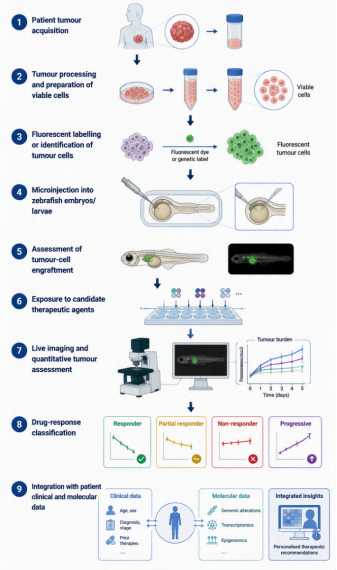
zAvatars are established by xenografting viable tumor cells obtained directly from an individual patient into zebrafish embryos or larvae, thereby creating an in vivo platform for functional assessment of tumor behavior and therapeutic response. The process begins with acquisition of a fresh tumor specimen, obtained, where clinically appropriate, from surgical resection or biopsy. To preserve the biological characteristics and viability of the tumor, samples should be processed promptly under sterile conditions. Tumor tissue is mechanically minced and may undergo enzymatic dissociation to generate a single-cell suspension, followed by removal of debris and assessment of cell viability . Depending on the experimental objective, tumor cells can be labelled with fluorescent dyes or derived from fluorescently tagged tumor material, allowing subsequent visualization and quantification by fluorescence microscopy . In some protocols, the tumor-cell population is further characterized using histological, immunophenotypic, or molecular markers to confirm its tumor origin and maintain correspondence with the patient's tumor profile . However, successful avatar generation may be limited by insufficient tissue, low cell viability, necrosis, or low tumor-cell yield, particularly when samples are small or obtained from limited biopsy material.
The resulting viable tumor-cell suspension is microinjected into appropriately staged zebrafish embryos or larvae . Xenotransplantation is commonly performed during early larval development because the animals are small, optically accessible, and amenable to relatively high-throughput manipulation. Depending on the biological question, tumor cells may be introduced into anatomical sites such as the yolk sac, perivitelline space, or other locations that support tumor-cell survival and dissemination. Following injection, embryos are maintained under controlled conditions to permit engraftment. Successful engraftment is typically confirmed by fluorescence or microscopic imaging, with exclusion or removal of embryos showing unsuccessful injection, excessive tissue damage, or inadequate tumor-cell establishment. Importantly, the number of injected cells, injection site, developmental stage, incubation period, drug exposure, and criteria for successful engraftment and response measurement should be standardized within a study because these variables can substantially influence xenograft growth and treatment-response measurements.
Once engraftment has been established, individual tumor xenografts can be exposed to candidate anticancer agents or clinically relevant drug combinations. Treatments may be administered through the water or other experimental routes appropriate to the compound and zebrafish developmental stage. Untreated or vehicle-treated xenografts are generally included as controls. Following treatment, serial live imaging is used to quantify changes in tumor burden, commonly through measurement of fluorescence intensity, fluorescent tumor area, or three-dimensional tumor volume where imaging resolution permits. Additional phenotypic endpoints may include tumor-cell proliferation, apoptosis, migration, invasion, vascular interactions, and dissemination to distant anatomical sites. Image-analysis software can be used to obtain quantitative measurements from individual larvae, enabling comparison of treatment effects across multiple patient-derived xenografts
The resulting phenotypes can be used to classify individual tumors as relatively sensitive or resistant to particular therapeutic agents or combinations. Importantly, the value of the zAvatar lies not simply in demonstrating that a drug inhibits tumor growth, but in comparing the functional response of a patient's tumor-derived xenograft with the patient's molecular characteristics and, where available, subsequent clinical response. Such integration can combine functional drug-response information with conventional biomarkers, including hormone-receptor and HER2 status, genomic alterations, transcriptomic profiles, and other molecular features. For clinical application, the workflow must also generate treatment-response information within a timeframe compatible with the patient's treatment-decision window.
The development of a patient-specific zebrafish model can be conceptualized as a multistep process outlined in the figure below.
Figure 1. Schematic illustration of the zebrafish avatar model from patient tumor acquisition, Tumor processing and preparation of viable cells, fluorescent labelling or identification of tumor cells, Microinjection into zebrafish embryos/larvae, Assessment of tumor-cell engraftment, Exposure to candidate therapeutic agents, Live imaging and quantitative tumor assessment, Drug-response classification and Integration with patient clinical and molecular data.
7. Frontier in the Utilization of zAvatars in Breast Cancer
The field of zAvatars is transitioning from proof-of-concept xenografting toward their potential use as functional models for individualized therapeutic decision-making. Current studies demonstrate that patient-derived breast tumor cells can be established in zebrafish and evaluated for treatment-associated changes in tumor burden, proliferation, apoptosis, migration, invasion, angiogenesis, and metastatic dissemination. However, the major frontier is no longer simply whether a patient tumor can engraft and respond to a drug in zebrafish, but whether these functional responses can be measured reproducibly and translated into clinically meaningful predictions of therapeutic benefit. This distinction is critical because a measurable phenotype in a zebrafish model does not necessarily represent clinical sensitivity or resistance in the corresponding patient.
A major gap concerns the standardization and validation of therapeutic-response monitoring. Differences in tumor-cell viability, sample composition, engraftment efficiency, implantation site, tumor burden, imaging procedures, treatment exposure, and criteria used to define response can introduce substantial variability between studies. In addition, patient tumors are heterogeneous, and small biopsy-derived samples may not fully represent the clonal and spatial diversity of the primary tumor or metastatic disease . Future zAvatar platforms should therefore establish standardized criteria for tumor viability, engraftment, baseline tumor burden, treatment conditions, imaging, quantitative endpoint measurement, and response classification. Response thresholds should be defined prospectively and, where possible, evaluated against clinical outcomes such as objective response, recurrence, progression, or pathological response. Such validation would allow zAvatars to progress from descriptive drug-screening models toward quantitatively interpretable systems for therapeutic monitoring.
The next developmental frontier for zAvatars centers on overcoming the microenvironmental limitations of conventional larval models. Despite providing a rapid, high-throughput platform for live imaging and drug screening, larval systems fail to fully replicate the human tumor microenvironment (TME). Engrafted patient-derived tumor cells interact primarily with host zebrafish stroma, endothelium, and immune cells. This introduces critical cross-species variations—particularly in cytokine signaling, extracellular matrix dynamics, vascularization, and pharmacokinetics—that ultimately compromise the fidelity of translational drug responses . Early larval zebrafish possess functional innate immune components, whereas adaptive immunity is incompletely developed at commonly used xenografting stages. Consequently, conventional zAvatars have limited capacity to reproduce human T-cell- and B-cell-mediated responses and may be particularly constrained for evaluating immunotherapies that depend on human antigen presentation, immune checkpoints, or tumor-specific immune recognition . Emerging approaches involving later developmental stages, immune-modified or immune-deficient fish, and humanized immune systems may help address these limitations, but their ability to reproduce the human tumor microenvironment remains to be established. Thus, future development should focus not on creating a single model that reproduces every component of human breast cancer, but on defining which biological questions zAvatars can answer reliably and integrating them with complementary organoid, mammalian, and molecular approaches.
A closely related frontier is the integration of zAvatar functional phenotypes with multi-omics data. Genomic, transcriptomic, proteomic, metabolomic, single-cell, and spatial approaches can provide increasingly detailed descriptions of tumor heterogeneity and the molecular states associated with treatment response . However, molecular characterization alone does not establish whether a tumor will functionally respond to a particular therapy . Conversely, zAvatars can provide a rapid functional phenotype but do not, by themselves, explain the molecular mechanisms underlying sensitivity or resistance. Integrating these two dimensions could therefore establish a complementary framework in which molecular profiling identifies candidate determinants of therapeutic response, while zAvatars provide an experimentally testable functional readout of those predictions. The emerging framework can be conceptualized as patient tumor profiling → multi-omics characterization → zAvatar-based functional drug testing → integration with clinical response. Genomic and transcriptomic data may identify mutations, copy-number alterations, gene-expression programs, or molecular pathways associated with treatment response, while single-cell and spatial approaches could resolve resistant cellular populations and spatially restricted tumor or microenvironmental states. The corresponding zAvatar could then be used to test whether these molecular features translate into an observable phenotype following exposure to individual drugs or drug combinations. This functional-molecular integration may be particularly valuable for heterogeneous breast cancers, in which bulk molecular measurements can obscure clinically important subpopulations. Nevertheless, systematic integration of multi-omics profiles with zAvatar therapeutic phenotypes remains an emerging area rather than an established clinical approach.
Integration of artificial intelligence (AI) with zAvatar platforms represents an emerging frontier for improving the quantitative analysis of zebrafish xenograft responses. AI-based image-analysis approaches have already been applied to zebrafish tumor xenografts to quantify dynamic phenotypes such as tumor growth, invasion, migration, and survival from high-content fluorescence imaging. Such approaches could potentially be extended to patient-derived breast cancer zAvatars, where large numbers of individual xenografts generate multidimensional imaging data following treatment . However, such applications remain constrained by the limited size and heterogeneity of available zAvatar datasets, differences in experimental protocols, and the lack of independently validated predictive models. Future studies should therefore prioritize standardized, clinically annotated datasets and external validation rather than developing models solely from individual experimental cohorts.
Ultimately, the frontier of zAvatar research lies in moving from the question “Can a patient tumor grow and respond to treatment in a zebrafish?” to the more clinically relevant question “Does sthe functional response observed in a standardized zAvatar provide predictive information beyond established pathological and molecular biomarkers?” Addressing this question will require larger and molecularly diverse patient cohorts, harmonized methods for tumor establishment and response quantification, improved representation of the tumor microenvironment, integration of longitudinal clinical and molecular data, and prospective validation against patient outcomes. If these challenges are addressed, zAvatars could evolve from rapid experimental drug-screening platforms into complementary precision-oncology tools that connect tumor molecular characteristics with functional therapeutic response. At present, however, their clinical predictive value remains promising but insufficiently validated, and their future utility will depend on demonstrating reproducibility, biological relevance, and incremental value over existing approaches to treatment selection.
8. Conclusion
The evidence reviewed supports the emerging feasibility of zebrafish patient-derived xenografts as functional models for investigating breast cancer biology and treatment-associated responses. Some studies have progressed beyond model establishment to compare zAvatar responses with outcomes in matched patients, providing preliminary evidence of patient-level response concordance. However, successful engraftment, interpretable assay results, and agreement with another preclinical model should not be equated with validated clinical predictive performance. The available patient-matched evidence remains limited, and further studies using predefined response criteria, larger prospective cohorts, blinded outcome assessment, and independent validation are needed to establish the reliability and generalizability of zAvatar-based predictions. Clinical utility represents a further, distinct stage of translation. It requires prospective evidence that using zAvatar results to guide treatment improves clinical decision-making or patient outcomes compared with standard approaches. Such benefit has not yet been established by the evidence reviewed. Therefore, zAvatars should presently be considered promising investigational platforms that may complement molecular profiling and conventional clinicopathological assessment, rather than validated tools for routine treatment selection. Future research should evaluate feasibility, predictive performance, and clinical utility as separate outcomes, with explicit criteria and appropriate study designs for each.
Abbreviations

PDX

Patient-derived Xenograft

TME

Tumor Microenvironment

PR

Progesterone Receptor

ER

Estrogen Receptor

TNBC

Triple Negative Breast Cancer

HER-2

Human Epidermal Growth Factor Receptor 2

ITH

Intratumor Heterogeneity

Author Contributions
Adams Kiplimo Maru: Conceptualization, Methodology, Writing – original draft, Writing – review & editing
Esther Njagi: Writing – review & editing
Walter Lelei: Writing – review & editing
Abraham Kittur: Writing – review & editing
Ogot Amy: Writing – review & editing
Conflicts of Interest
The authors declare no conflicts of interest.
References
[1] Chen S, Cao Z, Prettner K, Kuhn M, Yang J, Jiao L, et al. Estimates and Projections of the Global Economic Cost of 29 Cancers in 204 Countries and Territories from 2020 to 2050. JAMA Oncol. 2023 Apr 20; 9(4): 465–72.
[2] Sung H, Filho AM, Laversanne M, Ferlay J, Siegel RL, Soerjomataram I, et al. Global cancer statistics 2024: GLOBOCAN estimates of incidence and mortality worldwide for 34 cancers in 186 countries. Ca. 2026 Jul 8; 76(4): e70090.
[3] Kotsifaki A, Kalouda G, Karalexis E, Stathaki M, Metaxas G, Armakolas A. Emerging Breast Cancer Subpopulations: Functional Heterogeneity Beyond the Classical Subtypes. Int J Mol Sci. 2025 Dec 1; 26(23): 11599.
[4] Brandão M, Aftimos P, Azim HA, Sotiriou C. Molecular biology of breast cancer. Essential Concepts in Molecular Pathology. 2020 Jan 1; 449–61.
[5] Testa U, Castelli G, Pelosi E. Breast Cancer: A Molecularly Heterogenous Disease Needing Subtype-Specific Treatments. Medical Sciences. 2020 Mar 23; 8(1): 18.
[6] Jamal-Hanjani M, Quezada SA, Larkin J, Swanton C. Translational implications of tumor heterogeneity. Clinical Cancer Research. 2015 Mar 15; 21(6): 1258–66.
[7] Ramón y Cajal S, Sesé M, Capdevila C, Aasen T, De Mattos-Arruda L, Diaz-Cano SJ, et al. Clinical implications of intratumor heterogeneity: challenges and opportunities. J Mol Med (Berl). 2020 Feb 1; 98(2): 161.
[8] Molla G, Bitew M. Revolutionizing Personalized Medicine: Synergy with Multi-Omics Data Generation, Main Hurdles, and Future Perspectives. Biomedicines. 2024 Dec 1; 12(12): 2750.
[9] Hidalgo M, Amant F, Biankin A V., Budinská E, Byrne AT, Caldas C, et al. Patient Derived Xenograft Models: An Emerging Platform for Translational Cancer Research. Cancer Discov. 2014; 4(9): 998.
[10] Di Franco G, Usai A, Piccardi M, Cateni P, Palmeri M, Pollina LE, et al. Zebrafish Patient-Derived Xenograft Model to Predict Treatment Outcomes of Colorectal Cancer Patients. Biomedicines. 2022 Jul 1; 10(7): 1474.
[11] Fieuws C, Bek JW, Parton B, De Neef E, De Wever O, Hoorne M, et al. Zebrafish Avatars: Toward Functional Precision Medicine in Low-Grade Serous Ovarian Cancer. Cancers (Basel). 2024 May 1; 16(10): 1812.
[12] Costa B, Estrada MF, Gomes A, Fernandez LM, Azevedo JM, Póvoa V, et al. Zebrafish Avatar-test forecasts clinical response to chemotherapy in patients with colorectal cancer. Nat Commun. 2024 Dec 1; 15(1): 4771.
[13] Mendes R V., Ribeiro JM, Gouveia H, Rebelo de Almeida C, Castillo-Martin M, Brito MJ, et al. Zebrafish Avatar testing preclinical study predicts chemotherapy response in breast cancer. npj Precision Oncology 2025 9: 1. 2025 Apr 1; 9(1): 94-.
[14] Ravishankar MC, Toi PC, Dubashi B, Dharanipragada K. Changes in ER, PR, HER2, and Their Association With Disease Outcome in Invasive Breast Carcinoma (IBC) Patients Post-Neo Adjuvant Chemotherapy (NAC) and Surgery. Breast Cancer (Auckl). 2025 Jan 1; 19: 11782234251342464.
[15] Dai X, Li T, Bai Z, Yang Y, Liu X, Zhan J, et al. Breast cancer intrinsic subtype classification, clinical use and future trends. Am J Cancer Res. 2015; 5(10): 2929.
[16] Prat A, Pascual T, Adamo B. Intrinsic molecular subtypes of HER2+ breast cancer. Oncotarget. 2017; 8(43): 73362.
[17] Chikarmane SA, Tirumani SH, Howard SA, Jagannathan JP, Dipiro PJ. Metastatic patterns of breast cancer subtypes: What radiologists should know in the era of personalized cancer medicine. Clin Radiol. 2015 Jan 1; 70(1): 1–10.
[18] Orrantia-Borunda E, Anchondo-Nuñez P, Acuña-Aguilar LE, Gómez-Valles FO, Ramírez-Valdespino CA. Subtypes of Breast Cancer. Breast Cancer. 2022 Jan 1; 31–42.
[19] Cheng X. A Comprehensive Review of HER2 in Cancer Biology and Therapeutics. Genes 2024, Vol 15, Page 903. 2024 Jul 11; 15(7): 903.
[20] Pekarek L, García-Montero C, Casanova-Martin C, Ortega MA, Fraile-Martínez Ó. Beyond Molecular Classification in Metastatic Triple-Negative Breast Cancer: Toward Subtype-Guided Precision Oncology. International Journal of Molecular Sciences 2026, Vol 27, Page 5040. 2026 Jun 2; 27(11): 5040.
[21] Thakur S, Haider S, Natrajan R. Implications of tumour heterogeneity on cancer evolution and therapy resistance: lessons from breast cancer. Journal of Pathology. 2023 Aug 1; 260(5): 621–36.
[22] Guo L, Kong D, Liu J, Zhan L, Luo L, Zheng W, et al. Breast cancer heterogeneity and its implication in personalized precision therapy. Exp Hematol Oncol. 2023 Dec 1; 12(1): 3.
[23] Turner KM, Yeo SK, Holm TM, Shaughnessy E, Guan JL. Heterogeneity within molecular subtypes of breast cancer. Am J Physiol Cell Physiol. 2021 Aug 1; 321(2): C343.
[24] Hou Y, Nitta H, Li Z. HER2 Intratumoral Heterogeneity in Breast Cancer, an Evolving Concept. Cancers (Basel). 2023 May 1; 15(10): 2664.
[25] Akinsipe T, Mohamedelhassan R, Akinpelu A, Pondugula SR, Mistriotis P, Avila LA, et al. Cellular interactions in tumor microenvironment during breast cancer progression: new frontiers and implications for novel therapeutics. Front Immunol. 2024; 15.
[26] Moura T, Laranjeira P, Caramelo O, Gil AM, Paiva A. Breast Cancer and Tumor Microenvironment: The Crucial Role of Immune Cells. Current Oncology 2025, Vol 32, Page 143. 2025 Feb 28; 32(3): 143.
[27] Çakal S, Urgancı BE, Şimşek S. Key players in the breast cancer microenvironment: From fibroblasts to immune cells. World J Clin Oncol. 2025 Jul 24; 16(7): 107339.
[28] Naeemaee R, Harris K, Cross N, Griffin J, Quayle LA. Innovations in biomarker stratification for precision oncology. Clinical and Experimental Medicine 2026 26:1. 2026 Apr 28; 26(1): 235.
[29] Singh D, Dhiman VK, Pandey M, Dhiman VK, Sharma A, Pandey H, et al. Personalized medicine: An alternative for cancer treatment. Cancer Treat Res Commun. 2024 Jan 1; 42:100860.
[30] Singla P, Musyuni P, Aggarwal G, Singh H. Precision Medicine: An Emerging Paradigm for Improved Diagnosis and Safe Therapy in Pediatric Oncology. Cureus. 2021 Jul 19; 13(7): e16489.
[31] Swain SM, Shastry M, Hamilton E. Targeting HER2-positive breast cancer: advances and future directions. Nat Rev Drug Discov. 2022 Feb 1; 22(2): 101.
[32] Cao LQ, Sun H, Xie Y, Patel H, Bo L, Lin H, et al. Therapeutic evolution in HR+/HER2- breast cancer: from targeted therapy to endocrine therapy. Front Pharmacol. 2024; 15: 1340764.
[33] Srivastava R, Eswar K, Ramesh SSR, Prajapati A, Sonpipare T, Basa A, et al. Zebrafish as a Versatile Model Organism: From Tanks to Treatment. MedComm - Future Medicine. 2025 Sep 1; 4(3): e70028.
[34] Barbosa GR, de Souza AM, Silva PF, Fávero CS, de Oliveira JL, Carvalho HF, et al. Zebrafish as a Model for Translational Immuno-Oncology. Journal of Personalized Medicine 2025, Vol 15, Page 304. 2025 Jul 11; 15(7): 304.
[35] Bedell VM, Dubey P, Lee HB, Bailey DS, Anderson JL, Jamieson-Lucy A, et al. Zebrafishology, study design guidelines for rigorous and reproducible data using zebrafish. Communications Biology 2025 8:1. 2025 May 13; 8(1): 739-.
[36] Gamble JT, Elson DJ, Greenwood JA, Tanguay RL, Kolluri SK. The Zebrafish Xenograft Models for Investigating Cancer and Cancer Therapeutics. Biology (Basel). 2021 Apr 1; 10(4): 252.
[37] Hill D, Chen L, Snaar-Jagalska E, Chaudhry B. Embryonic zebrafish xenograft assay of human cancer metastasis. F1000Res. 2018; 7: 1682.
[38] Li X, Li M. The application of zebrafish patient-derived xenograft tumor models in the development of antitumor agents. Med Res Rev. 2023 Jan 1; 43(1): 212–36.
[39] Ignatius MS, Langenau DM. Fluorescent Imaging of Cancer in Zebrafish. Methods Cell Biol. 2011; 105: 437.
[40] Rong X, Chen H, Guo X, Sun X, Li L, Ye Y, et al. Zebrafish xenografts in breast cancer research. Front Immunol. 2025 Jul 10; 16: 1540610.
[41] Lee HC, Lin CY, Tsai HJ. Zebrafish, an In Vivo Platform to Screen Drugs and Proteins for Biomedical Use. Pharmaceuticals. 2021; 14(6): 500.
[42] Siddiqui S, Siddiqui H, Riguene E, Nomikos M. Zebrafish: A Versatile and Powerful Model for Biomedical Research. Bioessays. 2025 Dec 1; 47(12):e70080.
[43] Fuentes R, Letelier J, Tajer B, Valdivia LE, Mullins MC. Fishing forward and reverse: Advances in zebrafish phenomics. Mech Dev. 2018 Dec 1; 154:296–308.
[44] Raby L, Völkel P, Le Bourhis X, Angrand PO. Genetic Engineering of Zebrafish in Cancer Research. Cancers (Basel). 2020 Aug 1; 12(8): 2168.
[45] Nissa M un, Feng Y, Ali S, Bao B. Precision, Reproducibility, and Validation in Zebrafish Genome Editing: A Critical Review of CRISPR, Base, and Prime Editing Technologies. Fishes 2026, Vol 11, Page 41. 2026 Jan 9; 11(1): 41.
[46] Xiao J, Glasgow E, Agarwal S. Zebrafish Xenografts for Drug Discovery and Personalized Medicine. Trends Cancer. 2020 Jul 1; 6(7): 569–79.
[47] Fior R, Póvoa V, Mendes R V., Carvalho T, Gomes A, Figueiredo N, et al. Single-cell functional and chemosensitive profiling of combinatorial colorectal therapy in zebrafish xenografts. Proc Natl Acad Sci U S A. 2017 Sep 26; 114(39): E8234–43.
[48] Sugino Y, Bao X, Kageyama T, Sekito S, Miyachi S, Sasaki T, et al. Narrative Review of Patient Cancer Tissue–Derived Zebrafish Xenograft Models for Evaluating Drug Sensitivity as an Avatar Model for Clinical Application. Cancer Med. 2025 Jul 1; 14(13): e70942.
[49] Cascallar M, Alijas S, Pensado-López A, Vázquez-Ríos AJ, Sánchez L, Piñeiro R, et al. What Zebrafish and Nanotechnology Can Offer for Cancer Treatments in the Age of Personalized Medicine. Cancers 2022, Vol 14, Page 2238. 2022 Apr 30; 14(9): 2238.
[50] Carlino-Costa C, Belo MA de A. Experimental Fish Models in the Post-Genomic Era: Tools for Multidisciplinary Science. J 2025, Vol 8, Page 39. 2025 Oct 2;8(4): 39.
[51] Cani O, Zannotti L, De Vita A, Liverani C, Vanni S, Miserocchi G. Zebrafish tumor xenograft models for drug-screening: a systematic review of methods for treatment assessment. J Transl Med. 2026 Dec 1; 24(1): 417.
[52] Cabezas-Sáinz P, Pensado-López A, Sáinz B, Sánchez L. Modeling Cancer Using Zebrafish Xenografts: Drawbacks for Mimicking the Human Microenvironment. Cells. 2020 Sep 1; 9(9): 1978.
[53] Huo KG, D’Arcangelo E, Tsao MS. Patient-derived cell line, xenograft and organoid models in lung cancer therapy. Transl Lung Cancer Res. 2020 Oct 1;9(5): 2214.
[54] Gu A, Li J, Li MY, Liu Y. Patient-derived xenograft model in cancer: establishment and applications. MedComm (Beijing). 2025 Feb 1; 6(2): e70059.
[55] Idrisova KF, Simon HU, Gomzikova MO. Role of Patient-Derived Models of Cancer in Translational Oncology. Cancers (Basel). 2022 Jan 1; 15(1): 139.
[56] Liu Y, Wu W, Cai C, Zhang H, Shen H, Han Y. Patient-derived xenograft models in cancer therapy: technologies and applications. Signal Transduction and Targeted Therapy 2023 8:1. 2023 Apr 12; 8(1): 160.
[57] Drabsch Y, He S, Zhang L, Snaar-Jagalska BE, ten Dijke P. Transforming growth factor-β signalling controls human breast cancer metastasis in a zebrafish xenograft model. Breast Cancer Res. 2013 Nov 7; 15(6).
[58] Rebelo de Almeida C, Mendes RV, Pezzarossa A, Gago J, Carvalho C, Alves A, et al. Zebrafish xenografts as a fast screening platform for bevacizumab cancer therapy. Communications Biology 2020 3:1. 2020 Jun 10; 3(1): 299-.
[59] Varanda AB, Martins-Logrado A, Ferreira MG, Fior R. Zebrafish Xenografts Unveil Sensitivity to Olaparib beyond BRCA Status. Cancers (Basel). 2020 Jul 1; 12(7): 1769.
[60] Li Y, Huang W, Huang S, Du J, Huang C. Screening of anti-cancer agent using zebrafish: Comparison with the MTT assay. Biochem Biophys Res Commun. 2012 May 25; 422(1): 85–90.
[61] Cesaro B, Iaiza A, Piscopo F, Tarullo M, Cesari E, Rotili D, et al. Enhancing sensitivity of triple‐negative breast cancer to DNA‐damaging therapy through chemical inhibition of the m6A methyltransferase METTL3. Cancer Commun. 2023 Feb 1; 44(2): 282.
[62] Wawruszak A, Okoń E, Dudziak K. Advancements in Zebrafish Models for Breast Cancer Research: Unveiling Biomarkers, Targeted Therapies, and Personalized Medicine. Med Sci Monit. 2023; 29: e940550-1.
[63] Rodriguez GV, Ali Z, Erkstam A, Schueler J, Fahlgren A, Jensen LD. Abstract 2995: Zebrafish tumor-derived xenografts established from breast cancer pdx models predict treatment outcome and dissemination risk with superior sensitivity and specificity. Cancer Res. 2021 Jul 1; 81(13_Supplement): 2995–2995.
[64] Reinhardt F, Coen L, Rivandi M, Franken A, Setyono ESA, Lindenberg T, et al. DanioCTC: Analysis of Circulating Tumor Cells from Metastatic Breast Cancer Patients in Zebrafish Xenografts. Cancers (Basel). 2023 Nov 1; 15(22): 5411.
[65] Corsinovi D, Usai A, Sarlo M De, Giannaccini M, Ori M. Zebrafish Avatar to Develop Precision Breast Cancer Therapies. Anticancer Agents Med Chem. 2022 Apr 2; 22(4): 748–59.
[66] Costa B, Estrada MF, Mendes R V., Fior R. Zebrafish Avatars towards Personalized Medicine—A Comparative Review between Avatar Models. Cells. 2020 Feb 1; 9(2): 293.
[67] Tazawa H, Shigeyasu K, Noma K, Kagawa S, Sakurai F, Mizuguchi H, et al. Tumor‐targeted fluorescence labeling systems for cancer diagnosis and treatment. Cancer Sci. 2022 Jun 1; 113(6): 1919.
[68] Kobar K, Collett K, Prykhozhij S V., Berman JN. Zebrafish Cancer Predisposition Models. Front Cell Dev Biol. 2021 Apr 27; 9: 660069.
[69] Zhao Y, Sun H, Sha X, Gu L, Zhan Z, Li WJ. A Review of Automated Microinjection of Zebrafish Embryos. Micromachines 2019, Vol 10, Page 7. 2018 Dec 24; 10(1): 7.
[70] Ding Y, van der Kolk KJ, van der Ent W, Scotto di Mase M, Kowald S, Huizing J, et al. Automated microinjection for zebrafish xenograft models. npj Biomedical Innovations. 2025 Apr 23; 2(1): 13.
[71] Usai A, Di Franco G, Gabellini C, Morelli L, Raffa V. Establishment of Zebrafish Patient-derived Xenografts from Pancreatic Cancer for Chemosensitivity Testing. J Vis Exp. 2023 May 1; 2023(195).
[72] Vargas-Patron LA, Agudelo-Dueñãs N, Madrid-Wolff J, Venegas JA, González JM, Forero-Shelton M, et al. Xenotransplantation of human glioblastoma in Zebrafish larvae: In vivo imaging and proliferation assessment. Biol Open. 2019; 8(5): bio043257.
[73] Estrada MF, Amorim F, Ferreira da Silva F, Rebelo de Almeida C, Fontes M, Coelho R, et al. zAvatar-test—A functional precision model to personalize ovarian cancer treatments: Results from a co-clinical study. Cell Rep Med. 2025 Jan 20; 7(1): 102530.
[74] Loveless R, Shay C, Teng Y. Unveiling Tumor Microenvironment Interactions Using Zebrafish Models. Front Mol Biosci. 2021 Jan 14; 7: 611847.
[75] Li J, Hu J, Yang Y, Zhang H, Liu Y, Fang Y, et al. Drug resistance in cancer: molecular mechanisms and emerging treatment strategies. Molecular Biomedicine. 2025 Dec 1; 6(1): 111.
[76] Albuquerque C, Vanneschi L, Henriques R, Castelli M, Póvoa V, Fior R, et al. Object detection for automatic cancer cell counting in zebrafish xenografts. PLoS One. 2021 Nov 1; 16(11): e0260609.
[77] Sturtzel C, Grissenberger S, Bozatzi P, Scheuringer E, Wenninger-Weinzierl A, Zajec Z, et al. Refined high-content imaging-based phenotypic drug screening in zebrafish xenografts. npj Precision Oncology 2023 7:1. 2023 May 18; 7(1): 44.
Cite This Article
  • APA Style

    Maru, A. K., Njagi, E., Lelei, W., Kittur, A., Amy, O. (2026). Zebrafish in Personalized Breast Cancer Medicine. Frontiers, 6(4), 190-205. https://doi.org/10.11648/j.frontiers.20260604.16

    Copy | Download

    ACS Style

    Maru, A. K.; Njagi, E.; Lelei, W.; Kittur, A.; Amy, O. Zebrafish in Personalized Breast Cancer Medicine. Frontiers. 2026, 6(4), 190-205. doi: 10.11648/j.frontiers.20260604.16

    Copy | Download

    AMA Style

    Maru AK, Njagi E, Lelei W, Kittur A, Amy O. Zebrafish in Personalized Breast Cancer Medicine. Frontiers. 2026;6(4):190-205. doi: 10.11648/j.frontiers.20260604.16

    Copy | Download

  • @article{10.11648/j.frontiers.20260604.16,
      author = {Adams Kiplimo Maru and Esther Njagi and Walter Lelei and Abraham Kittur and Ogot Amy},
      title = {Zebrafish in Personalized Breast Cancer Medicine},
      journal = {Frontiers},
      volume = {6},
      number = {4},
      pages = {190-205},
      doi = {10.11648/j.frontiers.20260604.16},
      url = {https://doi.org/10.11648/j.frontiers.20260604.16},
      eprint = {https://article.sciencepublishinggroup.com/pdf/10.11648.j.frontiers.20260604.16},
      abstract = {Breast cancer is a biologically heterogeneous disease in which substantial variation in molecular characteristics, tumor evolution, and treatment response presents major challenges for personalized therapeutic decision-making. Although genomic and molecular profiling have improved patient stratification, these approaches do not always accurately predict the functional response of an individual tumor to specific therapies. Consequently, there is increasing interest in functional precision-oncology models that can complement molecular profiling by directly assessing tumor behavior and drug sensitivity. Zebrafish (Danio rerio) have emerged as a promising platform for this purpose because of their rapid development, optical transparency, genetic tractability, relatively low experimental requirements, and suitability for medium- to high-throughput in vivo drug screening. In particular, patient-derived xenografts established in zebrafish, commonly referred to as zebrafish avatars (zAvatars), enable tumor cells obtained from individual patients to be engrafted into zebrafish embryos or larvae and subsequently challenged with candidate anticancer therapies. This review examines the development and application of zebrafish models in personalized breast cancer medicine, with emphasis on the transition from established breast cancer cell-line xenografts to patient-derived zAvatars. zAvatars show promising feasibility and preliminary patient-response concordance, but their predictive performance requires independent validation. Their clinical utility remains unestablished, and they should currently be considered investigational tools rather than validated guides for routine treatment selection. Future integration of zebrafish avatars with complementary precision-medicine technologies and larger prospective clinical studies may establish their role as rapid and scalable platforms for individualized breast cancer therapeutic selection. However, challenges including variable engraftment, incomplete representation of the human tumor microenvironment, drug pharmacokinetic differences, limited clinical validation, and the need for standardized response criteria currently constrain routine clinical implementation.},
     year = {2026}
    }
    

    Copy | Download

  • TY  - JOUR
    T1  - Zebrafish in Personalized Breast Cancer Medicine
    AU  - Adams Kiplimo Maru
    AU  - Esther Njagi
    AU  - Walter Lelei
    AU  - Abraham Kittur
    AU  - Ogot Amy
    Y1  - 2026/11/06
    PY  - 2026
    N1  - https://doi.org/10.11648/j.frontiers.20260604.16
    DO  - 10.11648/j.frontiers.20260604.16
    T2  - Frontiers
    JF  - Frontiers
    JO  - Frontiers
    SP  - 190
    EP  - 205
    PB  - Science Publishing Group
    SN  - 2994-7197
    UR  - https://doi.org/10.11648/j.frontiers.20260604.16
    AB  - Breast cancer is a biologically heterogeneous disease in which substantial variation in molecular characteristics, tumor evolution, and treatment response presents major challenges for personalized therapeutic decision-making. Although genomic and molecular profiling have improved patient stratification, these approaches do not always accurately predict the functional response of an individual tumor to specific therapies. Consequently, there is increasing interest in functional precision-oncology models that can complement molecular profiling by directly assessing tumor behavior and drug sensitivity. Zebrafish (Danio rerio) have emerged as a promising platform for this purpose because of their rapid development, optical transparency, genetic tractability, relatively low experimental requirements, and suitability for medium- to high-throughput in vivo drug screening. In particular, patient-derived xenografts established in zebrafish, commonly referred to as zebrafish avatars (zAvatars), enable tumor cells obtained from individual patients to be engrafted into zebrafish embryos or larvae and subsequently challenged with candidate anticancer therapies. This review examines the development and application of zebrafish models in personalized breast cancer medicine, with emphasis on the transition from established breast cancer cell-line xenografts to patient-derived zAvatars. zAvatars show promising feasibility and preliminary patient-response concordance, but their predictive performance requires independent validation. Their clinical utility remains unestablished, and they should currently be considered investigational tools rather than validated guides for routine treatment selection. Future integration of zebrafish avatars with complementary precision-medicine technologies and larger prospective clinical studies may establish their role as rapid and scalable platforms for individualized breast cancer therapeutic selection. However, challenges including variable engraftment, incomplete representation of the human tumor microenvironment, drug pharmacokinetic differences, limited clinical validation, and the need for standardized response criteria currently constrain routine clinical implementation.
    VL  - 6
    IS  - 4
    ER  - 

    Copy | Download

Author Information