The Shroud of Turin is an important archaeological artifact that has been suggested to represent either the burial cloth of the historical Jesus of Nazareth or a clever hoax created during the medieval era. Previous studies have established that authentic blood components are present within the wound areas and shown that numerous serum borders exist, indicative of clotted blood being transferred to the cloth. Controversy exists regarding whether the body would have been washed prior to envelopment in the burial shroud. It has been suggested that Jewish burial customs would have prohibited washing of the body under these circumstances; in this case, resultant blood transfer would likely have occurred either when the blood was relatively fresh and in a liquid or gelatinous state, or after having dried and being remoistened via a high humidity, cave tomb environment. Alternatively, it has been proposed that the body was washed, followed by post-mortem emission from wounds, which were then transferred onto the cloth. Indeed, post-mortem blood has been found to rarely coagulate under these conditions, making such transfer possible even hours after death. The current study demonstrates that inhibition of coagulation precludes the formation of serum borders in bloodstains, an observation which is incompatible with what is observed on the Shroud. These data indicate that it is unlikely that the primary blood transfer mechanism involved washing of the body and subsequent post-mortem emission from wounds.
| Published in | International Journal of Archaeology (Volume 13, Issue 2) |
| DOI | 10.11648/j.ija.20251302.12 |
| Page(s) | 152-156 |
| Creative Commons |
This is an Open Access article, distributed under the terms of the Creative Commons Attribution 4.0 International License (http://creativecommons.org/licenses/by/4.0/), which permits unrestricted use, distribution and reproduction in any medium or format, provided the original work is properly cited. |
| Copyright |
Copyright © The Author(s), 2025. Published by Science Publishing Group |
Turin Shroud, Blood, Washing, Serum Halos
CTL | Control |
AC | Anti-coagulant |
VL | Visible Light |
UV | Ultraviolet Light |
| [1] | Schwalbe LA, Rogers RN. Physics and chemistry of the Shroud of Turin, a summary of the 1978 investigation. Anal. Chem Acta. 1982; 135: 4-49. |
| [2] | Jackson, J. Critical Summary of Shroud Observations, Data and Hypotheses. Colorado Springs, CO: Turin Shroud Center of Colorado Publishing; 2019. |
| [3] | Jumper E. J., Adler A. D., et al. A comprehensive examination of the various stains and images on the Shroud of Turin. Archaeological Chemistry III, ACS Advances in Chemistry 205, 22, American Chemical Society, Washington DC; p. 447-476 (1984). |
| [4] | Adler, A. The Orphaned Manuscript, Effata’ Editrice, (2002). |
| [5] | Kearse, K. P. Ultraviolet 365 as an Alternative Light Source for Detection of Blood Serum. J Forensic Sci. Sep; 65(5): 1716-1721 (2020). |
| [6] | Miller V. D. and Pellicori SF. Ultraviolet fluorescence photography of the Shroud of Turin. J Biol Photographers Assoc. Jul; 49, (3): 71-85 (1981). |
| [7] | Lavoie, G. The Shroud of Jesus, Sophia Institute Press, Manchester, NH, (2023). |
| [8] | Zugibe, F. The Crucifixion of Jesus: A Forensic Inquiry, M. Evans and Company, Inc. (2005). |
| [9] | Mole, R. H. Fibrinolysis and the fluidity of the blood post mortem. J of Pathology and Bacteriology 60(3): 413-427 (1948). |
| [10] | Haba, K., et al. Post mortem activation of human blood fibrinolytic enzyme in sudden and natural deaths. Acta Med. Okayma 17, 279-288 (1963), |
| [11] | Kearse, K. P. A Simple, Natural Mechanism for the Transfer of Dry Bloodstains onto the Sroud of Turin. Int. J. Archaeol. 2023, 11(2), 17-21 (2023). |
| [12] | Kearse K. P. Environmental influence on blood serum detection using ultraviolet 365. J Forensic Sci Res., 5: 030-036 (2021). |
| [13] | Kearse, K. P. The Effect of Humidity on Blood Serum Pattern Formation and Blood Transfer. J Forensic Sci Res, 7: 040-048 (2023). |
| [14] | Kearse, K. P. Visualization of Plasma and Serum Pattern Formation in Drying Drops of Blood ARC Journal of Forensic Science, 6, 2456-0049 (2022). |
| [15] | Donaldson AE, and Lamont IL. Biochemistry changes that occur after death: potential markers for determining post-mortem interval. PLoS One. 8(11): e82011 (2013). |
| [16] | Barbet, P. A doctor at Calvary, Roman Catholic Books, Ft. Collins, CO (1953). |
| [17] | Rucker, R. The Mysteries of the Shroud of Turin. Materials Evaluation, (80)2: 24-36 (2022). |
| [18] | Shroud Photos – The Site of Vernon D. Miller's 1978 Photographs of The Shroud of Turin accessed June 2025. |
APA Style
Kearse, K. (2025). Blood Transfer to the Shroud of Turin: The Washing Hypothesis Revisited. International Journal of Archaeology, 13(2), 152-156. https://doi.org/10.11648/j.ija.20251302.12
ACS Style
Kearse, K. Blood Transfer to the Shroud of Turin: The Washing Hypothesis Revisited. Int. J. Archaeol. 2025, 13(2), 152-156. doi: 10.11648/j.ija.20251302.12
@article{10.11648/j.ija.20251302.12,
  author = {Kelly Kearse},
  title = {Blood Transfer to the Shroud of Turin: The Washing Hypothesis Revisited
},
  journal = {International Journal of Archaeology},
  volume = {13},
  number = {2},
  pages = {152-156},
  doi = {10.11648/j.ija.20251302.12},
  url = {https://doi.org/10.11648/j.ija.20251302.12},
  eprint = {https://article.sciencepublishinggroup.com/pdf/10.11648.j.ija.20251302.12},
  abstract = {The Shroud of Turin is an important archaeological artifact that has been suggested to represent either the burial cloth of the historical Jesus of Nazareth or a clever hoax created during the medieval era. Previous studies have established that authentic blood components are present within the wound areas and shown that numerous serum borders exist, indicative of clotted blood being transferred to the cloth. Controversy exists regarding whether the body would have been washed prior to envelopment in the burial shroud. It has been suggested that Jewish burial customs would have prohibited washing of the body under these circumstances; in this case, resultant blood transfer would likely have occurred either when the blood was relatively fresh and in a liquid or gelatinous state, or after having dried and being remoistened via a high humidity, cave tomb environment. Alternatively, it has been proposed that the body was washed, followed by post-mortem emission from wounds, which were then transferred onto the cloth. Indeed, post-mortem blood has been found to rarely coagulate under these conditions, making such transfer possible even hours after death. The current study demonstrates that inhibition of coagulation precludes the formation of serum borders in bloodstains, an observation which is incompatible with what is observed on the Shroud. These data indicate that it is unlikely that the primary blood transfer mechanism involved washing of the body and subsequent post-mortem emission from wounds.},
 year = {2025}
}
TY  - JOUR
T1  - Blood Transfer to the Shroud of Turin: The Washing Hypothesis Revisited

AU  - Kelly Kearse
Y1  - 2025/07/16
PY  - 2025
N1  - https://doi.org/10.11648/j.ija.20251302.12
DO  - 10.11648/j.ija.20251302.12
T2  - International Journal of Archaeology
JF  - International Journal of Archaeology
JO  - International Journal of Archaeology
SP  - 152
EP  - 156
PB  - Science Publishing Group
SN  - 2330-7595
UR  - https://doi.org/10.11648/j.ija.20251302.12
AB  - The Shroud of Turin is an important archaeological artifact that has been suggested to represent either the burial cloth of the historical Jesus of Nazareth or a clever hoax created during the medieval era. Previous studies have established that authentic blood components are present within the wound areas and shown that numerous serum borders exist, indicative of clotted blood being transferred to the cloth. Controversy exists regarding whether the body would have been washed prior to envelopment in the burial shroud. It has been suggested that Jewish burial customs would have prohibited washing of the body under these circumstances; in this case, resultant blood transfer would likely have occurred either when the blood was relatively fresh and in a liquid or gelatinous state, or after having dried and being remoistened via a high humidity, cave tomb environment. Alternatively, it has been proposed that the body was washed, followed by post-mortem emission from wounds, which were then transferred onto the cloth. Indeed, post-mortem blood has been found to rarely coagulate under these conditions, making such transfer possible even hours after death. The current study demonstrates that inhibition of coagulation precludes the formation of serum borders in bloodstains, an observation which is incompatible with what is observed on the Shroud. These data indicate that it is unlikely that the primary blood transfer mechanism involved washing of the body and subsequent post-mortem emission from wounds.
VL  - 13
IS  - 2
ER  -
Science Department, Knoxville Catholic High School, Knoxville, Tennessee
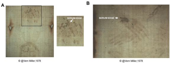
Figure 1. (A) Image of the Shroud taken with ultraviolet photography showing a partial view of the top of the ventral face region (bottom) and the dorsal area corresponding to the back of the head (top). An enlargement of the area indicated by the rectangle is shown to the right and fluorescent serum edges of bloodstains are indicated (white arrow). (B) Image of the Shroud taken with ultraviolet photography showing a view of the wrist wound. The fluorescent serum edges of bloodstains are indicated (white arrow).
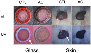
Figure 2. Control (CTL) and anti-coagulant (AC) treated blood were placed on glass or skin, allowed to dry, and observed under visible light (VL) or ultraviolet light (UV). The position of the serum halo is indicated by the white arrow.
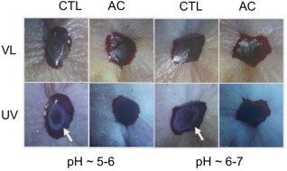
Figure 3. The pH of (CTL) and anti-coagulant (AC) treated blood (AC) was adjusted to the indicated range, placed on skin, allowed to dry, and observed under visible light (VL) or ultraviolet light (UV). See Materials and Methods section for specific details. The position of the serum halo is indicated by the white arrow.
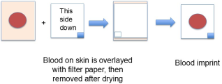
Figure 4. Schematic showing the method of blood imprinting. Blood is added onto skin and then after 20-25 minutes is overlayed with filter paper, which is removed after drying. Samples are then examined under visible and ultraviolet light to monitor the formation and transfer of serum edges.
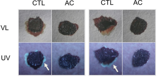
Figure 5. Blood from control (CTL) and anti-coagulant (AC) treated samples were added onto skin and then overlayed with filter paper after 20-25 minutes. After drying, the filter paper was removed and examined under visible light (VL) or ultraviolet light (UV). The position of the fluorescent serum borders is indicated by the arrow. The small fluorescent specks observed in both groups results from minor skin transfer from the immature mice cadavers used in these studies.
Information

