The rise of antimicrobial resistance has driven the search for alternative antibacterial agents, including plant-based compounds. This study evaluates the antimicrobial potential of selected herbal extracts against Streptobacillus moniliformis using the agar well diffusion method. The tested extracts included Basil leaves (Ocimum sanctum), Neem leaves (Azadirachta indica), Bael leaves (Aegle marmelos), Ginger (Zingiber officinale), Moringa seeds and leaves (Moringa oleifera), Dalchini (Cinnamomum verum), Lemon/Orange peels (Citrus limon and Citrus sinensis), and Ginger peels (Zingiber officinale). Among these, Bael leaves (Ocimum sanctum), and Lemon (Citrus limon) peels demonstrated significant antibacterial activity, forming distinct zones of inhibition. In contrast, Neem (Azadirachta indica) and Moringa (Moringa oleifera), extracts did not inhibit bacterial growth. The observed antimicrobial activity is likely due to the presence of bioactive compounds such as flavonoids, tannins, and essential oils, which may disrupt bacterial cell walls and metabolic processes. Notably, S. moniliformis exhibited limited survival in culture, while other bacterial strains showed minimal resistance. These findings suggest that certain herbal extracts, particularly Bael leaves and Lemon peels, may serve as natural antimicrobial agents against S. moniliformis. Among the tested extracts, Bael leaves (Aegle marmelos) and Lemon peels (Citrus limon) demonstrated significant antibacterial activity, with zones of inhibition measuring approximately 124mm and 23mm, respectively. Further studies are required to isolate and characterize the active compounds responsible for this antibacterial activity to explore their potential in developing alternative antimicrobial therapies.
| Published in | International Journal of Ecotoxicology and Ecobiology (Volume 10, Issue 3) |
| DOI | 10.11648/j.ijee.20251003.11 |
| Page(s) | 40-49 |
| Creative Commons |
This is an Open Access article, distributed under the terms of the Creative Commons Attribution 4.0 International License (http://creativecommons.org/licenses/by/4.0/), which permits unrestricted use, distribution and reproduction in any medium or format, provided the original work is properly cited. |
| Copyright |
Copyright © The Author(s), 2025. Published by Science Publishing Group |
Antimicrobial Resistance, Herbal Extracts, Streptobacillus moniliformis, Bioactive Compounds, Natural Antimicrobial Agents
S. No. | Herbs Name | Family Name | Part Used | Ayurvedic/Traditional Uses |
|---|---|---|---|---|
1 | Neem (Azadirachta indica) | Meliaceae | Leaves | Antibacterial, Antifungal, Skin Disorders |
2 | Amla (Phyllanthus emblica) | Phyllanthaceae | Leaves & Fruit | Immunity Booster, Antioxidant, Digestive Health |
3 | Bael (Aegle marmelos) | Rutaceae | Leaves | Digestive Aid, Anti-inflammatory, Respiratory Health |
4 | Moringa (Moringa oleifera) | Moringaceae | Leaves | Nutrient-Rich, Anti-inflammatory, Antioxidant |
5 | Lemon (Citrus limon) | Rutaceae | Fruit | Detoxification, Digestive Health, Skin Care |
6 | Garlic (Allium sativum) | Amaryllidaceae | Bulb | Antibacterial, Antiviral, Heart Health |
7 | Ginger (Zingiber officinale) | Zingiberaceae | Rhizome | Digestive Aid, Anti-inflammatory, Nausea Relief |
8 | Basil (Ocimum basilicum) | Lamiaceae | Leaves | Antioxidant, Anti-inflammatory, Digestive Health |
Herbal Extract | Inhibition zone (mm) (Mean ± SD) |
|---|---|
Neem (Azadirachta indica) | 14.5 ± 1.22 |
Amla (Phyllanthus emblica) Leaf& Fruit | 22.7±1.44 & 23.5±1.22 |
Bael (Aegle marmelos) | 28.17±1.44 |
Moringa (Moringa oleifera) | 17.0±0.62 |
Lemon (Citrus limon) | 27.0±1.63 |
Garlic (Allium sativum) | 23.2±1.13 |
Ginger (Zingiber officinale) | 21.02±1.05 |
Basil (Ocimum basilicum) | 26.33±1.24 |
AMR | Antimicrobial Resistance |
SM | Streptobacillus moniliformis |
EO | Essential Oils |
CFU | Colony Forming Units |
ZDI | Zone of Diffusion Inhibition |
MIC | Minimum Inhibitory Concentration |
MHA | Mueller-hinton Agar |
DMSO | Dimethyl Sulfoxide |
UV | Ultraviolet |
RPM | Revolutions per Minute |
| [1] | Nascimento, G. G., Locatelli, J., Freitas, P. C., & Silva, G. L. (2000). Antibacterial activity of plant extracts and phytochemicals on antibiotic-resistant bacteria. Brazilian journal of microbiology, 31, 247-256. |
| [2] | Ulloa-Urizar, G., Aguilar-Luis, M. A., De Lama-Odría, M. D. C., Camarena-Lizarzaburu, J., & del Valle Mendoza, J. (2015). Antibacterial activity of five Peruvian medicinal plants against Pseudomonas aeruginosa. Asian Pacific Journal of Tropical Biomedicine, 5(11), 928-931. |
| [3] | Akrayi, H. F. S., & Abdulrahman, Z. F. A. (2013). Evaluation of the antibacterial efficacy and the phytochemical analysis of some plant extracts against human pathogenic bacteria. Journal of Pharmacy and Clinical Sciences, 7, 29-39. |
| [4] | Okeke, M. I., Iroegbu, C. U., Eze, E. N., Okoli, A. S., & Esimone, C. O. (2001). Evaluation of extracts of the root of Landolphia owerrience for antibacterial activity. Journal of ethnopharmacology, 78(2-3), 119-127. |
| [5] | Zameer, F., Rukmangada, M. S., Chauhan, J. B., Khanum, S. A., Kumar, P., Devi, A. T., & Dhananjaya, B. L. (2016). Evaluation of adhesive and anti-adhesive properties of Pseudomonas aeruginosa biofilms and their inhibition by herbal plants. Iranian journal of microbiology, 8(2), 108. |
| [6] | Narayanan, A. S., Raja, S. S. S., Ponmurugan, K., Kandekar, S. C., Natarajaseenivasan, K., Maripandi, A., & Mandeel, Q. A. (2011). Antibacterial activity of selected medicinal plants against multiple antibiotic resistant uropathogens: a study from Kolli Hills, Tamil Nadu, India. Beneficial Microbes, 2(3), 235-244. |
| [7] | Mostafa, A. A., Al-Askar, A. A., Almaary, K. S., Dawoud, T. M., Sholkamy, E. N., & Bakri, M. M. (2018). Antimicrobial activity of some plant extracts against bacterial strains causing food poisoning diseases. Saudi journal of biological sciences, 25(2), 361-366. |
| [8] | Abalaka, M. E., Daniyan, S. Y., Oyeleke, S. B., & Adeyemo, S. O. (2012). The antibacterial evaluation of Moringa oleifera leaf extracts on selected bacterial pathogens. Journal of Microbiology research, 2(2), 1-4. |
| [9] | Hsieh, P. C., Mau, J. L., & Huang, S. H. (2001). Antimicrobial effect of various combinations of plant extracts. Food Microbiology, 18(1), 35-43. |
| [10] | Karuppiah, P., & Rajaram, S. (2012). Antibacterial effect of Allium sativum cloves and Zingiber officinale rhizomes against multiple-drug resistant clinical pathogens. Asian Pacific journal of tropical biomedicine, 2(8), 597-601. |
| [11] | Prabuseenivasan, S., Jayakumar, M., & Ignacimuthu, S. (2006). In vitro antibacterial activity of some plant essential oils. BMC complementary and alternative medicine, 6, 1-8. |
| [12] | Nostro, A., Cellini, L., Bartolomeo, S. D., Campli, E. D., Grande, R., Cannatelli, M. A., & Alonzo, V. (2005). Antibacterial effect of plant extracts against Helicobacter pylori. Phytotherapy Research: An International Journal Devoted to Pharmacological and Toxicological Evaluation of Natural Product Derivatives, 19(3), 198-202. |
| [13] | Mummed, B., Abraha, A., Feyera, T., Nigusse, A., & Assefa, S. (2018). In vitro antibacterial activity of selected medicinal plants in the traditional treatment of skin and wound infections in eastern Ethiopia. BioMed research international, 2018(1), 1862401. |
| [14] | Zaidan, M. R., Noor Rain, A., Badrul, A. R., Adlin, A., Norazah, A., & Zakiah, I. (2005). In vitro screening of five local medicinal plants for antibacterial activity using disc diffusion method. Trop biomed, 22(2), 165-170. |
| [15] | Bhalodia, N. R., & Shukla, V. J. (2011). Antibacterial and antifungal activities from leaf extracts of Cassia fistula l.: An ethnomedicinal plant. Journal of advanced pharmaceutical technology & research, 2(2), 104-109. |
| [16] | Behbahani, B. A., Yazdi, F. T., Mortazavi, A., Gholian, M. M., Zendeboodi, F., & Vasiee, A. (2014). Antimicrobial effect of Carboxy Methyl Cellulose (CMC) containing aqueous and ethanolic Eucalyptus camaldulensis L. leaves extract against Streptococcus pyogenes, Pseudomonas aeruginosa and Staphylococcus epidermidis. Archives of Advances in Biosciences, 5(2). |
| [17] | Kayser, O., & Kolodziej, H. (1997). Antibacterial activity of extracts and constituents of Pelargonium sidoides and Pelargonium reniforme. Planta medica, 63(06), 508-510. |
| [18] | Bonjar, S. (2004). Evaluation of antibacterial properties of some medicinal plants used in Iran. Journal of ethnopharmacology, 94(2-3), 301-305. |
| [19] | Shahrajabian, M. H., & Sun, W. (2023). Survey on medicinal plants and herbs in traditional Iranian medicine with anti-oxidant, anti-viral, anti-microbial, and anti-inflammation properties. Letters in Drug Design & Discovery, 20(11), 1707-1743. |
| [20] | Adunola, A. T., Chidimma, A. L., Olatunde, D. S., & Peter, O. A. (2015). Antibacterial activity of watermelon (Citrullus lanatus) seed against selected microorganisms. African Journal of Biotechnology, 14(14), 1224-1229. |
| [21] | Andoğan, B. C., Baydar, H., Kaya, S., Demirci, M., Özbaşar, D., & Mumcu, E. (2002). Antimicrobial activity and chemical composition of some essential oils. Archives of pharmacal research, 25, 860-864. |
| [22] | Bose, D., & Chatterjee, S. (2016). Biogenic synthesis of silver nanoparticles using guava (Psidium guajava) leaf extract and its antibacterial activity against Pseudomonas aeruginosa. Applied Nanoscience, 6(6), 895-901. |
| [23] | Wood, S. J., Kuzel, T. M., & Shafikhani, S. H. (2023). Pseudomonas aeruginosa: infections, animal modeling, and therapeutics. Cells, 12(1), 199. |
| [24] | Elfadadny, A., Ragab, R. F., AlHarbi, M., Badshah, F., Ibáñez-Arancibia, E., Farag, A., & Nageeb, W. M. (2024). Antimicrobial resistance of Pseudomonas aeruginosa: navigating clinical impacts, current resistance trends, and innovations in breaking therapies. Frontiers in Microbiology, 15, 1374466. |
| [25] | El Zowalaty, M. E., Al Thani, A. A., Webster, T. J., El Zowalaty, A. E., Schweizer, H. P., Nasrallah, G. K.,... & Ashour, H. M. (2015). Pseudomonas aeruginosa: arsenal of resistance mechanisms, decades of changing resistance profiles, and future antimicrobial therapies. Future Microbiology, 10(10), 1683-1706. |
| [26] | El Chakhtoura, N. G., Saade, E., Iovleva, A., Yasmin, M., Wilson, B., Perez, F., & Bonomo, R. A. (2018). Therapies for multidrug resistant and extensively drug-resistant non-fermenting gram-negative bacteria causing nosocomial infections: a perilous journey toward ‘molecularly targeted’therapy. Expert review of anti-infective therapy, 16(2), 89-110. |
| [27] | Sahu, M. C., Dubey, D., Rath, S., Debata, N. K., & Padhy, R. N. (2012). Multidrug resistance of Pseudomonas aeruginosa as known from surveillance of nosocomial and community infections in an Indian teaching hospital. Journal of Public Health, 20, 413-423. |
| [28] | Proctor, L. L., Ward, W. L., Roggy, C. S., Koontz, A. G., Clark, K. M., Quinn, A. P., & Brooks, B. D. (2021). Potential therapeutic targets for combination antibody therapy against Pseudomonas aeruginosa infections. Antibiotics, 10(12), 1530. |
| [29] | Giovagnorio, F., De Vito, A., Madeddu, G., Parisi, S. G., & Geremia, N. (2023). Resistance in Pseudomonas aeruginosa: a narrative review of antibiogram interpretation and emerging treatments. Antibiotics, 12(11), 1621. |
| [30] | Navon-Venezia, S., Ben-Ami, R., & Carmeli, Y. (2005). Update on Pseudomonas aeruginosa and Acinetobacter baumannii infections in the healthcare setting. Current opinion in infectious diseases, 18(4), 306-313. |
| [31] | Peykov, S., & Strateva, T. (2023). Whole-genome sequencing-based resistome analysis of nosocomial multidrug-resistant non-fermenting Gram-negative pathogens from the Balkans. Microorganisms, 11(3), 651. |
| [32] | de la Fuente-Nunez, C., Cesaro, A., & Hancock, R. E. (2023). Antibiotic failure: Beyond antimicrobial resistance. Drug Resistance Updates, 101012. |
| [33] | Malhotra, S., Hayes Jr, D., & Wozniak, D. J. (2019). Cystic fibrosis and Pseudomonas aeruginosa: the host-microbe interface. Clinical microbiology reviews, 32(3), 10-1128. |
| [34] | Demain, A. L., & Sanchez, S. (2009). Microbial drug discovery: 80 years of progress. The Journal of antibiotics, 62(1), 5-16. |
| [35] | Lopes, B. S., Hanafiah, A., Nachimuthu, R., Muthupandian, S., Md Nesran, Z. N., & Patil, S. (2022). The role of antimicrobial peptides as antimicrobial and antibiofilm agents in tackling the silent pandemic of antimicrobial resistance. Molecules, 27(9), 2995. |
| [36] | Lauman, P., & Dennis, J. J. (2021). Advances in phage therapy: targeting the Burkholderia cepacia complex. Viruses, 13(7), 1331. |
| [37] | Reece, E., Bettio, P. H. D. A., & Renwick, J. (2021). Polymicrobial interactions in the cystic fibrosis airway microbiome impact the antimicrobial susceptibility of Pseudomonas aeruginosa. Antibiotics, 10(7), 827. |
| [38] | Anju, V. T., Busi, S., Imchen, M., Kumavath, R., Mohan, M. S., Salim, S. A., & Dyavaiah, M. (2022). Polymicrobial infections and biofilms: clinical significance and eradication strategies. Antibiotics, 11(12), 1731. |
| [39] | Batoni, G., Maisetta, G., & Esin, S. (2021). Therapeutic potential of antimicrobial peptides in polymicrobial biofilm-associated infections. International Journal of Molecular Sciences, 22(2), 482. |
| [40] | Rudilla Mateo, H. (2019). Synthetic Polymyxin-based Peptides Against Multidrug Resistant Bacteria: A Therapeutic Option. |
| [41] | Ali, A., Zahra, A., Kamthan, M., Husain, F. M., Albalawi, T., Zubair, M., & Noorani, M. S. (2023). Microbial biofilms: applications, clinical consequences, and alternative therapies. Microorganisms, 11(8), 1934. |
| [42] | Ruffin, M., & Brochiero, E. (2019). Repair process impairment by Pseudomonas aeruginosa in epithelial tissues: major features and potential therapeutic avenues. Frontiers in cellular and infection microbiology, 9, 182. |
| [43] | Bush, K., & Bradford, P. A. (2020). Epidemiology of β-lactamase-producing pathogens. Clinical microbiology reviews, 33(2), 10-1128. |
| [44] | Fodor, A., Abate, B. A., Deák, P., Fodor, L., Gyenge, E., Klein, M. G., & Makrai, L. (2020). Multidrug resistance (MDR) and collateral sensitivity in bacteria, with special attention to genetic and evolutionary aspects and to the perspectives of antimicrobial peptides-a review. Pathogens, 9(7), 522. |
| [45] | De Sousa, T., Hébraud, M., Dapkevicius, M. L. E., Maltez, L., Pereira, J. E., Capita, R., & Poeta, P. (2021). Genomic and Metabolic Characteristics of the Pathogenicity in Pseudomonas aeruginosa. International journal of molecular sciences, 22(23), 12892. |
| [46] | Da Cruz Nizer, W. S., Inkovskiy, V., Versey, Z., Strempel, N., Cassol, E., & Overhage, J. (2021). Oxidative stress response in Pseudomonas aeruginosa. Pathogens, 10(9), 1187. |
| [47] | Sun, H., Pulakat, L., & Anderson, D. W. (2020). Challenges and new therapeutic approaches in the management of chronic wounds. Current drug targets, 21(12), 1264-1275. |
| [48] | Hammoudi Halat, D., & Ayoub Moubareck, C. (2020). The current burden of carbapenemases: review of significant properties and dissemination among gram-negative bacteria. Antibiotics, 9(4), 186. |
| [49] | Reza, A., Sutton, J. M., & Rahman, K. M. (2019). Effectiveness of efflux pump inhibitors as biofilm disruptors and resistance breakers in gram-negative (ESKAPEE) bacteria. Antibiotics, 8(4), 229. |
| [50] | Bassetti, M., Poulakou, G., Ruppe, E., Bouza, E., Van Hal, S. J., & Brink, A. (2017). Antimicrobial resistance in the next 30 years, humankind, bugs and drugs: a visionary approach. Intensive care medicine, 43, 1464-1475. |
| [51] | Mubeen, B., Ansar, A. N., Rasool, R., Ullah, I., Imam, S. S., Alshehri, S., & Kazmi, I. (2021). Nanotechnology as a novel approach in combating microbes providing an alternative to antibiotics. Antibiotics, 10(12), 1473. |
| [52] | Sultan, M., Arya, R., & Kim, K. K. (2021). Roles of two-component systems in Pseudomonas aeruginosa virulence. International journal of molecular sciences, 22(22), 12152. |
| [53] | Siegel, J. D., Rhinehart, E., Jackson, M., Chiarello, L., & Healthcare Infection Control Practices Advisory Committee. (2019, May). 2007 Guideline for Isolation Precautions: Preventing Transmission of Infectious Agents in Health Care Settings. |
| [54] | Scoffone, V. C., Trespidi, G., Chiarelli, L. R., Barbieri, G., & Buroni, S. (2019). Quorum sensing as antivirulence target in cystic fibrosis pathogens. International journal of molecular sciences, 20(8), 1838. |
| [55] | Mba, I. E., & Nweze, E. I. (2022). Focus: antimicrobial resistance: antimicrobial peptides therapy: an emerging alternative for treating drug-resistant bacteria. The Yale journal of biology and medicine, 95(4), 445. |
| [56] | Rudilla, H., Fusté, E., Cajal, Y., Rabanal, F., Vinuesa, T., & Viñas, M. (2016). Synergistic antipseudomonal effects of synthetic peptide AMP38 and carbapenems. Molecules, 21(9), 1223. |
| [57] | Minandri, F., Bonchi, C., Frangipani, E., Imperi, F., & Visca, P. (2014). Promises and failures of gallium as an antibacterial agent. Future microbiology, 9(3), 379-397. |
| [58] | Lee, S. H., Teo, J., Heng, D., Ng, W. K., Zhao, Y., & Tan, R. B. (2016). Tailored antibiotic combination powders for inhaled rotational antibiotic therapy. Journal of Pharmaceutical Sciences, 105(4), 1501-1512. |
APA Style
Singh, K. K., Saini, M., Prakash, D. (2025). Screening of Natural Plant Extracts for Antimicrobial Activity Against Streptobacillus moniliformis. International Journal of Ecotoxicology and Ecobiology, 10(3), 40-49. https://doi.org/10.11648/j.ijee.20251003.11
ACS Style
Singh, K. K.; Saini, M.; Prakash, D. Screening of Natural Plant Extracts for Antimicrobial Activity Against Streptobacillus moniliformis. Int. J. Ecotoxicol. Ecobiol. 2025, 10(3), 40-49. doi: 10.11648/j.ijee.20251003.11
@article{10.11648/j.ijee.20251003.11,
  author = {Kishlay Kant Singh and Mansi Saini and Divya Prakash},
  title = {Screening of Natural Plant Extracts for Antimicrobial Activity Against Streptobacillus moniliformis
},
  journal = {International Journal of Ecotoxicology and Ecobiology},
  volume = {10},
  number = {3},
  pages = {40-49},
  doi = {10.11648/j.ijee.20251003.11},
  url = {https://doi.org/10.11648/j.ijee.20251003.11},
  eprint = {https://article.sciencepublishinggroup.com/pdf/10.11648.j.ijee.20251003.11},
  abstract = {The rise of antimicrobial resistance has driven the search for alternative antibacterial agents, including plant-based compounds. This study evaluates the antimicrobial potential of selected herbal extracts against Streptobacillus moniliformis using the agar well diffusion method. The tested extracts included Basil leaves (Ocimum sanctum), Neem leaves (Azadirachta indica), Bael leaves (Aegle marmelos), Ginger (Zingiber officinale), Moringa seeds and leaves (Moringa oleifera), Dalchini (Cinnamomum verum), Lemon/Orange peels (Citrus limon and Citrus sinensis), and Ginger peels (Zingiber officinale). Among these, Bael leaves (Ocimum sanctum), and Lemon (Citrus limon) peels demonstrated significant antibacterial activity, forming distinct zones of inhibition. In contrast, Neem (Azadirachta indica) and Moringa (Moringa oleifera), extracts did not inhibit bacterial growth. The observed antimicrobial activity is likely due to the presence of bioactive compounds such as flavonoids, tannins, and essential oils, which may disrupt bacterial cell walls and metabolic processes. Notably, S. moniliformis exhibited limited survival in culture, while other bacterial strains showed minimal resistance. These findings suggest that certain herbal extracts, particularly Bael leaves and Lemon peels, may serve as natural antimicrobial agents against S. moniliformis. Among the tested extracts, Bael leaves (Aegle marmelos) and Lemon peels (Citrus limon) demonstrated significant antibacterial activity, with zones of inhibition measuring approximately 124mm and 23mm, respectively. Further studies are required to isolate and characterize the active compounds responsible for this antibacterial activity to explore their potential in developing alternative antimicrobial therapies.
},
 year = {2025}
}
TY  - JOUR
T1  - Screening of Natural Plant Extracts for Antimicrobial Activity Against Streptobacillus moniliformis

AU  - Kishlay Kant Singh
AU  - Mansi Saini
AU  - Divya Prakash
Y1  - 2025/09/02
PY  - 2025
N1  - https://doi.org/10.11648/j.ijee.20251003.11
DO  - 10.11648/j.ijee.20251003.11
T2  - International Journal of Ecotoxicology and Ecobiology
JF  - International Journal of Ecotoxicology and Ecobiology
JO  - International Journal of Ecotoxicology and Ecobiology
SP  - 40
EP  - 49
PB  - Science Publishing Group
SN  - 2575-1735
UR  - https://doi.org/10.11648/j.ijee.20251003.11
AB  - The rise of antimicrobial resistance has driven the search for alternative antibacterial agents, including plant-based compounds. This study evaluates the antimicrobial potential of selected herbal extracts against Streptobacillus moniliformis using the agar well diffusion method. The tested extracts included Basil leaves (Ocimum sanctum), Neem leaves (Azadirachta indica), Bael leaves (Aegle marmelos), Ginger (Zingiber officinale), Moringa seeds and leaves (Moringa oleifera), Dalchini (Cinnamomum verum), Lemon/Orange peels (Citrus limon and Citrus sinensis), and Ginger peels (Zingiber officinale). Among these, Bael leaves (Ocimum sanctum), and Lemon (Citrus limon) peels demonstrated significant antibacterial activity, forming distinct zones of inhibition. In contrast, Neem (Azadirachta indica) and Moringa (Moringa oleifera), extracts did not inhibit bacterial growth. The observed antimicrobial activity is likely due to the presence of bioactive compounds such as flavonoids, tannins, and essential oils, which may disrupt bacterial cell walls and metabolic processes. Notably, S. moniliformis exhibited limited survival in culture, while other bacterial strains showed minimal resistance. These findings suggest that certain herbal extracts, particularly Bael leaves and Lemon peels, may serve as natural antimicrobial agents against S. moniliformis. Among the tested extracts, Bael leaves (Aegle marmelos) and Lemon peels (Citrus limon) demonstrated significant antibacterial activity, with zones of inhibition measuring approximately 124mm and 23mm, respectively. Further studies are required to isolate and characterize the active compounds responsible for this antibacterial activity to explore their potential in developing alternative antimicrobial therapies.

VL  - 10
IS  - 3
ER  -
School of Biotechnology & Life Sciences, Shobhit Institute of Engineering & Technology, Meerut, India
School of Biological Engineering & Sciences, Shobhit University, Meerut, India
School of Biotechnology & Life Sciences, Shobhit Institute of Engineering & Technology, Meerut, India
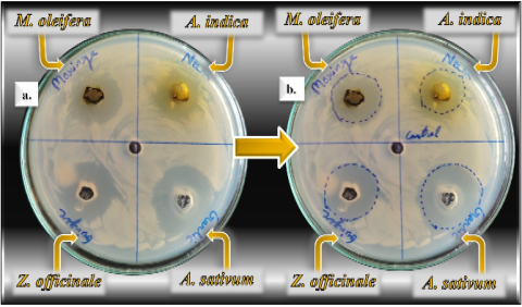
Figure 1. Growth inhibition of multidrug-resistant (MDR) Streptobacillus moniliformis by herbal extracts (i.e. Neem, Moringa, Ginger & Garlic); Without marking ZOI (a) & marking ZOI (b).
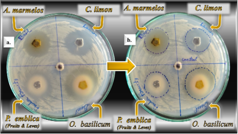
Figure 2. Growth inhibition of multidrug-resistant (MDR) Streptobacillus moniliformis by herbal extracts (i.e. Bael, Lemon, Basil, & Amla); Without marking ZOI (a) & marking ZOI (b).
Figure 3. Evaluation of the antibacterial properties extract of herbs against S. moniliformis.
Information

