Abstract
Background. Midfacial trauma (MT) is frequently accompanied by ophthalmological complications that may result in significant visual morbidity and long-term disability. Early recognition and timely management of ocular involvement in patients with fractures of the midface are essential for preserving visual function and improving overall outcomes. Aim of study. To evaluate the spectrum and frequency of ophthalmological complications associated with midfacial trauma and to analyze clinical factors influencing visual prognosis in patients treated at the National Medical Center (NMC) between 2023 and 2025. Materials and methods. A prospective clinical study was conducted on 120 patients with MT who were admitted to the NMC from January 2023 to June 2025. Demographic data, etiology and pattern of midfacial injury, clinical ophthalmological findings, imaging results, and treatment modalities were analyzed. All patients underwent a standardized ophthalmic examination including visual acuity assessment, anterior and posterior segment evaluation, ocular motility testing, and imaging studies when indicated. Results. The majority of patients were males (65.0%) with a mean age of 38.7±12.4 years. Road traffic accidents were the leading cause of MT (55.0%), followed by interpersonal violence (25.0%) and falls (15.0%). The most common ophthalmological manifestations were periorbital edema and ecchymosis (70.8%), subconjunctival hemorrhage (42.5%), diplopia (25.8%), limitation of ocular motility (21.7%), enophthalmos (18.3%), traumatic optic neuropathy (TON) (12.5%), and corneal injury (10.0%). Patients with zygomaticomaxillary complex (ZMC) and orbital floor fractures demonstrated a significantly higher risk of diplopia and enophthalmos. TON was strongly associated with high-energy mechanisms and delayed presentation. Conclusion. Ophthalmological complications are highly prevalent in patients with MT and often coexist with complex fracture patterns. A multidisciplinary approach involving maxillofacial surgeons and ophthalmologists is crucial for early diagnosis and optimal management. Routine comprehensive ophthalmic assessment is recommended in all patients with MT to prevent irreversible visual loss and to enhance functional and cosmetic outcomes.
Keywords
Midfacial Trauma, Orbital Fractures, Ophthalmic Complications, Traumatic Optic Neuropathy, Diplopia, Enophthalmos
1. Introduction
MT represents a significant subset of maxillofacial injuries and is frequently associated with ocular and orbital complications
| [4] | Johnson P, Ahmed Z. Diplopia and enophthalmos following orbital floor fractures: long-term outcomes after surgical repair. British Journal of Oral and Maxillofacial Surgery. 2019; 57(8): 780–787. |
[4]
. The midface comprises complex anatomical structures including the maxilla, zygomatic bone, nasal bones, and the bony orbit, which are closely related to the globe and optic nerve. High-energy trauma such as road traffic accidents, interpersonal violence, and falls can result in fractures of the midface and orbit, thereby predisposing patients to a wide range of ophthalmological manifestations
| [1] | Smith J, Rodriguez A, Patel K. Orbital complications of midfacial fractures: a clinical review. Journal of Craniofacial Trauma. 2022; 14(3): 145–156. |
[1]
. These complications range from mild soft tissue injuries to severe conditions such as globe rupture and TON, which may lead to permanent visual loss
| [6] | Fernandes R, Koerich L, Ponzoni D. Naso-orbito-ethmoidal fractures: clinical features and ophthalmic implications. Journal of Oral and Maxillofacial Surgery. 2018; 76(9): 1890. e1–1890. e9. |
| [7] | Singh V, Prasad R, Bhatia R. Ocular injuries associated with zygomaticomaxillary complex fractures. Journal of Ophthalmology. 2019; 2019: 1–7. |
[6, 7]
.
Epidemiological studies have shown that orbital involvement occurs in a substantial proportion of patients with midfacial fractures. The close proximity of the orbital walls to the paranasal sinuses and midfacial buttresses predisposes them to fracture lines that may compromise the structural integrity of the orbit and the function of extraocular muscles
| [2] | Chen L, Gupta R, Lee V. Traumatic optic neuropathy: current concepts and management strategies. Eye and Brain. 2021; 13: 25–36. |
| [10] | Park J, Lee J. Outcomes of orbital wall reconstruction using titanium mesh and resorbable plates. Journal of Cranio-Maxillofacial Surgery. 2020; 48(2): 123–130. |
[2, 10]
. Consequently, patients may present with enophthalmos, hypoglobus, diplopia, restriction of ocular movements, and cosmetic deformities. TON, although less frequent, remains one of the most devastating complications, often associated with high-energy impact and secondary compressive effects within the optic canal
| [9] | Reddy K, Thomas S. High-energy craniofacial trauma: incidence of orbital and ocular involvement. Journal of Neuro-Ophthalmology. 2022; 42(1): 37–44. |
[9]
.
Worldwide, the patterns of MT are influenced by socioeconomic, cultural, and environmental factors. In many developing regions, the rapid increase in motorization, inadequate use of protective equipment, and poor road safety contribute to the high incidence of road traffic-related facial injuries. Interpersonal violence is another major etiological factor, particularly in young adult males. Understanding the local epidemiology and etiological factors is important for developing preventive strategies and optimizing resource allocation
| [1] | Smith J, Rodriguez A, Patel K. Orbital complications of midfacial fractures: a clinical review. Journal of Craniofacial Trauma. 2022; 14(3): 145–156. |
| [9] | Reddy K, Thomas S. High-energy craniofacial trauma: incidence of orbital and ocular involvement. Journal of Neuro-Ophthalmology. 2022; 42(1): 37–44. |
[1, 9]
.
Despite progress in diagnostic imaging and surgical techniques, ophthalmological complications of midfacial trauma are still underrecognized and sometimes inadequately managed, especially in emergency settings
| [3] | Alvarez M, Kim S, Park JH. Patterns of midfacial fractures and associated ocular injuries in a tertiary trauma center. Ophthalmic Plastic and Reconstructive Surgery. 2020; 36(5): 412–419. |
[3]
. Delayed referral to ophthalmology and underestimation of subtle clinical signs may lead to missed diagnoses and irreversible visual sequelae. Therefore, comprehensive assessment protocols and close collaboration between maxillofacial surgeons, neurosurgeons, and ophthalmologists are essential
| [5] | Li Y, Nakamura N. Epidemiology of maxillofacial trauma in the era of increased motorization: a systematic review. International Journal of Oral and Maxillofacial Surgery. 2021; 50(3): 301–312. |
| [8] | Mustafa A, Karim M. Multidisciplinary management of orbital fractures: the role of the ophthalmologist. Clinical Ophthalmology. 2020; 14: 2321–2330. |
[5, 8]
.
The present study addresses the spectrum and frequency of ophthalmological complications in patients with midfacial trauma treated at a tertiary NMC. By analyzing clinical characteristics, injury patterns, and visual outcomes, we aim to highlight key risk factors and emphasize the importance of early and systematic ophthalmological evaluation in this patient population.
2. Aim of the Study
The aim of this study was to evaluate the spectrum and frequency of ophthalmological complications associated with midfacial trauma and to analyze clinical and etiological factors influencing visual outcomes in patients treated at the National Medical Center from 2023 to 2025.
3. Materials and Methods
Study Design and Setting. This was a prospective clinical observational study conducted at the NMC, Tashkent, Uzbekistan, from January 2023 to June 2025. The study adhered to the principles of the Declaration of Helsinki and was approved by the institutional ethics committee. Informed consent was obtained from all participants or their legal guardians.
Study Population. A total of 120 consecutive patients with radiologically confirmed midfacial fractures were included in the study. Inclusion criteria were: (1) age 18 years and older; (2) presence of fractures involving the midface (zygomaticomaxillary complex, maxilla, nasal bones, orbital walls, or Le Fort fractures); and (3) admission to the NMC within 14 days of injury. Exclusion criteria included isolated mandibular fractures, isolated soft tissue facial injuries without bony involvement, pre-existing severe ocular pathology unrelated to trauma, and incomplete clinical data.
Data Collection. Demographic data (age, sex), mechanism of injury (road traffic accident, interpersonal violence, fall, occupational injury, others), time from trauma to presentation, and associated systemic injuries were recorded. Maxillofacial assessment included clinical examination and imaging studies (computed tomography and/or radiography) to classify fracture patterns. Midfacial fractures were categorized into ZMC fractures, isolated orbital wall fractures, naso-orbito-ethmoidal fractures, Le Fort I–III fractures, and combined patterns.
Ophthalmological Examination. All patients underwent a standardized ophthalmological evaluation performed by an experienced ophthalmologist. The examination protocol included measurement of best-corrected visual acuity using a Snellen chart or equivalent, pupillary reflex testing, intraocular pressure measurement when not contraindicated, slit-lamp biomicroscopy of the anterior segment, and dilated fundus examination where feasible. Ocular motility was assessed in all cardinal positions of gaze. Exophthalmometry and Hess charting were performed in selected cases to quantify globe position and motility defects.
Radiological Assessment. High-resolution computed tomography of the facial skeleton and orbits was performed in axial and coronal planes with three-dimensional reconstruction when available. The extent and location of orbital wall defects, presence of bone fragments impinging on extraocular muscles, and optic canal involvement were documented. Imaging findings were correlated with clinical manifestations such as enophthalmos, diplopia, and TON.
Treatment Protocol. Management of midfacial fractures followed a multidisciplinary approach involving maxillofacial surgeons and ophthalmologists. Surgical repair of fractures was performed using open reduction and internal fixation techniques when indicated. Orbital wall reconstruction was carried out with titanium mesh, resorbable plates, or autologous grafts depending on defect size and surgeon preference. Medical management for ocular injuries included topical antibiotics and lubricants, systemic steroids in selected cases of traumatic optic neuropathy, and intraocular pressure-lowering agents when necessary. Patients with sight-threatening conditions such as globe rupture or severe intraocular hemorrhage were immediately referred for vitreoretinal or oculoplastic intervention.
Statistical Analysis. Collected data were entered into a database and analyzed using descriptive statistics. Categorical variables such as sex, etiology of trauma, and types of ophthalmological complications were presented as frequencies and percentages. Continuous variables such as age were expressed as mean ± standard deviation. Associations between fracture patterns and specific ophthalmological complications were explored using chi-square testing, with a significance level set at p < 0.05.
4. Results
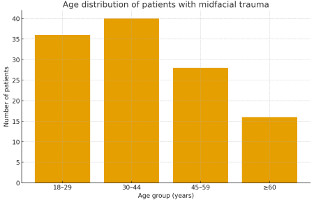
Demographic Characteristics. A total of 120 patients with midfacial trauma were included in the analysis. The mean age was 38.7±12.4 years (range 18–72 years). The majority of patients were male (n=78, 65.0%), while females accounted for 42 cases (35.0%). The most affected age group was 30–44 years, followed by 18–29 years (
Figure 1).
Etiology of Trauma. Road traffic accidents were the predominant cause of MT, observed in 66 patients (55.0%). Interpersonal violence accounted for 30 cases (25.0%), while falls were responsible for 18 cases (15.0%). Occupational injuries and other causes were less frequent, together comprising 6 cases (5.0%) (
Table 1).
Fracture Patterns. ZMC fractures were the most common pattern, identified in 52 patients (43.3%). Isolated orbital wall fractures were diagnosed in 28 patients (23.3%), naso-orbito-ethmoidal fractures in 14 patients (11.7%), and Le Fort fractures (I–III) in 16 patients (13.3%). Combined fracture patterns involving multiple midfacial structures were observed in 10 patients (8.3%) (
Table 2).
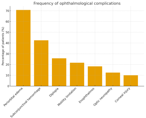
Spectrum of Ophthalmological Complications. Ophthalmological manifestations were documented in 102 out of 120 patients (85.0%). The most frequent findings were periorbital edema and ecchymosis (n=85, 70.8%), subconjunctival hemorrhage (n=51, 42.5%), and eyelid lacerations (n=28, 23.3%). Diplopia was present in 31 patients (25.8%), while limitation of ocular motility was observed in 26 patients (21.7%). Enophthalmos of ≥2 mm was detected in 22 patients (18.3%), predominantly in those with orbital floor and medial wall fractures. TON was diagnosed in 15 patients (12.5%), and corneal epithelial defects or stromal injuries were identified in 12 patients (10.0%).
Association Between Fracture Patterns and Ocular Findings. Patients with ZMC and isolated orbital wall fractures showed a significantly higher incidence of diplopia and enophthalmos compared to other fracture types (p < 0.05). TON was predominantly associated with high-energy mechanisms such as road traffic accidents and combined midfacial fracture patterns. Delayed presentation (>72 hours after trauma) was more common among patients with optic neuropathy and was associated with poorer visual outcomes (
Figure 2).
Visual Outcomes. At the final follow-up (3–6 months after injury), 78 patients (65.0%) maintained or regained good visual acuity (≥0.8), while 27 patients (22.5%) had moderate visual impairment (0.3–0.7). Severe visual loss (<0.3) or blindness in the affected eye was documented in 15 patients (12.5%), most of whom had sustained TON or severe globe injuries. Early diagnosis, prompt decompression in selected cases, and adequate rehabilitation were associated with better visual outcomes.
Figure 1. Age distribution of patients with midfacial trauma.
Figure 2. Frequency of ophthalmological complications in patients with midfacial trauma.
Table 1. Demographic and etiological characteristics of patients with midfacial trauma.
Parameter | Category | Number (n=120) | Percentage (%) |
Sex | Male | 78 | 65.0 |
Sex | Female | 42 | 35.0 |
Etiology | Road traffic accident | 66 | 55.0 |
Etiology | Interpersonal violence | 30 | 25.0 |
Etiology | Fall | 18 | 15.0 |
Etiology | Other/occupational | 6 | 5.0 |
Table 2. Distribution of fracture patterns and selected ophthalmological complications.
Fracture pattern | Number of patients | Diplopia (n) | Enophthalmos (n) | TON (n) |
Zygomaticomaxillary complex | 52 | 17 | 13 | 5 |
Isolated orbital wall | 28 | 8 | 9 | 2 |
Naso-orbito-ethmoidal | 14 | 3 | 2 | 2 |
Le Fort I–III | 16 | 3 | 2 | 3 |
Combined patterns | 10 | 3 | 2 | 3 |
5. Discussion
The present study provides a comprehensive overview of the ophthalmological complications associated with MT in a tertiary referral center over a two-and-a-half-year period. Our findings confirm that ocular and orbital involvement is highly prevalent in patients with midfacial fractures, with 85.0% of cases demonstrating at least one ophthalmological manifestation. This underscores the need for systematic ophthalmic screening in all patients presenting with midfacial injuries.
The demographic profile of our cohort, with a predominance of young to middle-aged males and road traffic accidents as the leading cause of injury, is consistent with reports from many regions undergoing rapid urbanization and motorization. The social and economic implications of facial and ocular trauma in this productive age group are considerable, particularly when permanent visual impairment occurs.
Periorbital edema, ecchymosis, and subconjunctival hemorrhage were the most common ocular findings in our study. Although these manifestations are often self-limiting and may be perceived as minor, they serve as important clinical indicators of underlying orbital injury. Subtle signs such as mild enophthalmos, minimal diplopia, or discrete motility restriction can easily be overlooked in the acute trauma setting, especially when systemic injuries are present. Our data demonstrate that enophthalmos and diplopia were significantly associated with ZMC and isolated orbital wall fractures, highlighting the importance of targeted imaging and timely orbital reconstruction in these patients.
TON, although less frequent, remains one of the most severe complications observed. In our series, 12.5% of patients developed evidence of optic nerve injury, most commonly in association with high-energy road traffic accidents and combined fracture patterns. The management of TON continues to be controversial, with ongoing debate regarding the role of high-dose steroids and surgical decompression. In our center, treatment decisions were individualized based on clinical and radiological findings, time since injury, and patient comorbidities. Nevertheless, a substantial proportion of patients with TON had poor visual outcomes at follow-up, emphasizing the devastating nature of this complication and the need for preventive strategies aimed at reducing high-energy facial trauma.
Comparing our results with Eric Zhong’s article
| [11] | Zhong E, Chou TY, Chaleff AJ, Scofield-Kaplan SM, Perzia BM, Naqvi J, Hou W. Orbital Fractures and Risk Factors for Ocular Injury. Clin Ophthalmol. 2022 Dec 14; 16: 4153-4161. https://doi.org/10.2147/OPTH.S391175 |
[11]
, the frequencies of diplopia, enophthalmos, and motility restriction are within the ranges reported by other tertiary centers. Variations between studies may reflect differences in injury mechanisms, fracture classification, referral patterns, and timing of ophthalmological evaluation. Some authors have highlighted the underdiagnosis of ocular injuries in non-specialist settings, particularly when standardized examination protocols are lacking. Our experience supports the implementation of structured ophthalmic assessment pathways for all MT patients, including documentation of visual acuity, pupillary responses, globe position, and ocular motility, as well as early imaging of the orbits when indicated.
The multidisciplinary approach adopted in our institution, with close collaboration between maxillofacial surgeons and ophthalmologists, facilitated early identification and management of sight-threatening conditions. Surgical repair of orbital fractures using contemporary fixation techniques enabled restoration of orbital volume and reduction of enophthalmos in many cases. However, residual diplopia and subtle motility disturbances persisted in a subset of patients, indicating the complexity of extraocular muscle entrapment and scarring.
Limitations of the current study include the single-center design and the moderate sample size, which may limit the generalizability of the findings. Additionally, long-term follow-up beyond six months was not available for all patients, potentially underestimating late complications such as progressive enophthalmos or post-traumatic glaucoma. Future studies with larger multicenter cohorts and longer follow-up are needed to better characterize risk factors for poor visual outcomes and to evaluate the effectiveness of specific treatment protocols.
Despite these limitations, our study contributes valuable data on the pattern and impact of ophthalmological complications in midfacial trauma and reinforces the importance of early, structured, and multidisciplinary care for affected patients.
6. Conclusion
Ophthalmological complications are common and clinically significant in patients with midfacial trauma. In this prospective clinical series, the majority of patients exhibited ocular or orbital manifestations, ranging from periorbital soft tissue injuries to severe conditions such as traumatic optic neuropathy. Zygomaticomaxillary complex and isolated orbital wall fractures were particularly associated with diplopia and enophthalmos, while high-energy mechanisms and combined fracture patterns predisposed to optic nerve damage.
Our findings support the routine implementation of comprehensive ophthalmological assessment in all patients with midfacial fractures and underscore the value of a multidisciplinary approach involving maxillofacial surgeons and ophthalmologists. Early identification and management of ocular complications can help prevent irreversible visual loss and improve both functional and aesthetic outcomes. Preventive measures aimed at reducing high-energy facial trauma, particularly road traffic accidents, remain a public health priority.
Abbreviations
MT | Midfacial Trauma |
NMC | National Medical Center |
TON | Traumatic Optic Neuropathy |
ZMC | Zygomaticomaxillary Complex |
Conflicts of Interest
The authors declare that there is no conflict of interest.
References
| [1] |
Smith J, Rodriguez A, Patel K. Orbital complications of midfacial fractures: a clinical review. Journal of Craniofacial Trauma. 2022; 14(3): 145–156.
|
| [2] |
Chen L, Gupta R, Lee V. Traumatic optic neuropathy: current concepts and management strategies. Eye and Brain. 2021; 13: 25–36.
|
| [3] |
Alvarez M, Kim S, Park JH. Patterns of midfacial fractures and associated ocular injuries in a tertiary trauma center. Ophthalmic Plastic and Reconstructive Surgery. 2020; 36(5): 412–419.
|
| [4] |
Johnson P, Ahmed Z. Diplopia and enophthalmos following orbital floor fractures: long-term outcomes after surgical repair. British Journal of Oral and Maxillofacial Surgery. 2019; 57(8): 780–787.
|
| [5] |
Li Y, Nakamura N. Epidemiology of maxillofacial trauma in the era of increased motorization: a systematic review. International Journal of Oral and Maxillofacial Surgery. 2021; 50(3): 301–312.
|
| [6] |
Fernandes R, Koerich L, Ponzoni D. Naso-orbito-ethmoidal fractures: clinical features and ophthalmic implications. Journal of Oral and Maxillofacial Surgery. 2018; 76(9): 1890. e1–1890. e9.
|
| [7] |
Singh V, Prasad R, Bhatia R. Ocular injuries associated with zygomaticomaxillary complex fractures. Journal of Ophthalmology. 2019; 2019: 1–7.
|
| [8] |
Mustafa A, Karim M. Multidisciplinary management of orbital fractures: the role of the ophthalmologist. Clinical Ophthalmology. 2020; 14: 2321–2330.
|
| [9] |
Reddy K, Thomas S. High-energy craniofacial trauma: incidence of orbital and ocular involvement. Journal of Neuro-Ophthalmology. 2022; 42(1): 37–44.
|
| [10] |
Park J, Lee J. Outcomes of orbital wall reconstruction using titanium mesh and resorbable plates. Journal of Cranio-Maxillofacial Surgery. 2020; 48(2): 123–130.
|
| [11] |
Zhong E, Chou TY, Chaleff AJ, Scofield-Kaplan SM, Perzia BM, Naqvi J, Hou W. Orbital Fractures and Risk Factors for Ocular Injury. Clin Ophthalmol. 2022 Dec 14; 16: 4153-4161.
https://doi.org/10.2147/OPTH.S391175
|
Cite This Article
-
APA Style
Saidaminovna, A. S., Nuraliyevich, K. M. (2026). Analysis of Ophthalmological Manifestations of Midface Injuries. International Journal of Ophthalmology & Visual Science, 11(2), 25-30. https://doi.org/10.11648/j.ijovs.20261102.12
 Copy
|
Copy
|
 Download
ACS Style
Saidaminovna, A. S.; Nuraliyevich, K. M. Analysis of Ophthalmological Manifestations of Midface Injuries. Int. J. Ophthalmol. Vis. Sci. 2026, 11(2), 25-30. doi: 10.11648/j.ijovs.20261102.12
Copy
|
Download
AMA Style
Saidaminovna AS, Nuraliyevich KM. Analysis of Ophthalmological Manifestations of Midface Injuries. Int J Ophthalmol Vis Sci. 2026;11(2):25-30. doi: 10.11648/j.ijovs.20261102.12
Copy
|
Download
-
@article{10.11648/j.ijovs.20261102.12,
  author = {Agzamova Sayyora Saidaminovna and Khikmatov Mirkamol Nuraliyevich},
  title = {Analysis of Ophthalmological Manifestations of Midface Injuries},
  journal = {International Journal of Ophthalmology & Visual Science},
  volume = {11},
  number = {2},
  pages = {25-30},
  doi = {10.11648/j.ijovs.20261102.12},
  url = {https://doi.org/10.11648/j.ijovs.20261102.12},
  eprint = {https://article.sciencepublishinggroup.com/pdf/10.11648.j.ijovs.20261102.12},
  abstract = {Background. Midfacial trauma (MT) is frequently accompanied by ophthalmological complications that may result in significant visual morbidity and long-term disability. Early recognition and timely management of ocular involvement in patients with fractures of the midface are essential for preserving visual function and improving overall outcomes. Aim of study. To evaluate the spectrum and frequency of ophthalmological complications associated with midfacial trauma and to analyze clinical factors influencing visual prognosis in patients treated at the National Medical Center (NMC) between 2023 and 2025. Materials and methods. A prospective clinical study was conducted on 120 patients with MT who were admitted to the NMC from January 2023 to June 2025. Demographic data, etiology and pattern of midfacial injury, clinical ophthalmological findings, imaging results, and treatment modalities were analyzed. All patients underwent a standardized ophthalmic examination including visual acuity assessment, anterior and posterior segment evaluation, ocular motility testing, and imaging studies when indicated. Results. The majority of patients were males (65.0%) with a mean age of 38.7±12.4 years. Road traffic accidents were the leading cause of MT (55.0%), followed by interpersonal violence (25.0%) and falls (15.0%). The most common ophthalmological manifestations were periorbital edema and ecchymosis (70.8%), subconjunctival hemorrhage (42.5%), diplopia (25.8%), limitation of ocular motility (21.7%), enophthalmos (18.3%), traumatic optic neuropathy (TON) (12.5%), and corneal injury (10.0%). Patients with zygomaticomaxillary complex (ZMC) and orbital floor fractures demonstrated a significantly higher risk of diplopia and enophthalmos. TON was strongly associated with high-energy mechanisms and delayed presentation. Conclusion. Ophthalmological complications are highly prevalent in patients with MT and often coexist with complex fracture patterns. A multidisciplinary approach involving maxillofacial surgeons and ophthalmologists is crucial for early diagnosis and optimal management. Routine comprehensive ophthalmic assessment is recommended in all patients with MT to prevent irreversible visual loss and to enhance functional and cosmetic outcomes.},
 year = {2026}
}
Copy
|
Download
-
TY  - JOUR
T1  - Analysis of Ophthalmological Manifestations of Midface Injuries
AU  - Agzamova Sayyora Saidaminovna
AU  - Khikmatov Mirkamol Nuraliyevich
Y1  - 2026/07/22
PY  - 2026
N1  - https://doi.org/10.11648/j.ijovs.20261102.12
DO  - 10.11648/j.ijovs.20261102.12
T2  - International Journal of Ophthalmology & Visual Science
JF  - International Journal of Ophthalmology & Visual Science
JO  - International Journal of Ophthalmology & Visual Science
SP  - 25
EP  - 30
PB  - Science Publishing Group
SN  - 2637-3858
UR  - https://doi.org/10.11648/j.ijovs.20261102.12
AB  - Background. Midfacial trauma (MT) is frequently accompanied by ophthalmological complications that may result in significant visual morbidity and long-term disability. Early recognition and timely management of ocular involvement in patients with fractures of the midface are essential for preserving visual function and improving overall outcomes. Aim of study. To evaluate the spectrum and frequency of ophthalmological complications associated with midfacial trauma and to analyze clinical factors influencing visual prognosis in patients treated at the National Medical Center (NMC) between 2023 and 2025. Materials and methods. A prospective clinical study was conducted on 120 patients with MT who were admitted to the NMC from January 2023 to June 2025. Demographic data, etiology and pattern of midfacial injury, clinical ophthalmological findings, imaging results, and treatment modalities were analyzed. All patients underwent a standardized ophthalmic examination including visual acuity assessment, anterior and posterior segment evaluation, ocular motility testing, and imaging studies when indicated. Results. The majority of patients were males (65.0%) with a mean age of 38.7±12.4 years. Road traffic accidents were the leading cause of MT (55.0%), followed by interpersonal violence (25.0%) and falls (15.0%). The most common ophthalmological manifestations were periorbital edema and ecchymosis (70.8%), subconjunctival hemorrhage (42.5%), diplopia (25.8%), limitation of ocular motility (21.7%), enophthalmos (18.3%), traumatic optic neuropathy (TON) (12.5%), and corneal injury (10.0%). Patients with zygomaticomaxillary complex (ZMC) and orbital floor fractures demonstrated a significantly higher risk of diplopia and enophthalmos. TON was strongly associated with high-energy mechanisms and delayed presentation. Conclusion. Ophthalmological complications are highly prevalent in patients with MT and often coexist with complex fracture patterns. A multidisciplinary approach involving maxillofacial surgeons and ophthalmologists is crucial for early diagnosis and optimal management. Routine comprehensive ophthalmic assessment is recommended in all patients with MT to prevent irreversible visual loss and to enhance functional and cosmetic outcomes.
VL  - 11
IS  - 2
ER  -
Copy
|
Download





 Share This Article
 Twitter
 Linked In
 Facebook