2. Anatomy of GPN
The facial nerve, also known as the intermediate facial nerve
, strictly speaking, refers to the special visceral motor nerves that originate from the facial nerve nucleus and innervate the facial muscles such as the expression muscles in the second gill arch. The intermediate nerve is the other motor and sensory fibers that make up the facial nerve. Therefore, the facial nerve refers to a mixed nerve composed of four components: special and general visceral efferent fibers, special visceral afferent fibers, and general somatic afferent fibers
| [2] | Angeles Fernández-Gil M, Palacios-Bote R, Leo-Barahona M, et al. Anatomy of the brainstem: a gaze into the stem of life [J]. Semin Ultrasound CT MR, 2010, 31 (3): 196-219.
https://doi.org/10.1053/j.sult.2010.03.006 |
[2]
.
GPN originates from the motor, parasympathetic and special sensory nuclei of the superior medulla oblongata, containing motor, sensory and autonomic nerve fibers, and is the IX pair of mixed cranial nerves
. The motor nucleus sends out motor fibers to innervate the pharyngeal muscles (stylopharyngeal muscles), and the visceral motor efferent fibers from the hypocrine salivary nucleus innervate the parotid gland. General visceral sensory information from the middle ear, Eustachian tube and pharyngeal mucosa enters the spinal trigeminal nerve bundle and terminates in the spinal trigeminal nerve nucleus. Parasympathetic afferent fibers from special sensory (taste) and carotid sinus baroreceptors and carotid body chemoreceptors in the posterior 1/3 of the tongue enter the solitary tract nucleus
| [2] | Angeles Fernández-Gil M, Palacios-Bote R, Leo-Barahona M, et al. Anatomy of the brainstem: a gaze into the stem of life [J]. Semin Ultrasound CT MR, 2010, 31 (3): 196-219.
https://doi.org/10.1053/j.sult.2010.03.006 |
[2]
.
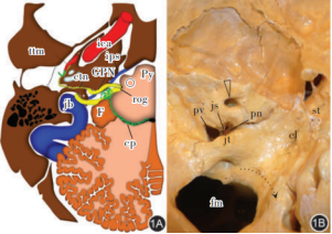
GPN occurs in the cerebellobulbar cistern directly below the medulla oblongata, at the level of the olive sulcus, between the olive nucleus and the inferior margin of the inferior foot of the cerebellum (
Figure 1)
| [3] | García Santos JM, Sánchez Jiménez S, Tovar Pérez M, et al. Tracking the glossopharyngeal nerve pathway through anatomical references in cross-sectional imaging techniques: a pictorial review [J]. Insights Imaging, 2018, 9 (4): 559-569.
https://doi.org/10.1007/s13244-018-0630-5 |
[3]
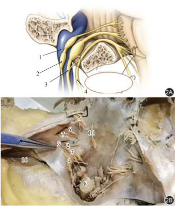
. At this level, it and the vagus and accessory nerves enter the jugular foramen at the posterior edge of the occipital jugular nodule, where the GPN enters the exclusive skull outlet (the jugular foramen nerve section) of the anterolateral part of the jugular foramen, vagus Nerve and accessory nerve accompanying internal jugular vein from the posteromedial part of the jugular foramen (the vascular part of the jugular foramen) out of the cranium (
Figure 2)
| [4] | Johal J, Iwanaga J, Tubbs K, et al. The accessory nerve: a comprehensive review of its anatomy, development, variations, landmarks and clinical considerations [J]. Anat Rec (Hoboken), 2019, 302 (4): 620-629. https://doi.org/10.1002/ar.23823 |
| [5] | Wang C, Kundaria S, Fernandez-Miranda J, et al. A description of the anatomy of the glossopharyngeal nerve as encountered in transoral surgery [J]. Laryngoscope, 2016, 126 (9): 2010-2015. https://doi.org/10.1002/lary.25706 |
| [6] | Prades JM, Gavid M, Asanau A, et al. Surgical anatomy of the styloid muscles and the extracranial glossopharyngeal nerve [J]. Surg Radiol Anat, 2014, 36 (2): 141-146.
https://doi.org/10.1007/s00276-013-1162-9 |
| [7] | Roldan-Valadez E, Martinez-Anda JJ, Corona-Cedillo R. 3T MRI and 128-slice dual-source CT cisternography images of the cranial nerves a brief pictorial review for clinicians [J]. Clin Anat, 2014, 27 (1): 31-45. https://doi.org/10.1002/ca.22311 |
[4-7]
, GPN has two enlarged ganglia in the jugular foramen: the upper ganglion processes general sensory information, and the lower ganglion processes visceral sensation: mainly the taste in the back 1/3 of the tongue and the innervation of the carotid sinus and body, and the tympanic nerve is divided from the lower ganglion, which enters the tympanic cavity at the level of the cochlear promontory in the medial wall through the tympanic tubule
| [3] | García Santos JM, Sánchez Jiménez S, Tovar Pérez M, et al. Tracking the glossopharyngeal nerve pathway through anatomical references in cross-sectional imaging techniques: a pictorial review [J]. Insights Imaging, 2018, 9 (4): 559-569.
https://doi.org/10.1007/s13244-018-0630-5 |
[3]
, forming a submucosal plexus that transmits sensory information, transmitting general sensation from the middle ear mucosa and the eustachian tube, and in the tympanic tube. The tendon level of tensor membranosus emits a small nerve branch (deep petrous nerve), which meets the facial nerve petrous nerve branch from the geniculate ganglion to synthesize the petrous nerve into the middle cranial fossa. After exiting the cranium, it is crosslinked with the mandibular branch of the trigeminal nerve through the auricular ganglion, and innervates the parotid gland through the auricular temporal branch of the trigeminal nerve
.
Note: GPN is the glossopharyngeal nerve. Figure 1A is a drawing of a transverse section at the medulla oblongata level (view from above). GPN is emitted from the olive sulcus (rog) and then passes through the cerebellobulbar cistern to the jugular foramen. Within the pool, it is located in the inferior and anterior fourth ventricle of the choroid plexus (cp) and the cerebellar flocculus (F). GPN leaves the medial part of the skull and the anterior part of the jugular foramen and is closely related to the inferior petrosal sinus (ips). Ctn is tympanic or Jacobson nerve; ica is the internal carotid artery; jb is the jugular bulb; ○ is the lower olive pit; Py is a pyramid. Figure 1B is a skull base (view from inside). The posterior cranial nerve is closely related to the jugular vein nodule (jt). The GPN (indicated by the black dashed arrow) exits the cranium through the nerve part (pn) of the jugular foramen, while the internal jugular vein and vagus nerve, accessory nerves exit the cranium from the vascular part (pv) of the jugular foramen, separated by a bone fibrous septum (js) between pn and pv. cl is a clivus; fm is a foramen magnum; st is the sella turcica; The triangular arrow is the inner auditory channel.
Figure 2. Schematic diagram of GPN, vagus nerve and accessory nerve exiting the jugular foramen.
Note: GPN is the glossopharyngeal nerve; Figure 2A is an anatomical schematic
| [4] | Johal J, Iwanaga J, Tubbs K, et al. The accessory nerve: a comprehensive review of its anatomy, development, variations, landmarks and clinical considerations [J]. Anat Rec (Hoboken), 2019, 302 (4): 620-629. https://doi.org/10.1002/ar.23823 |
[4]
, with GPN having two enlarged ganglia in the jugular foramen; Figure 2B is an anatomical view of the specimen
| [3] | García Santos JM, Sánchez Jiménez S, Tovar Pérez M, et al. Tracking the glossopharyngeal nerve pathway through anatomical references in cross-sectional imaging techniques: a pictorial review [J]. Insights Imaging, 2018, 9 (4): 559-569.
https://doi.org/10.1007/s13244-018-0630-5 |
[3]
. The GPN exits the cranium from the nerve part anterior and superior to the jugular foramen, and the vagus nerve and accessory nerves exit the cranium from the vascular part posterior and inferior to the jugular foramen. In the figure, 1 is the internal jugular vein, 2 is the GPN, 3 is the vagus nerve, 4 is the accessory nerve, 5 is the trigeminal nerve root, 6 is the facial nerve, 7 is the auditory nerve, 8 is the cervical spinal cord, 9 is the jugular foramen, 10 is the sigmoid sinus, and 11 is the abducens nerve

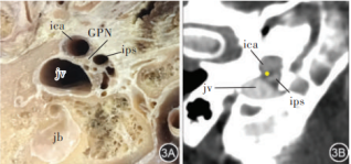
When GPN enters the jugular foramen, it is located in the GPN groove (the nerve part of the jugular foramen) in the petrous part of the jugular foramen. At the outer port of the jugular foramen, there is a dense connective tissue bundle to fix GPN to the bone margin, internal carotid artery and the initiation of internal carotid artery (
Figure 3). This connective tissue bundle helps to identify GPN
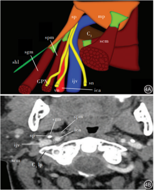
. After exiting the jugular foramen, the GPN is located medially of the internal jugular vein, laterally of the internal carotid artery and behind the styloid process, that is, in the internal jugular artery and venous space behind the styloid process
(
Figure 4)
| [3] | García Santos JM, Sánchez Jiménez S, Tovar Pérez M, et al. Tracking the glossopharyngeal nerve pathway through anatomical references in cross-sectional imaging techniques: a pictorial review [J]. Insights Imaging, 2018, 9 (4): 559-569.
https://doi.org/10.1007/s13244-018-0630-5 |
[3]
. Until it descends to the level of the C1 transverse process of the neck, GPN bypasses the carotid artery laterally, exits the internal carotid artery and vein space, and enters the posteromedial styloid process and behind the stylopharyngeal muscle
| [12] | Lai PF, Wu X, Lan SH, et al. Anatomical study of a surgical approach through the neck to the jugular foramen under endoscopy [J]. Surg Radiol Anat, 2021, 43 (2): 251-260.
https://doi.org/10.1007/S00276-020-02574-9 |
[12]
. Therefore, at the level of the C1 transverse process, the styloid process, the internal carotid artery and the stylopharyngeal muscle can be used as important localization markers to find GPN
| [6] | Prades JM, Gavid M, Asanau A, et al. Surgical anatomy of the styloid muscles and the extracranial glossopharyngeal nerve [J]. Surg Radiol Anat, 2014, 36 (2): 141-146.
https://doi.org/10.1007/s00276-013-1162-9 |
[6]
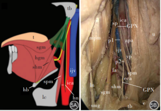
. Also at the level of the C1 transverse process, the carotid sinus and Hering's nerve emitted by GPN descend along the anterior wall of the internal carotid artery to the carotid sinus to participate in the decompression reflex. The main trunk of the GPN moves forward and down to the front of the styloid pharyngeal muscle, walking on a muscle tripod surrounded by the styloid-glossus, styloid-pharyngeal and styloid-hyoid muscles (styloid pyramid with facial artery at the base) (
Figure 5)
| [3] | García Santos JM, Sánchez Jiménez S, Tovar Pérez M, et al. Tracking the glossopharyngeal nerve pathway through anatomical references in cross-sectional imaging techniques: a pictorial review [J]. Insights Imaging, 2018, 9 (4): 559-569.
https://doi.org/10.1007/s13244-018-0630-5 |
[3]
inside. When it circles the exterior of the stylopharyngeal muscle to the medial side of its anterior surface and descends to the level of the middle tendon of the digastric muscle, the GPN advances into the oropharynx and divides into pharyngeal and lingual branches
| [7] | Roldan-Valadez E, Martinez-Anda JJ, Corona-Cedillo R. 3T MRI and 128-slice dual-source CT cisternography images of the cranial nerves a brief pictorial review for clinicians [J]. Clin Anat, 2014, 27 (1): 31-45. https://doi.org/10.1002/ca.22311 |
[7]
, the movement of the medial styloid pharyngeal muscle and the anterior styloid hyoid muscle are innervated, and the general sensation and taste of the oropharynx, soft palate, uvula, tonsil and the posterior 1/3 of the tongue are respectively.
Note: GPN is the glossopharyngeal nerve; Figure 3A The cadaveric portion is at the level of the skull base, and when viewed from below, the GPN exits the venous foramen near the inferior petrosal sinus (ips), the internal carotid artery (ica) and jugular vein (jv); jb is the jugular bulb; Figure 3B is a Computer tomography (CT) cross-sectional scan at the level of the outer orifice of the jugular foramen of the skull base; The GPN is located between the internal carotid artery (ica) and the jugular vein (jv), the position indicated by the yellow dot in the figure, and the ips is the inferior petrosal sinus.
Note: GPN is the glossopharyngeal nerve; In Figure 4A, GPN is located in the carotid space medial to the internal jugular vein (ijv) and lateral to the internal carotid artery (ica), showing the relationship between GPN and the internal carotid artery, internal carotid artery, styloid process, vagus nerve, and accessory nerves after leaving the left jugular foramen. The styloid process (sp) is its anterolateral reference point. In the first cervical vertebra (C1) Horizontal GPN surrounds to the anterolateral of the internal carotid artery and descends between the styloid process (sp) and the styloid pharyngeal muscle (spm). scm is sternocleidomastoid muscle; sgm is the styloglossus muscle; shl is the stylohyoid ligament; mp is the mastoid: sn is the accessory nerve; vn is the vagus nerve. Figure 4B is a right side of the neck, axial computed tomography angiogram (CTA) Soft tissue window at the level of the C1 transverse process (C1 lp) showing the positional relationship of internal carotid artery, internal jugular vein, styloid process and GPN at the level of the C1 transverse process. The GPN (arrow) is located anterolateral to the internal carotid artery, anteromedial to the internal jugular vein, and posteromedial to the styloid process. The GPN then circles to the anteromedial side of the internal carotid artery to the lateral side of the styloid pharyngeal muscle. At this time, the Hering nerve leaves the GPN and descends in front of the internal carotid artery to the carotid sinus and carotid body
Note: GPN is the glossopharyngeal nerve;
Figure 5A shows left GPN and styloid pyramid Positional relationship of a muscle tripod surrounded by styloid-glossus muscle (sgm), styloid-pharyngeal muscle (spm) and styloid-hyoid muscle (shm), with facial artery at the bottom, green vertebral shape: The main trunk of GPN bypasses anteriorly and internally at the level of the C1 transverse process to the front of the internal carotid artery (ica), branches out of Hering's nerve (hn), and then descends along the lateral side of styloid-pharyngeal muscle (spm) into the styloid pyramid. Figure 5B is an autopsy diagram of the right GPN walking in the neck. This figure exposes the upper and lower segments of the GPN, with the middle segment of the GPN located in the styloid pharyngeal muscle (spm) Posterolateral. apa is the ascending pharyngeal artery, e is the epiglottis, fa is the facial artery, ica is the internal carotid artery, ijv is the internal jugular vein, eca is the external carotid artery, ln is the lingual nerve, mpm is the internal pterygoid muscle, p1 is the upper low point of gpn, p2 is the lower apex of gpn, sgm is the styloid glossus muscle, shm is the styloid hyoid muscle, sp is the styloid process, smg is the submandibular gland, spm is the styloid pharyngeal muscle, tb is the base of the tongue, u is the uvula, and vp is the pharyngeal branch of the vagus nerve
It can be seen that GPN is emitted from the medulla oblongata and then passes transversely through the medulla oblongata cistern of the cerebellum to the nerve part on the anterior and medial side of the jugular foramen, giving off the sensation that the tympanic nerve innervates the tympanic cavity and the mucosa of the eustachian tube, and has branches that cross-link with the facial nerve to form lesser petrosal nerve, which enter the auricular temporal branch of the trigeminal nerve to innervate the parotid gland after substitution through the auricular ganglion; After exiting the jugular foramen, it descends along the internal carotid artery and venous space to the level of C1 transverse process to emit carotid sinus and carotid body nerve, and descends along the anterior wall of internal carotid artery to carotid sinus to participate in decompression reflex; The main trunk of GPN descends to the anteromedial side of the internal carotid artery, and the posterior medial side of the styloid process descends along the anterolateral side of the styloid pharyngeal muscle to the oropharynx to form pharyngeal branches and lingual branches, which govern the movement of pharyngeal muscle, the sensation of oropharyngeal area and the taste of the posterior 1/3 of the tongue. During the whole GPN journey, although there are clear bone markers in the jugular foramen, it is too close to the internal jugular artery, vein, vagus nerve and accessory nerve, so it is not suitable for puncture block target. At the level of C1 transverse process, GPN gradually shifts out from the internal carotid artery and venous space, and travels to the posteromedial styloid process, the anterolateral internal carotid artery, and the anteromedial styloid pharyngeal muscle. There are both C1 transverse process and styloid process as bone markers, internal carotid artery as vascular markers, and styloid pharyngeal muscle as soft tissue markers
| [3] | García Santos JM, Sánchez Jiménez S, Tovar Pérez M, et al. Tracking the glossopharyngeal nerve pathway through anatomical references in cross-sectional imaging techniques: a pictorial review [J]. Insights Imaging, 2018, 9 (4): 559-569.
https://doi.org/10.1007/s13244-018-0630-5 |
| [5] | Wang C, Kundaria S, Fernandez-Miranda J, et al. A description of the anatomy of the glossopharyngeal nerve as encountered in transoral surgery [J]. Laryngoscope, 2016, 126 (9): 2010-2015. https://doi.org/10.1002/lary.25706 |
| [6] | Prades JM, Gavid M, Asanau A, et al. Surgical anatomy of the styloid muscles and the extracranial glossopharyngeal nerve [J]. Surg Radiol Anat, 2014, 36 (2): 141-146.
https://doi.org/10.1007/s00276-013-1162-9 |
| [7] | Roldan-Valadez E, Martinez-Anda JJ, Corona-Cedillo R. 3T MRI and 128-slice dual-source CT cisternography images of the cranial nerves a brief pictorial review for clinicians [J]. Clin Anat, 2014, 27 (1): 31-45. https://doi.org/10.1002/ca.22311 |
| [13] | Ozveren MF, Türe U, Ozek MM, et al. Anatomic landmarks of the glossopharyngeal nerve: a microsurgical anatomic study [J]. Neurosurgery, 2003, 52 (6): 1400-1410; discussion 1410.
https://doi.org/10.1227/01.neu.0000064807.62571.02 |
[3, 5-7, 13]
. Regardless of CT or ultrasound guidance, this should be the best target choice for GPN block or radiofrequency. When GPN enters the oropharynx, it can barely be used as nerve block here because of its small and diffuse branches
| [5] | Wang C, Kundaria S, Fernandez-Miranda J, et al. A description of the anatomy of the glossopharyngeal nerve as encountered in transoral surgery [J]. Laryngoscope, 2016, 126 (9): 2010-2015. https://doi.org/10.1002/lary.25706 |
[5]
, but it should not be used as a target for radiofrequency therapy.
3. Diagnosis and Treatment of Glossopharyngeal Nerve Dysfunction
3.1. Clinical Manifestations of GPN Dysfunction
Although isolated GPN involvement is rare, the main clinical manifestations of clinical GPN dysfunction are GPN stimulation (GPN pain) and GPN disruption. The clinical manifestation of GPN pain is paroxysmal severe pain in the area of GPN distribution. Pain is generally induced by swallowing, chewing, coughing or speaking, mostly unilateral pain, and very few patients have bilateral pain. The pain is distributed in the pharynx, tonsil area, base of the tongue, front of the ear and deep in the ear. It feels like tear, electric shock, acupuncture, knife cut or burning, and it is more common in middle-aged and elderly people over 40 years old. In few cases, pain is also accompanied by bradycardia, and some patients can be combined with fainting. The incidence of GPN pain years was approximately 0.7 per 100 000 patients
| [14] | Katusic S, Williams DB, Beard CM, et al. Incidence and clinical features of glossopharyngeal neuralgia, Rochester, Minnesota, 1945-1984 [J]. Neuroepidemiology 1991, 10 (5-6): 266-275. https://doi.org/10.1159/000110283 |
[14]
. Its etiology and pathogenesis are similar to those of trigeminal neuralgia, which are mostly caused by the demyelination of nerve roots caused by the compression of GPN roots in the cerebellobulbar cistern of the posterior fossa by the posterior inferior cerebellar artery. If GPN is damaged, it will be manifested as hypoesthesia in the oropharyngeal area, disappearance of taste in the posterior 1/3 of the tongue and dysphagia. At this time, the uvula will bias to the healthy side, and mild atrophy of parotid denervation and carotid sinus dysfunction may also occur: such as instability of heart rhythm and blood pressure
| [3] | García Santos JM, Sánchez Jiménez S, Tovar Pérez M, et al. Tracking the glossopharyngeal nerve pathway through anatomical references in cross-sectional imaging techniques: a pictorial review [J]. Insights Imaging, 2018, 9 (4): 559-569.
https://doi.org/10.1007/s13244-018-0630-5 |
[3]
.
3.2. Diagnosis and Differential Diagnosis of GPN Dysfunction
According to the clinical manifestations of GPN dysfunction, it is often possible to preliminarily judge whether it is GPN pain or GPN destructive injury. Patients with initial diagnosis of GPN pain must undergo cranial Magnetic Resonance Imaging(MRI) and skull base CT scan to further rule out intracranial or skull base space-occupying lesions and determine whether the nerve roots of GPN are accompanied by vascular compression
| [15] | Functional Neurosurgery Group of Neurosurgery Branch of Chinese Medical Association, Expert Committee of Functional Neurosurgery of Neurosurgeon Branch of Chinese Medical Physicians Association, Neurosurgery Branch of Beijing Medical Association, etc. Chinese expert consensus on microvascular decompression for trigeminal neuralgia and glossopharyngeal neuralgia (2015) [J]. Chinese Journal of Neurosurgery, 2015, 31(3): 217⁃220.
https://doi.org/10.3760/cma.j.issn.1001-2346.2015.03.001 |
[15]
. At the same time, it needs to be distinguished from the pain of the mandibular branch of the trigeminal nerve: the nature of the pain attack is the same, but the "trigger point" of the former is mostly located in the lower lip, lower gum or mandibular skin; The latter "trigger point" is mostly located in the pharyngeal mucosa, that is, the former touches the mandible, lower gum or anterior side of the tongue and easily induces pain attacks, while the latter often induces pain attacks when swallowing. At this time, local anesthetic topical anesthesia test or GPN block can be used for differential diagnosis on the mucosa of the throat: if the pain cannot be induced after topical anesthesia or GPN block of the throat, it is GPN pain, otherwise it is pain in the mandibular branch of trigeminal nerve.
If there are clinical manifestations of GPN damage, in addition to MRI of the skull and plain CT scan of the skull base to find out the cause, it should also be asked whether there is a history of tumor radiotherapy or surgery of the mandibular region of the skull base to further confirm the diagnosis.
3.3. Indications and Contraindications for GPN Block
Indications for GPN block include, (1) to diagnose GPN pain, need Differential diagnosis from trigeminal neuralgia; (2) those with confirmed GPN pain, poor conservative treatment with oral analgesics or unable to tolerate adverse drug reactions; (3) those who are unwilling to undergo craniotomy and microvascular decompression treatment for confirmed GPN pain without conservative treatment; (4) Patients with ineffective or recurrent craniotomy and microvascular decompression treatment for confirmed GPN pain. However, those with severe local or systemic infection at the site to be punctured, those with severe coagulation dysfunction, those who cannot communicate and cooperate, and those who are unwilling to accept GPN block are all contraindications for GPN block.
3.4. Operation Techniques of GPN Block
Generalized GPN block includes four methods: (1) topical anesthesia of GPN lingual branch and pharyngeal branch nerve endings in pharyngeal mucosa; (2) Submucosal injection area block of throat; (3) GPN trunk block; (4) GPN trunk radiofrequency therapy. Because the first two techniques are extremely simple, they can only be used for diagnostic block for differential diagnosis or temporary analgesia; GPN stem block can be used for differential diagnosis by local anesthetic block, and long-acting nerve damaging drugs (absolute ethanol, phenolic glycerol, methylene blue, doxorubicin) can be used to achieve the therapeutic effect on GPN pain. However, GPN is accompanied by vagus nerve and accessory nerve from the cranium, and the three of them exit the cranial cavity from the jugular foramen together, and travel together in the internal jugular artery and venous sulcus, and gradually separate until they travel to the level of C1 transverse process. Therefore, it is difficult to avoid injury to the accompanying accessory nerve and vagus nerve by using chemical destructive drugs to block GPN pain. Although accessory nerve injury does not seem to cause serious consequences, hoarseness and cough caused by vagus nerve injury are often catastrophic consequences. Therefore, GPN block technology used for GPN pain treatment actually refers to GPN trunk radio-frequency ablation (RFA).
3.4.1. Preoperative Preparation
The diagnosis and differential diagnosis were confirmed again, and the indications for GPN block treatment were confirmed, and contraindications were excluded. Inform patients about the advantages and disadvantages of various GPN blocks and alternatives, and sign informed consent. Prepare nerve block medication, emergency medication, radio frequency instrument, monitor, emergency endotracheal intubation kit and simple respirator. Fast for 6 h before treatment and open upper extremity intravenous fluid channel for later use.
3.4.2. Topical Anesthesia of GPN Lingual and Pharyngeal Branch Nerve Endings in Pharyngeal Mucosa
Instruct the patient to tilt his head back and open his mouth to the maximum extent. Take a topical anesthesia spray gun and spray 1% tetracaine or 2% lidocaine 2~3 ml into the pharynx. Instruct the patient to keep his mouth open and tilt back, and not swallow the sprayed local anesthetic. After 5~10 minutes, spit out the pharyngeal liquid. Ask if the oropharynx is numb. If there is no numbness, spray again, but the total amount should not exceed 5 ml. When the patient complains of pharyngeal numbness, observe whether the original pain disappears. If so, let the patient swallow or touch with a cotton swab to test whether it can induce the pain.
3.4.3. Submucosal Regional Block of Throat
Instruct the patient to sit under the bright light (shadowless lamp or headlamp or flashlight source), tilt his head back and open his mouth to the maximum extent. After sterilizing the pharynx with iodophor, hold a syringe with a No. 5 needle to puncture the submucosa of the affected pharyngeal wall. After withdrawing without blood, inject 5ml of 2% lidocaine into the mucosal bulge (multiple injections can be made, but the depth is limited to the submucosa). Fill sterile dry cotton balls or gauze in the pharynx and close the mouth to prevent bleeding from the puncture point. After 5 to 10 minutes, the pharyngeal tamponade was removed and asked if the original pain had disappeared. If so, test whether pharyngeal touch can still induce the original pain. It is not advisable to inject chemically damaging drugs such as absolute ethanol or phenolic glycerol to prevent pharyngeal mucosal necrosis.
3.4.4. GPN Trunk Block
From the anatomy of GPN, it can be seen that although the jugular foramen is the cranial outlet of GPN, GPN is too adhered to the internal jugular vein, vagus nerve, accessory nerve and internal carotid artery here, so it is not suitable for block or radiofrequency therapy target. At the level of the C1 transverse process, the distance between the three cranial nerves begins to open. At this level, the GPN trunk is located posteromedially of the styloid process, in front of the internal carotid artery, and posterolaterally of the styloid pharyngeal muscle (see
Figures 4 and 5). There are both bone markers and vascular and muscular markers here, which should be the best puncture target for GPN trunk block or radiofrequency. Although the distance from the target to the skin is shallow, because its rear is adjacent to the internal jugular vein and internal carotid artery, in order to improve the puncture accuracy, reduce puncture complications, and achieve the purpose of precision medicine, expert consensus recommends that GPN trunk block or radiofrequency puncture should be completed under the guidance of accurate and definite ultrasound or CT, and blind puncture with bare hands should be resolutely abandoned.
3.4.5. Ultrasound-guided GPN Trunk block
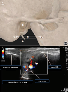
The patient is instructed to lie on his side on the operating table, and the high-frequency linear array probe is placed between the mandible and the mastoid process. Under ultrasound, three highly bright bone echoes of the mastoid process, styloid process and mandible and the external carotid artery on the superficial surface of the styloid process can be seen (
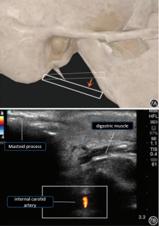
Figure 6). Then, with the mastoid as the origin, the mandibular end probe is rotated to the caudal side until the styloid process disappears, and the internal carotid artery in the digastric muscle and the deep surface of the styloid process is visible (
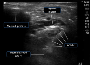
Figure 7). Subsequently, ultrasound-guided in-plane puncture was performed, the needle tip was punctured to the superficial surface of the internal carotid artery (
Figure 8), and the nerve localization stimulator assisted in positioning the GPN. The needle tip was fine-tuned on the superficial surface of the internal carotid artery until the GPN was determined, and the drug was injected to complete the GPN block.
Figure 6. Schematic diagram of high-frequency probe placement position and ultrasound Doppler findings.
Note: Figure 6A is a schematic diagram of the placement position of the high-frequency probe, where A is the styloid process, B is the mastoid process, C is the mandible, D is the ultrasound probe, and E is the external auditory canal. Figure 6B is an ultrasound Doppler image, in which the red dots in the box are arteries, the blue and green dots are veins, and the white dots are bone
Figure 7. Rotating a high-frequency probe to find the internal carotid artery.
Note: Figure 7A is a schematic diagram of the position of the ultrasound probe, and Figure 7B is an ultrasound view at this position; B-ultrasound-guided glossopharyngeal nerve block puncture, puncture needle shadow is blurry, not as clear as that guided by CT.
Figure 8. In-plane puncture to superficial surface of internal carotid artery.
3.4.6. CT-Guided GPN Trunk Block
(i). Posture and Puncture Path Design
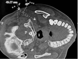
Figure 9. CT-guided GPN trunk block puncture pathway design.
Note: GPN is the glossopharyngeal nerve; In the figure, A is the styloid process, B is the transverse process of the atlas, C is the vertebral artery, D is the atlas, E is the odontoid process, F is the cervical spinal cord, G is the cervical occipital condyle, H is the occipital bone, I is the pharyngeal cavity, J is the mandible, K teeth, L mastoid process, O is the puncture point, 42.17 mm is the puncture depth (distance from the puncture point to the target), and an angle of 49°is the puncture Angle (the angle between the puncture route and the sagittal plane), the 8 small circles at the top of the figure are the positioning grids placed on the body surface
The patient entered the CT treatment room, lying on his side on the CT operating table, routinely monitoring blood pressure, electrocardiogram, blood oxygen saturation, and giving nasal tube oxygen inhalation, and placing a CT positioning grid in the area below the ear lobe. The cranial localization image was taken in the paranasal sinus mode
| [16] | Xing W, Huang B, Chu DT, et al. Radiation dose and protection measures during simulating CT guided percutaneous radiofrequency puncture in patients with trigeminal neuralgia [J]. Chinese Journal of Painology, 2020, 16 (3): 220-224.
https://doi.org/10.3760/cma.j.cn101379-20200108⁃00004 |
[16]
, and the axial scan of the mastoid styloid process region was performed with a layer thickness of 2 mm, and the resulting picture was played back. The layer containing C1 transverse process and styloid process was selected as the puncture layer, and the area where the posteromedial styloid process, the anterior internal carotid artery and the lateral pharyngeal muscle of styloid process were located in this layer is the puncture target. Use CT self-contained measurement tool software to draw a straight line from the target point upward to avoid the internal jugular vein and bone obstruction. The intersection point between this line and the skin in the lateral cervical region is the puncture point. Measure the distance from puncture point to target point (puncture depth) and the angle between the puncture route and the sagittal plane (puncture angle) (
Figure 9).
(ii). Puncture and Block Operation
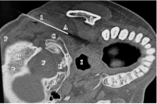
Figure 10. CT images of CT-guided GPN trunk block puncture in place with the needle tip at the level of the transverse process of the atlas and the posteromedial styloid process.
Note: GPN is the glossopharyngeal nerve; In the
Figure 1 is the puncture needle, A is the styloid process, C is the vertebral artery, D is the atlas, E is the odontoid process, F is the cervical spinal cord, G is the cervical occipital condyle, H is the occipital bone, I is the pharyngeal cavity, and J is the mandible
The puncture point was subjected to local anesthesia with 2% lidocaine 2 ml, and the 5 cm-long gauge needle was used to puncture slowly and forcefully to the target point (the area where the posteromedial styloid process, the anterior internal carotid artery and the lateral styloid pharyngeal muscle are located) under the guidance of CT according to the designed puncture path (
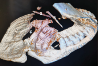
Figure 10), and the three-dimensional reconstruction observation of CT scan was confirmed (
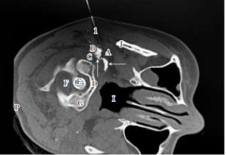
Figure 11). Slowly inject 2-3 ml of a local anesthetic compound solution containing 3% iohexol, after aspiration shows no blood (for diagnostic block, only 2 ml of 2% lidocaine 0.4 ml of 3% iohexol, diluted with saline to 4 ml).Iohexol is a nonionic contrast agent, and iohexol is routinely added in diagnostic block and treatment to observe the distribution of drug solution. For the treatment of GPN pain, preparation of liquid: 2% lidocaine (or 0.75% ropivacaine) 2 ml + vitamin B12 (Mecobalamin 0.5 mg) 1 ml +3% iohexol 0.4 ml, an appropriate amount of water-soluble glucocorticoid can be added as appropriate, diluted to 4 ml with normal saline. CT scan again, observe the distribution of the injected drug solution (
Figure 12) and observe the blocking effect.
Figure 11. CT 3D reconstruction of GPN trunk block puncture in place.
Note: GPN is the glossopharyngeal nerve; 1 is the puncture needle, 2 is the styloid process, 3 is the transverse process of the atlas, 4 is the atlas of the mandible, 5 is the occipital bone, and 6 is the foramen magnum of the occipital bone
Figure 12. CT images after GPN trunk block.
Note: GPN is the glossopharyngeal nerve; 1 is the puncture needle, A is the styloid process, B is the transverse process of the atlas, C is the vertebral artery, D is the atlas, E is the odontoid process, F is the cervical spinal cord, G is the cervical occipital condyle, I is the pharyngeal cavity, and J is the mandible; The white irregular shape indicated by the arrow is GPN blocking solution (containing 3% iohexol); The insulating layer of the needle body is white, the printing depth mark is printed, and the needle tip is round and blunt, which can avoid cutting damage to tissue, thereby reducing hematoma formation and uncontrollable nerve damage
3.4.7. GPN Trunk RFA
The posture and puncture path design of the patient for puncture and electrophysiological test are the same as before. The puncture needle is replaced with a special radiofrequency needle for blunt facial nerve radiofrequency with a length of 10 cm and a naked end length of 5 mm (patent number ZL20212 2310301.2, produced by Henan Tuoren Medical Equipment Co., Ltd.), and the puncture is guided by ultrasound or CT to the target (posteromedial styloid process at the level of C1 transverse process, front of internal carotid artery and front of styloid pharyngeal muscle), Perform sensory and motor electrical stimulation tests with a current of 0.5-1.0 mA at a frequency of 50 Hz and a pulse width 0.1 mS, and a frequency of 2 Hz and a pulse width of 1 ms, respectively. If the former can induce a tingling sensation in the original pain area of the ipsilateral pharynx, and the patient has no nausea, cough, and sudden changes in heart rate, and the latter has no rhythmic jitter of neck and shoulder muscles, then radio frequency therapy is feasible. Otherwise, the position of the needle tip is adjusted before the electrical stimulation test is performed, and the radiofrequency treatment can only be performed when the pharyngeal paresthesia is only positive.
After the electrophysiological test of treatment operation in different RF modes confirms that the RF needle tip is close to the GPN, standard pulse RF (42 ℃, frequency 2 Hz, pulse width 20 ms, voltage 37~41 V, time 120~360 s)
| [17] | Bharti N, Chattopadhyay S, Singla N, et al. Pulsed radiofrequency ablation for the treatment of glossopharyngeal neuralgia secondary to oropharyngeal carcinoma [J]. Pain Physician, 2018, 21 (3): 295-302. https://doi.org/10.36076/ppj.2018.3.295 |
| [18] | Jia Y, Shrestha N, Wang X, et al. The Long-Term outcome of CT-Guided pulsed radiofrequency in the treatment of idiopathic glossopharyngeal neuralgia: A retrospective Multi-Center case series [J]. J Pain Res, 2020, 13: 2093-2102.
https://doi.org/10.2147/JPR.S259994 |
[17, 18]
or high voltage long-range pulse RF (42-46 ℃, frequency 2 Hz, pulse width 20 ms, voltage 50~90 V, time 900 s) can be selected
| [19] | Jia Donglin, Yi Duan, Li Shuiqing. Analysis of therapeutic effect of high voltage long-duration pulsed radiofrequency in the treatment of neuralgia after lumbar spine surgery [J]. National Medical Journal of China, 2021, 101 (43): 3569-3574.
https://doi.org/10.3760/cma.j.cn112137-20210407-00828 |
| [20] | Pain Branch of Chinese Medical Association. Consensus of experts in pain department of radiofrequency therapy technology [J]. National Medical Journal of China, 2019, 99 (45): 3547-3553. https://doi.org/10.3760/cma.j.issn.0376⁃2491.2019.45.004 |
[19, 20]
; Standard continuous radio frequency (temperature 70~90 ℃, time 180~300 s) can also be selected
| [21] | Ren Y E, Du Y M, Cong H J, et al.Efficacy of radiofrequency thermocoagulation guided by three-dimensional computer tomography for treatment of severe recurrent glossopharyngeal neuralgia[J]. Chinese Journal of Anesthesiology, 2017, 37(11): 1368⁃1370. https://doi.org/10.3760/cma.j.issn.0254-1416.2017.11.022 |
| [22] | Wang Xiaoping, Zuo Xinlu, Zeng Yuanjie, et al. Long-term efficacy of CT-guided radiofrequency thermocoagulation in the treatment of glossopharyngeal neuralgia [J]. China General Practice, 2016, 19 (12): 1379-1382.
https://doi.org/10.3969/j.issn.1007-9572.2016.12.005 |
| [23] | Huang Bing, Wu Dan, Du Xindan, et al. The observation of CT guided bipolar radiofrequency thermocoagulation behind styloid process in the treatment of patients with glossopharyngeal neuralgia[J]. Chinese Journal of Painology, 2021, 17 (2): 178-185. https://doi.org/10.3760/cma.j.cn101658-20200226-00027 |
| [24] | Telischak NA, Heit JJ, Campos LW, et al. Fluoroscopic C-arm and CT-guided selective radiofrequency ablation for trigeminal and glossopharyngeal facial pain syndromes [J]. Pain Med, 2018, 19(1): 130-141.
https://doi.org/10.1093/pm/pnx088 |
| [25] | Song L, He L, Pei Q, et al. CT-guided percutaneous radiofrequency thermocoagulation for glossopharyngeal neuralgia: a retrospective clinical study of 117 cases [J]. Clin Neurol Neurosurg, 2019, 178: 42-45.
https://doi.org/10.1016/j.clineuro.2019.01.013 |
| [26] | Zhu Q, Wang S, Chen R, et al. Continuous radiofrequency thermocoagulation under CT-guidance for glossopharyngeal neuralgia: two case reports [J]. Medicine (Baltimore), 2018, 97(24): e11079. https://doi.org/10.1097/MD.0000000000011079 |
[21-26]
. The standard pulsed radiofrequency temperature is generally no higher than 42℃, which will not cause protein denaturation, and the curative effect is poor (46.2% ~93.0% of patients with immediate pain relief of 50% after surgery, and the time effect is short for 2~7 months)
| [17] | Bharti N, Chattopadhyay S, Singla N, et al. Pulsed radiofrequency ablation for the treatment of glossopharyngeal neuralgia secondary to oropharyngeal carcinoma [J]. Pain Physician, 2018, 21 (3): 295-302. https://doi.org/10.36076/ppj.2018.3.295 |
| [18] | Jia Y, Shrestha N, Wang X, et al. The Long-Term outcome of CT-Guided pulsed radiofrequency in the treatment of idiopathic glossopharyngeal neuralgia: A retrospective Multi-Center case series [J]. J Pain Res, 2020, 13: 2093-2102.
https://doi.org/10.2147/JPR.S259994 |
[17, 18]
, but it is relatively safe and will not cause long-term nerve damage; Standard continuous radiofrequency can directly damage the target nerve by high-temperature thermocoagulation, so the curative effect is definite (the rate of complete disappearance of pain immediately after operation is 78.8% ~97.0%), and the duration is long-lasting (1~10 years)
| [21] | Ren Y E, Du Y M, Cong H J, et al.Efficacy of radiofrequency thermocoagulation guided by three-dimensional computer tomography for treatment of severe recurrent glossopharyngeal neuralgia[J]. Chinese Journal of Anesthesiology, 2017, 37(11): 1368⁃1370. https://doi.org/10.3760/cma.j.issn.0254-1416.2017.11.022 |
| [22] | Wang Xiaoping, Zuo Xinlu, Zeng Yuanjie, et al. Long-term efficacy of CT-guided radiofrequency thermocoagulation in the treatment of glossopharyngeal neuralgia [J]. China General Practice, 2016, 19 (12): 1379-1382.
https://doi.org/10.3969/j.issn.1007-9572.2016.12.005 |
| [23] | Huang Bing, Wu Dan, Du Xindan, et al. The observation of CT guided bipolar radiofrequency thermocoagulation behind styloid process in the treatment of patients with glossopharyngeal neuralgia[J]. Chinese Journal of Painology, 2021, 17 (2): 178-185. https://doi.org/10.3760/cma.j.cn101658-20200226-00027 |
| [24] | Telischak NA, Heit JJ, Campos LW, et al. Fluoroscopic C-arm and CT-guided selective radiofrequency ablation for trigeminal and glossopharyngeal facial pain syndromes [J]. Pain Med, 2018, 19(1): 130-141.
https://doi.org/10.1093/pm/pnx088 |
| [25] | Song L, He L, Pei Q, et al. CT-guided percutaneous radiofrequency thermocoagulation for glossopharyngeal neuralgia: a retrospective clinical study of 117 cases [J]. Clin Neurol Neurosurg, 2019, 178: 42-45.
https://doi.org/10.1016/j.clineuro.2019.01.013 |
| [26] | Zhu Q, Wang S, Chen R, et al. Continuous radiofrequency thermocoagulation under CT-guidance for glossopharyngeal neuralgia: two case reports [J]. Medicine (Baltimore), 2018, 97(24): e11079. https://doi.org/10.1097/MD.0000000000011079 |
[21-26]
. However, once the vagus nerve is involved, it often causes weeks, months or even permanent dysphagia and hoarseness. The efficacy of high voltage long-range pulsed radio frequency is better than that of standard pulsed radio frequency, although it is still far lower than that of standard continuous radio frequency, but its safety is higher than that of standard continuous radio frequency. For doctors who are new to GPN RFA, this consensus recommends high-voltage long-duration pulsed RF therapy. Only after accumulating rich clinical experience can the standard continuous radiofrequency mode be prudently applied to the treatment of GPN pain.
During the implementation of standard radiofrequency, the patient should be instructed to continuously make the same sound (for example, "My name is Zhang San"), and the patient's heart rate changes should be closely monitored. Once nausea, a sudden increase or decrease in heart rate, or a change in voice tone (becoming low-pitched or hoarse) occurs, the radiofrequency procedure must be terminated immediately, and several milliliters of saline should be injected into the patient's mouth and instructed to be swallowed. Observe the original pain can still be induced and whether there is dysphagia or a choking reaction. If swallowing can no longer induce a pain attack, the treatment is ended; if it can induce a pain attack and there is no choking or hoarseness, the needle tip position should be appropriately adjusted before performing standard radiofrequency again; if it can still induce a pain attack and there is choking and/or hoarseness, the treatment is considered failed, and the patient should be returned to the ward for symptomatic treatment. After the vagus nerve injury recovers, GPN radiofrequency procedure or other treatment methods (such as microvascular decompression surgery, etc.) can be sought. For the specific operation of GPN radiofrequency, see Video 2 and 3.
4. GPN Block or RFA Precautions
4.1. Intraoperative Monitoring and Prohibition of Bilateral GPN Block
Closely monitor the patient's vital signs during GPN trunk block or radiofrequency therapy, carefully observe the patient's response during puncture and radiofrequency, and do not perform bilateral GPN block at the same time. Mainly observe the possible vascular injury during puncture (mistakenly puncture of internal jugular artery and vein) to form hematoma and whether there are complications such as hypertension, arrhythmia, nausea, choking, hoarseness, weakness of tongue extension and facial paralysis during radiofrequency treatment. Moreover, GPN and vagus nerve are relatively adjacent in the posterior styloid space. If bilateral GPN block is performed, it may block bilateral vagus nerve at the same time, resulting in severe hoarseness and dysphagia, and even severe arrhythmia, which is life-threatening. Therefore, bilateral retrostyloid GPN blocks should not be performed. During block or radiofrequency therapy, the methods reported by Ren Xiaomei et al.
should also be used for monitoring and management, and when necessary, the methods described by Pan Lijuan et al.
| [28] | Pan Lijuan, Huang Bing, Ren Xiaoshu, et al. Management and prevention of critical events in the treatment of trigeminal neuralgia with extracranial non-semilunar radiofrequency thermocoagulation [J]. China Journal of Emergency Resuscitation and Disaster Medicine, 2018, 13 (10): 963-965.
https://doi.org/10.3969/j.issn.1673⁃6966.2018.10.009 |
[28]
should be used to handle critical events to ensure the safe conduct of GPN block/RFA therapy.
4.2. Local Anesthetic Poisoning Should be Prevented During GPN Block
When performing GPN block, whether topical anesthesia or submucosal injection area block or nerve trunk block, the single dose of local anesthetic should be less than 100 mg converted into lidocaine, so as to prevent toxic reactions caused by mistaken injection of local anesthetic into the internal carotid artery, vein or other blood vessels in the neck. If glucocorticoids need to be used in the blocking solution, the use of suspension preparations should be prohibited to avoid microembolism of cerebral blood vessels caused by injection into the internal carotid artery.
4.3. GPN Blocking Try to Avoid the Use of Chemically Destructive Drugs
During GPN block, because the distribution of the injected drug is uncontrollable
, chemical neurolytic drugs should not be used to prevent accumulation and damage the adjacent vagus, accessory, and facial nerves. More controlled GPN trunk RFA technology should be used instead of injecting chemically destructive drugs to avoid non-responsible nerve injury complications. Although various imaging-guided use of anhydrous alcohol, phenolic glycerol, and doxorubicin alone or in combination with radiofrequency has been reported for chemodestructive block treatment of GPN pain
| [30] | Cheng Xinzhi, Wu Haisheng. Efficacy of Glossopharyngeal Nerve Injection of Doxorubicin Guided by Neurostimulator in the Treatment of Glossopharyngeal Neuralgia [J]. Pain Clinic Journal, 2019, 15 (5): 348-351.
https://doi.org/10.3760/cma.j.issn.1672-9633.2019.05.006 |
| [31] | Song Yinghao, Ling Bingyu, Guan Jingjing, et al. Therapeutic effect of CT-guided radiofrequency thermocoagulation combined with doxorubicin injection in the treatment of primary glossopharyngeal neuralgia [J]. Chinese Journal of Painology, 2022, 18 (1): 72-76.
https://doi.org/10.3760/cma.j.CN101658-20200225-00026 |
[30, 31]
, it may spread to the vagus nerve and cause hoarseness and drinking water choking cough, which takes several months and is difficult to recover. The accumulated lung infection and inflammation that follows often have catastrophic consequences and should be discarded.
4.4. A Blunt Puncture Needle Should be Used in GPN RFA
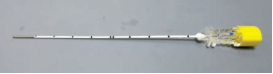
When performing GPN block, whether it is injection or RFA therapy, the use of sharp puncture needles should be avoided. It is recommended to use the special blunt-tip radiofrequency puncture needle (
Figure 13, patent number ZL2017 1635823.7) developed by the author. The insulating layer of the needle body is white, the printed depth mark, and the needle tip is round and blunt, It can avoid cutting nerves or blood vessels along the puncture path, thereby reducing complications from accidental vagus nerve injury and the formation of neck hematomas caused by the of the internal carotid artery or vein. When performing extracranial nerve radiofrequency treatment for hemifacial spasm
| [32] | Huang B, Yao M, Chen Q, et al. Awake CT-guided percutaneous stylomastoid foramen puncture and radiofrequency ablation of facial nerve for treatment of hemifacial spasm [J]. J Neurosurg, 2021: 135(5):1459-1465
https://doi.org/10.3171/2020.10.JNS203209 |
[32]
and Meige syndrome
| [33] | Huang B, Du XD, Yao M, et al. CT-guided radiofrequency ablation of the extracranial cranial nerve for the treatment of Meige's syndrome [J]. Front Neurosci, 2022, 16: 1013555.
https://doi.org/10.3389/fnins.2022.1013555 |
[33]
, it was also found that the sharp puncture needle can cause uncontrollable facial nerve puncture and cutting injury, which makes it difficult to control the degree of facial paralysis. Therefore, it is proposed that the blunt radiofrequency needle of this patent should be used in cranial nerve block or radiofrequency, so as to avoid the cutting injury caused by the sharp puncture needle tip mistakenly hitting the accessory nerve or vagus nerve
| [34] | Huang Bing, Yan Min, Lin Huidan, et al. Consensus of Chinese pain experts on the application of facial nerve block technique in facial nerve dysfunction diseases(2022 edition) [J]. Chinese Journal of Painology, 2022, 18 (2): 152-159.
https://doi.org/10.3760/cma.j.cn101658-20220227-00034 |
| [35] | Huang Bing, Du Xindan, Huang Hao, et al. Technique and efficacy of CT-guided radiofrequency puncture of stylomastoid foramen for primary hemifacial spasm [J]. Chinese Journal of Painology, 2020, 16 (5): 386-393.
https://doi.org/10.3760/cma.j.cn101658-20200108-00005 |
| [36] | Head and face pain group of pain branch of Chinese medical association. Consensus of Chinese experts on technical specifications for radiofrequency treatment of hemifacial spasm with stylomastoid facial nerve guided by CT [J]. National Medical Journal of China, 2022, 102 (17): 1267-1271.
https://doi.org/10.3760/cma.j.cn112137-20211222-02852 |
[34-36]
.
Figure 13. Special cranial nerve radiofrequency puncture needle, which is characterized by a blunt needle tip, white insulation layer of needle body and depth mark (it has been converted and produced by Henan Tuoren Medical Equipment Co., Ltd.).
4.5.Preoperative Neck CTA and Addition of Iohexol to the Block Solution
In order to reduce mistaken puncture of internal carotid artery and internal jugular vein, CTA of the neck should be performed before CT-guided GPN trunk block / RFA puncture, and the contrast agent iohexol should be added to the injected drug solution. Unlike ultrasound-guided puncture, the internal jugular artery and vein could not be clearly developed during CT-guided GPN puncture. If CTA examination is done before operation, the positional relationship between the styloid process and the internal jugular artery and vein at the puncture level of the patient can be grasped, so as to avoid blood vessels when designing the puncture path, so as to reduce the risk of neck hematoma caused by accidental puncture of blood vessels. If ultrasound-guided puncture is performed, preoperative CTA is not required. Therefore, when performing GPN trunk block or RFA puncture procedures, ultrasound and Combined guidance of CT was performed
. Moreover, when CT-guided descending GPN trunk block, it is advisable to add nonionic contrast agent (iohexol concentration is 3%) to the block drug solution, and CT scan immediately after injection can not only observe the distribution of the injected drug solution, but also know whether the injected drug solution has been injected into the blood vessel (if the injected drug solution cannot be found on CT scan or only has a small volume after injection, it indicates that the injected drug solution has been completely or partially injected into the blood vessel), so as to take timely countermeasures.
4.6. Puncture Target at the Level of the C1 Transverse Process
The puncture target of GPN block should be at the level of the lower edge of the transverse process of C1 vertebral body, and the puncture needle tip should be at the posteromedial side of the styloid process. In the 1920s, when Sicard and Robineau first proposed the use of extracranial nerve ablation to treat GPN, the treatment target was the ganglion of GPN in the jugular foramen
. However, it was not easy to avoid the internal jugular vein and internal carotid artery by using the jugular foramen as the puncture target, and it was also difficult to distinguish GPN from the vagus nerve and accessory nerves in the jugular foramen for highly selective GPN radiofrequency treatment. Complications such as hoarseness and drinking water choking and cough caused by neck hematoma and vagus nerve injury were many and serious, so radiofrequency treatment of GPN pain was not popularized at that time, and was basically replaced by craniotomy and microvascular decompression
. In recent years, with the continuous in-depth research on GPN and jugular foramen anatomy
| [39] | Li Guangfeng, Zhang Li, Yu Yanbing. Press in surgical treatment of primary glossopharyngeal neuralgia [J]. Chinese Journal of Neurosurgery, 2014, 30 (9): 953-955.
https://doi.org/10.3760/cma.j.issn.1001-2346.2014.09.027 |
| [40] | Hou Wenzhong, Jiang Xiaodan, Wang Xiangyu, et al. Microscopic anatomy of jugular foramen and its clinical significance [J]. Chinese Journal of Neuromedicine, 2008, 7 (4): 347-349, 356.
https://doi.org/10.3760/cma.j.issn.1671-8925.2008.04.006 |
| [41] | Rubinstein D, Burton BS, Walker AL. The anatomy of the inferior petrosal sinus, glossopharyngeal nerve, vagus nerve, and accessory nerve in the jugular foramen [J]. AJNR Am J Neuroradiol, 1995, 16 (1): 185-194. |
| [42] | Ozveren MF, Türe U. The microsurgical anatomy of the glossopharyngeal nerve with respect to the jugular foramen lesions [J]. Neurosurg Focus, 2004, 17 (2): E3.
https://doi.org/10.3171/foc.2004.17.2.3 |
[39-42]
, especially the development of tomographic anatomy, people have fully understood the movement of GPN at different levels of the neck, and found that at the level of the transverse process of the C1 vertebral body, the GPN trunk is located in the back and inside of the styloid process, in front of the internal carotid artery and on the outside of the styloid pharyngeal muscle, with both bone markers and vascular and muscular markers. The best puncture target with multiple references has been found for GPN trunk block or radiofrequency guided by CT or ultrasound, and the radiofrequency treatment technology of GPN pain may have new vitality.
4.7. Strict Indications and Informed Consent
Strictly follow the indications of GPN block, especially nerve trunk block or RFA, and repeatedly explain the advantages and disadvantages of block or RFA therapy to patients and their families before doing GPN trunk block or RFA: especially the inaccuracy and indurability of nerve block and pulsed radiofrequency therapy; Although the efficacy of standard radiofrequency therapy is definite and long-lasting, complications such as hoarseness and choking and cough after drinking water may occur. The patient must be fully communicated before surgery. If the patient and his family cannot accept it, standard continuous radiofrequency therapy should not be performed for him. The patient can be recommended to choose other alternative treatment options such as craniotomy microvascular decompression or gamma knife
| [43] | Pommier B, Touzet G, Lucas C, et al. Glossopharyngeal neuralgia treated by Gamma knife radiosurgery: safety and efficacy through long-term follow-up [J]. J Neurosurg, 2018, 128 (5): 1372-1379.
https://doi.org/10.3171/2017.3.JNS162542 |
| [44] | Martínez-Álvarez R, Martínez-Moreno N, Kusak ME, et al. Glossopharyngeal neuralgia and radiosurgery [J]. J Neurosurg, 2014, 121(suppl 2): 222-225.
https://doi.org/10.3171/2014.8.GKS141273 |
[43, 44]
.
Author Contributions
Bing Huang: Conceptualization, Project administration, Writing – original draft, Writing – review & editing
Jianguo Guo: Data curation, Funding acquisition, Investigation, Methodology
Xin Dan Du: Data curation, Funding acquisition, Investigation, Methodology
Shuangshuang Xu: Data curation, Funding acquisition, Investigation, Methodology
Yong Fei: Resources, Visualization
Ling Jie Xia: Resources, Visualization
Ke Ma: Resources, Visualization
Xian Zhao: Resources, Visualization
Dasheng Wu: Resources, Visualization
Baishan Wu: Resources, Visualization
Guangjian Zhang: Resources, Visualization
Xue Li: Data curation, Funding acquisition, Investigation, Methodology
Daying Zhang: Supervision, Writing – review & editing
Bifa Fan: Supervision, Writing – review & editing
Ming Yao: Supervision, Writing – review & editing





 Share This Article
 Twitter
 Linked In
 Facebook

 Copy
 Download