Research Article | | Peer-Reviewed

Efficacy of Antibiotics Against Pseudomonas aeruginosa Isolated from Respiratory Devices in Tanzania

Received: 2 March 2026     Accepted: 13 March 2026     Published: 26 March 2026
Views:       Downloads:
Abstract

Pseudomonas aeruginosa is an opportunistic pathogen responsible for severe hospital-acquired infections, including ventilator-associated pneumonia. Due to its biofilm-forming ability and antimicrobial resistance, it presents a significant public health challenge. This study assessed the efficacy of selected antibiotics against P. aeruginosa isolates associated with respiratory devices from regional referral hospitals in mainland Tanzania. A cross-sectional study was conducted in 2024, in which samples were collected from January to March in Emergency wards, Intensive care units, and Medical wards. Samples collected included water from the Oxygen humidification container, swab samples from reusable masks and the connectors. Laboratory analysis using standard microbiological techniques and PCR were employed for isolation and confirmation of P. aeruginosa. Antimicrobial susceptibility testing was performed using the Kirby-Bauer disc diffusion method. Out of the collected samples (N=231), P. aeruginosa was detected at an overall prevalence of 14.7% (n=34). The analysis of prevalence by sample type revealed that water for humidification had the highest prevalence of 30.6%, followed by respirators at 8.2% and the least in connectors at 3.5%. P. aeruginosa showed a notable resistance towards gentamycin, followed by meropenem, and the least resistance was shown in ceftazidime. On the other hand, P. aeruginosa were fully susceptible to piperacillin-tazobactam combination. A very small proportion of isolates demonstrated multidrug resistance (MDR). Despite the noted resistance majority of the antibiotics used to treat respiratory tract infection (RTIs) in this study showed significant efficacy.

Published in Medicine and Health Sciences (Volume 2, Issue 2)
DOI 10.11648/j.mhs.20260202.13
Page(s) 86-96
Creative Commons

This is an Open Access article, distributed under the terms of the Creative Commons Attribution 4.0 International License (http://creativecommons.org/licenses/by/4.0/), which permits unrestricted use, distribution and reproduction in any medium or format, provided the original work is properly cited.

Copyright

Copyright © The Author(s), 2026. Published by Science Publishing Group

Keywords

Pseudomonas aeruginosa Detection, Respiratory Tract Infection, Antibiotic Resistance, Oxygen Therapy Devices

1. Introduction
Pseudomonas aeruginosa is a prevalent opportunistic bacterium that leads to acute nosocomial necrotizing pneumonia and is the primary cause of persistent lung infections in immunocompromised individuals . They can grow well at 25°C to 37°C under aerobic conditions and can be isolated from several environments such as water, soil, and mammal tissue . Gram-negative bacteria like P. aeruginosa, amongst the Enterococcus faecium, Staphylococcus aureus, Klebsiella pneumoniae, Acinetobacter baumannii, Pseudomonas aeruginosa and Enterobacter spp. (ESKAPE) group members are highly characterised for their antimicrobial resistance due to their ability to form a biofilm . Effective healthcare organizations like the Center for Disease Control (CDC) and the World Health Organization (WHO) have identified this concern and recommended a response to the crisis . A study in 2019 on antimicrobial resistance (AMR) revealed that out of the estimated 4.95 million deaths associated with AMR, 1.5 million deaths accounted for respiratory infections, which include among others contaminations in the respiratory devices by the P. aeruginosa which is a member of the ESKAPE group of pathogens . Healthcare-associated infections pose significant challenges to patient safety and healthcare quality globally, with respiratory infections being particularly prevalent and severe. Studies across Europe, Asia, and America have reported that P. aeruginosa has been the main cause of several Hospital-acquired infections (HAIs), such as Ventilator Associated Pneumonia, while accounting for 20% of all HAIs in Europe and the United States .
Studies conducted in India reported that P. aeruginosa is the fourth most common cause of lower respiratory infections . Immunocompromised patients, such as HIV/AIDS patients, are the most affected individuals with bacterial pneumonia caused by P. aeruginosa which is more prevalent in hospital settings, this situation leads to an increased rate of mortality and morbidity in this group . Factors like medical conditions, such as HIV/AIDS, cancer, malnutrition, old age, genetic disorders, and medications like immunosuppressants, as used in organ transplants, act as causative agents to immunocompromised individuals . In a study done between April and May 2023 at Muhimbili National Hospital in Tanzania, bacterial isolates from 856 patients with bloodstream infections (BSI) were obtained, and 43.28% of all isolates were ESKAPE pathogens which are predominant in hospitals and P. aeruginosa accounting for 4.77% . It has been reported that P. aeruginosa contribute up to 8% of all HAIs, like pulmonary infections (tracheobronchitis), urinary tract infections (UTI), skin and soft tissue (burn wounds), bacteremia and endocarditis . To ensure the safety and well-being of patients undergoing treatment in referral hospitals with the help of respiratory devices, it is critical to determine that the devices do not contribute to HAIs.
Tanzania Medicines and Medical Devices Authority (TMDA), is regulatory authority in Tanzania, mandated to ensure the quality, safety and effectiveness of medicines and medical devices used in clinical practices. Therefore, this study aimed to isolate P. aeruginosa in hospital settings and assess the efficacy of selected antibiotics against the isolates.
2. Methodology
2.1. Study Area
The study covered all 28 Regional Referral Hospitals (RRH) and (1) one zone hospital in mainland Tanzania. The location of each of the visited referral hospital is shown in Figure 1.
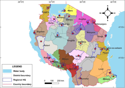
Figure 1. A map of Tanzania showing the locations of all RRH where samples were collected.
2.2. Study Design and Sampling Strategy
A cross-section study type and purposive sampling design (experiential criterion) was employed in this study between January and March 2024. The list of hospitals was obtained from the website of the Ministry of Health, Tanzania, https://hfrs.moh.go.tz. Names of all RRH visited were given a specific code for ease of tabulation and results presentation.
2.3. Sample Size and Determination
A total of 261 samples were expected from all RRH in Tanzania. These samples were to be collected from humidifiers, connectors, and reusable masks. Samples were gathered from three hospital departments namely; Emergency Units, Intensive Care Units, and Medical Wards.
2.4. Sample Collection
2.4.1. Water Samples from the Humidifier
Sterile Pasteur pipettes were used to aseptically collect water samples from a container used for oxygen humidification and then transferred aseptically to 15 mL sterile Falcon tubes. The falcon tubes were securely sealed and labelled, ready for transportation to the TMDA laboratory at a cool temperature of 2-8°C. A total of 85 water samples were obtained, consisting of 29 from the Emergency unit, 27 from the ICU and 29 from the medical wards.
2.4.2. Swab Samples
Swab samples from humidifiers, connectors and from masks or respirator or respirators were obtained by aseptically swabbing around the connector areas of the humidifiers and reusable oxygen masks. The swabs were then placed into transport media, sealed, labelled, maintained in cool temperature of 2-8°C and transported to TMDA laboratory for analyses within 24hrs.
2.5. Laboratory Analysis
2.5.1. Enrichment of Bacteria for the Isolation and Identification of P. aeruginosa
Collected samples were inoculated into nutrient broths to promote the multiplication of the microorganisms. Tryptic Soy Broth (TSB) (Oxoid UK) was used for water samples from humidifiers at a ratio of 1: 10 serial dilutions. The swab samples also inoculated separately into TSB. The inoculated broths were incubated at 37℃ for 24 hrs. as previously described .
2.5.2. Macro Morphology Identification
The enriched inoculums were then sub-cultured onto Cetrimide agar (CA) (Oxoid-UK), a selective medium used for the isolation of P. aeruginosa. The inoculated media were then incubated in a calibrated incubator (Memmert Incubator, Germany) at 32-35℃ for 18-24 hrs. After incubation time plates were observed for round, bluish-green with smooth surfaces to imply presumptive P. aeruginosa colonies. The vivid single colony were sub-cultured onto Nutrient agar (NA), (Oxoid UK) to obtain a pure culture for subsequent analytical tests.
2.5.3. Micro Morphology Identification
Overnight pure cultures of suspected isolates were subjected to Gram staining procedure whereby single pure colonies were smeared on the slides, heat-fixed, and stained by Gram stain as per the manufacturer's procedure. The stained slides were observed under microscope at x100 oil immersion for Gram-negative rod bacilli. Isolates were then subjected to a biochemical test for species identification.
2.5.4. Biochemical Identification
Presumptive P. aeruginosa isolates were then tested for presence of Cytochrome oxidase enzyme. Briefly, a sterile applicator sticks was used to pick single colony from an overnight pure culture onto the surface of the oxidase test strip and observed for deep blue coloration to indicate a positive oxidase test, which is a biochemical characteristic of P. aeruginosa.
2.5.5. Confirmation of P. aeruginosa by Molecular Methods
(i). Genomic DNA Extraction
The presumptive P. aeruginosa isolates that showed green color on Cetrimide agar and were oxidase-positive were then subjected to PCR confirmation. Boiling method was employed to extract extract genomic DNA from presumptive P. aeruginosa isolates. Briefly, four pure colonies were suspended in Eppendorf tube containing 100µL of nuclease-free water as described . The tubes were heated at 95℃ inside the water bath for 5 minutes and then transferred into the -20℃ freezer for 10 minutes to lyse the cells. The tubes were then centrifuged at 10,000 rpm for one-minute, Sterile Dnase free tips and calibrated micropipette was used to draw 80µL of the supernatant, which contained genomic DNA (gDNA), and transferred it into a clean PCR tube.
(ii). PCR Analysis
Confirmation of P. aeruginosa was performed using a molecular technique for detecting the presence of the P. aeruginosa Oprl gene with 504 bp in the presumptive isolates. The forward (Oprl F-5’ ATG GAA ATG CTG AAA TTC GGC-3’), and the reverse primers (3’-CTT CTT CAG CTC GAC GCG ACG-5’) were used as previously described . Master Mix was prepared, each PCR tube contained 16µL of nuclease-free water, 0.5µL of forward primer, 0.5µL of reverse primer, and 5µL of premix. Finally, 3µL of DNA template was added, making a total of 25µL PCR reaction mixtures. The PCR amplification conditions shown in Table 1 were used for this study.
Table 1. PCR amplification conditions.

Condition

Temperature (℃)

Time (minutes)

No. of cycles

Initial denaturation

96

5

1

Denaturation

96

1

40

Annealing

55

1

40

Extension

72

1

40

Final extension

72

10

1

The PCR products were analyzed by gel electrophoresis whereby 1% agarose gel stained with ethidium bromide (0.5 µg/mL) was used. The presence and absence of DNA bands were visualized using a UV trans-illumination machine (UVP Bio-Doc-It imaging system, Cambridge-UK).
2.6. Antimicrobial Susceptibility Test
The antimicrobial susceptibility of the confirmed P. aeruginosa isolates was performed by using the Kirby-Bauer disc diffusion method. Mueller Hinton Agar (MHA) (Oxoid UK) lot no. 2114574 was used as described in the Clinical and Laboratory Standards Institute (CLSI) 34th Edition of performance standards for antimicrobial susceptibility testing . Pure colonies were subjected to antimicrobial susceptibility test against antibiotics agent from five groups of a namely Cephalosporins, Fluoroquinolones, Aminoglycosides, Carbapenems, and β-lactam combination. Overnight pure culture of P. aeruginosa were suspended in Phosphate-Buffered Saline (PBS) to obtain a solution with a turbidity equivalent to a 0.5 McFarland standard solution corresponding to about 1.5 × 10^8 CFU/mL of bacterial concentration. Using a sterile swab, the suspended colonies were streaked onto Muller Hinton agar plates and allowed to dry. Antibiotic discs of Ceftazidime 30µg, Ciprofloxacin 5µg, Gentamicin 10µg Meropenem 10µg- Piperacillin/Tazobactam 100/10µg- (Liofichem, Italy) were placed on the inoculated agar surface, kept for 1 hour at room temperature to allow diffusion to take place and incubated at 37°C for 24 hrs. After incubation, the zones of inhibition were measured using calibrated vernier calliper and measurements were recorded and interpreted as resistant, intermediate or susceptible according to CLSI Guidelines 34th Edition. MDR strains of P. aeruginosa were identified when isolates found to be resistant to more than three different classes of antibiotics. P. aeruginosa ATCC 27853 was used as a positive control for all stages requiring positive controls.
2.7. Quality Control
Sterility and verification procedures were employed as per materials such as Falcon tubes and Pasteur pipettes were checked for sterility before use, similarly all culture media were also verified. Positive and negative controls were used for all samples received from the field to ensure the reliability of the results as previously described .
2.8. Statistical Analysis
Statistical analysis was conducted using Statistical Product and Service Solutions (SPSS) version 20.0. The Chi-square (χ²) test was used to assess associations between categorical variables. A p-value <0.05 was considered statistically significant in the multivariate analysis.
3. Results
3.1. Sample Collection Responses
The collected samples amounted to 88.5% (n=231) of the expected samples (N=261) from all regional referral hospitals in Tanzania mainland. These samples included 85 samples from water for oxygen humidification, 85 from connectors and 61 from reusable masks. Samples were gathered from three hospital departments namely Emergency Units, Intensive Care Units and Medical Wards. The variation observed in sample numbers, specifically in reusable masks, was due to some hospitals use only disposable oxygen masks which allows only to be used once and then dispose of, few hospital facilities still use reusable masks after disinfecting them.
3.2. PCR Identification of P. aeruginosa
PCR amplification of the Oprl gene target was successfully attained for most of the presumptive positive tested P. aeruginosa isolates, as demonstrated by the agarose gel electrophoresis image. Clear and distinct single bands of uniform molecular size (504bp) were observed across the majority of presumptive positive P. aeruginosa sample lanes. The absence of non-specific bands and smearing indicates high specificity of the primers and good DNA quality. Lanes that showed an absent band, suggests the absence of the P. aeruginosa Oprl gene in the samples, as shown in Figure 2.
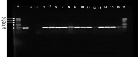
Figure 2. Agarose gel electrophoresis of PCR-amplified Oprl gene from P. aeruginosa isolates, lane M; markers, lane 1; positive control, lane 2; negative control and lane 3-16; isolate samples.
3.3. Prevalence of P. aeruginosa Across Hospitals
Among the 231 samples collected from regional referral hospitals, P. aeruginosa was detected in 34 of them, corresponding to an overall prevalence of 14.7%. The analysis of χ2 and p-value revealed no significant difference in prevalence of P. aeruginosa across the hospitals (χ2 = 25.637, p = 0.375) as shown in Table 2. Similar trends in prevalence were also observed in other studies when the isolation was performed on clinical samples from critically ill patients . Looking at individual facilities where samples were collected for this study, the highest prevalence per hospital was observed at 44% (n=4) out of the collected samples (n=9) per hospital facility. On the other hand, P. aeruginosa was not detected in samples from 9 hospitals, which is 31% of all hospitals, while 20 (69%) of the hospitals had at least one P. aeruginosa-positive sample. Results from other studies have shown the association of P. aeruginosa and respiratory devices used in hospital settings, which underlines the importance of these bacteria in treatment protocols . These results suggest a varied approach and intensity of infections control and hospital disinfection approaches, and call for continued effort in the same direction to attain a unified HAIs control.
Table 2. Prevalence of P. aeruginosa across Hospitals.

Hospital codes

P. aeruginosa

χ2

p-value

Absent

Present

n (%)

n (%)

RRH001

6 (85.7)

1 (14.3)

25.637

0.375

RRH002

8 (88.9)

1 (11.1)

RRH003

7 (100)

0 (0)

RRH004

5 (62.5)

3 (37.5)

RRH005

6 (100)

0 (0)

RRH006

6 (75)

2 (25)

RRH007

9 (100)

0 (0)

RRH008

6 (75)

2 (25)

RRH009

8 (88.9)

1 (11.1)

RRH010

5 (62.5)

3 (37.5)

RRH011

4 (66.7)

2 (33.3)

RRH012

7 (87.5)

1 (12.5)

RRH013

9 (100)

0 (0)

RRH014

9 (100)

0 (0)

RRH015

6 (85.7)

1 (14.3)

RRH016

9 (100)

0 (0)

RRH017

6 (75)

2 (25)

RRH018

7 (77.8)

2 (22.2)

RRH019

8 (88.9)

1 (11.1)

RRH020

8 (88.9)

1 (11.1)

RRH021

7 (87.5)

1 (12.5)

RRH022

7 (87.5)

1 (12.5)

RRH023

7 (77.8)

2 (22.2)

RRH024

6 (85.7)

1 (14.3)

RRH025

5 (55.6)

4 (44.4)

RRH026

5 (100.0)

0 (0)

RRH027

7 (77.8)

2 (22.2)

RRH028

6 (100)

0 (0)

RRH029

8 (100)

0 (0)

Total

197 (85.3)

34 (14.7)

3.4. Distribution of P. aeruginosa Across the Hospital Units
The assessment of the isolation of P. aeruginosa revealed that, among the collected samples (N=231), 85.3% (n=197) were negative, while 14.7% (n=34) were positive for P. aeruginosa. This low positive rate specifically for P. aeruginosa signifies the effectiveness of hygiene and infection control in hospital settings as shown in Table 3. However, despite the low detection rate, the association of P. aeruginosa with respiratory devices in some facilities suggests inadequacy in infection control, which may be attributed to the types of disinfectants used and the frequency of disinfection. These results call for a detailed study on the efficacy of hospital disinfectants used. Studies elsewhere reported the association of P. aeruginosa having ability to form biofilm, thriving in harsh and wet environments, such as the oxygen humidification process which ultimately led to spreads of opportunistic infections and hence raises concerns .
Table 3. Distribution of P. aeruginosa across the hospital units.

Unit name

P. aeruginosa isolation [n (%)]

Positive

Negative

χ2

p-value

EMD

11 (13.9)

68 (86.1)

1.371

0.523

ICU

14 (18.4)

62 (81.6)

WARD

9 (11.8)

67 (88.2)

TOTAL

34 (14.7)

197 (85.3)

3.5. Distribution of the Positivity Rate of P. aeruginosa Across the Hospital Units
The distribution of P. aeruginosa across the hospital units showed that among the total positive isolates (N=34), 41.2%(n=14) were detected from the ICUs, while the EMD ward accounted for 32.4% and 26.5% (n=9) in medical wards, respectively. Despite the slightly high prevalence in the ICU, generally, there was no significant difference in prevalence across the wards (χ2 = 1.37, p = 0.5) as shown in Table 4. The overall low prevalence could be explained by the effectiveness of decontamination implementation for infection control measures in most sampled hospital environments. Additionally, isolation of P. aeruginosa in emergency wards suggests that HAIs with P. aeruginosa can begin as early as arrival in the EMD. These results depict that hospital hygiene and infections control need more stringent control since there is a very high interaction between the hospital environment and the incoming patients and their relatives.
Table 4. Distribution of positivity rate of P. aeruginosa across the hospital units.

Unit name

P. aeruginosa isolates

n

%

χ2

P-value

EMD

11

32.3

1.371

0.523

ICU

14

41.2

WARD

9

26.5

TOTAL (N)

34

100

3.6. Prevalence of P. aeruginosa per Sample Type
Prevalence of P. aeruginosa across sample types, namely connector (CS), water for humidification (WS) and reusable masks samples (RS), revealed that P. aeruginosa prevalence was notably high in WS 30.6% (n=26), followed by CS at 8.2% (n=5) and in RS 3.5% (n=3). However, the overall undetected rate was at 85.3% for all samples pooled together as shown in Table 5. These findings were predicted due to the ability of P. aeruginosa to thrive in wet environments such as long-standing water or non-sterile water when used to humidify oxygen. This may present a high risk of introducing bacteria into already sick patients, resulting in complications to the treatment regimen. To make the matter worse, once bacteria are present in water for humidification, they may be distributed throughout the oxygen therapy system, including valves and the connector and possibly form biofilms which make them difficult to sanitize. Findings from another study conducted in Italy revealed a similar tendency, which shows bacterial contamination of the water from the humidifier from the medical ward, surgical, and emergency units were 83%, 77% and 50%, respectively .
Table 5. Prevalence of P. aeruginosa across sample types.

Sample type

P. aeruginosa

Absent n (%)

Present n (%)

X2

p= value

Connector (CS)

82 (96.5)

3 (3.5)

27.599

<0.0001

Water for humidification (WS)

59 (69.4)

26 (30.6)

Respirator/reusable Masks (RS)

56 (91.8)

5 (8.2)

3.7. Antimicrobial Susceptibility Test
The antimicrobial susceptibility of P. aeruginosa isolates against six different antibiotics revealed that among the confirmed P. aeruginosa isolates (n=34) majority were susceptible to the six antibiotics commonly prescribed for treatment of RTIs in Tanzania. A small proportion were resistant to Gentamycin (CN) and Meropenem (MRP) and to a lesser extent Ciprofloxacin (CIP) and Ceftazidime (CAZ). Piperacillin/Tazobactam (TZE) combination showed the highest efficacy against the clinical isolates used in this study as shown in Figure 3. Recent studies have highlighted the difficulties in treating critically ill patients. This study revealed P. aeruginosa being isolated in respiratory devices, in the ICU, where critically ill patients receive oxygen therapy, hence suggesting difficulties or treatment burden endured by patients. Furthermore, this bacterium has been reported to have a high mortality rate compared to other bacteria . When linking to the current findings in this study, it strongly calls for thorough hospital hygiene and sanitation, especially in low-income settings where access to medications is limited.
These results suggest the need to address the observed resistance, but generally highlight that the majority of currently available antibiotics recommended for the treatment of respiratory tract infection in Tanzania are still effective. Henceforth, we recommend ongoing rational use and AMR stewardship to protect these critically important medicines.
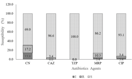
Figure 3. The Antimicrobial susceptibility profile of P. aeruginosa against six different antibiotics for treatment of RTIs in Tanzania.
3.8. Prevalence of Multidrug Resistance Among Isolates
Overall, the findings indicated a low prevalence of MDR among P. aeruginosa isolates across all regional referral hospitals, with only 11% (n = 4) from a single hospital identified as AMR, as shown in Table 6. This unique case presumes that it could be the same isolate circulating in the same hospital. A bigger study with wide coverage to establish the extent of MDR, if any, at district hospitals. The difficulties in treating MDR have been reported in other studies , thus strict measures are required to limit their spread to other facilities. This finding further necessitates future studies to be conducted to isolate resistant genes and establish phylogenetic relationships among the isolates.
Table 6. Antibiotic Resistance Profiles and Status of Pseudomonas Isolates across Various Hospitals.

Hospital code

Susceptible (S) (n)

Intermediate (I) (n)

Resistance (R) (n)

MDR status

RRH001

5

0

1

Not MDR

RRH002

5

1

0

Not MDR

RRH003

2

0

4

MDR

RRH006

11

0

1

Not MDR

RRH008

5

1

0

Not MDR

RRH009

6

0

0

Not MDR

RRH010

24

0

0

Not MDR

RRH011

10

0

2

Not MDR

RRH012

6

0

0

Not MDR

RRH015

6

0

0

Not MDR

RRH018

12

0

0

Not MDR

RRH017

11

1

0

Not MDR

RRH020

6

0

0

Not MDR

RRH021

6

0

0

Not MDR

RRH022

4

0

2

Not MDR

RRH023

6

0

0

Not MDR

RRH024

5

1

0

Not MDR

RRH025

23

1

0

Not MDR

RRH027

5

1

0

Not MDR

4. Conclusion
The current study highlighted several critical findings; first most antibiotics used to treat RTIs are still efficacious in case of HAIs by the P. aeruginosa, second the antibiotic resistance was low in percentage, but worth noting. The presence of MDR isolates in one of the RRHs accentuates the challenges of managing infections with antimicrobial therapies and water for humidification showed significant bacterial colonization. The prevalence of these pathogens varied across different hospital units, with P. aeruginosa showing a higher presence in ICUs and EMDs and thus strict infection control measures are warranted.
Abbreviations

AMR

Antimicrobial Resistance

ATCC

American Type Culture Collection

CA

Cetrimide Agar

CDC

Center for Disease Control

CLSI

Clinical and Laboratory Standards Institute

EMD

Emergence Department

ESKAPE

Enterococcus faecium, Staphylococcus aureus, Klebsiella pneumoniae, Acinetobacter baumannii, Pseudomonas aeruginosa and Enterobacter Spp.

HAI

Hospital Acquired Infections

ICU

Intensive Care Unit

MDR

Multi Drug Resistance

MHA

Mueller Hinton Agar

PCR

Polymerase Chain Reaction

RRH

Regional Referral Hospital

NA

Nutrient Agar

RTI

Respiratory Tract Infection

SPP

Species

SPSS

Statistical Product and Service Solutions

TAE

Tris-acetate-EDTA

TMDA

Tanzania Medicines and Medical Devices Authority

TSB

Tryptose Soy Broth

USP

United State Pharmacopoeia

USPNF

United States Pharmacopoeia-National Formulary

Acknowledgments
We would like to acknowledge the contribution of Mr. John Mwingira, who supported the statistical analysis of the data from this study to produce scientifically meaningful results.
Author Contributions
Adelard Bartholomew Mtenga: Conceptualization, Data curation, Formal Analysis, Methodology, Supervision, Writing – original draft, Writing – review & editing
Elizabeth Erasto Kasekwa: Data curation, Investigation, Methodology, Writing – review & editing
Adam Mitangu Fimbo: Project administration, Resources, Supervision, Writing – review & editing
Saxon Joseph Mwambene: Data curation, Investigation, Methodology
Revocatus Evarist Makonope: Data curation, Investigation, Methodology
Raphael Zozimus Sangeda: Writing – review & editing
Danstan Hipolite Shewiyo: Project administration, Supervision, Writing – review & editing
Data Availability Statement
The data supporting the outcome of this research work have been reported in this manuscript.
Conflicts of Interest
Authors declare no conflicts of interest.
References
[1] Gellatly SL, Hancock REW. Pseudomonas aeruginosa: New insights into pathogenesis and host defenses. Pathog Dis. 2013; 67(3): 159–73.
[2] Stover CK, Pham XQ, Erwin AL, Mizoguchi SD, Warrener P, Hickey MJ, et al. Complete genome sequence of Pseudomonas aeruginosa PAO1, an opportunistic pathogen. Nature. 2000; 406(6799): 959–64.
[3] Denissen J, Reyneke B, Waso-Reyneke M, Havenga B, Barnard T, Khan S, et al. Prevalence of ESKAPE pathogens in the environment: Antibiotic resistance status, community-acquired infection and risk to human health. Int J Hyg Environ Health. 2022; 244(July): 114006.
[4] Abushaheen MA, Muzaheed, Fatani AJ, Alosaimi M, Mansy W, George M, et al. Antimicrobial resistance, mechanisms and its clinical significance. Disease-a-Month. 2020; 66(6): 1–21.
[5] Augustine FF, Mgaya XM, Yahya SA, Niccodem EM, Matee MI. An alarming prevalence of multidrug-resistant (MDR) ESKAPE pathogens and other drug-resistant bacteria isolated from patients with bloodstream infections hospitalized at Muhimbili National Hospital in Dar es Salaam, Tanzania. Ger J Microbiol. 2023; 3(3): 7–15.
[6] Cole SJ, Records AR, Orr MW, Linden SB, Lee VT. Catheter-associated urinary tract infection by Pseudomonas aeruginosa is mediated by exopolysaccharide-independent biofilms. Infect Immun. 2014; 82(5): 2048–58.
[7] Kollef MH, Chastre J, Fagon JY, François B, Niederman MS, Rello J, et al. Global prospective epidemiologic and surveillance study of ventilator-associated pneumonia due to pseudomonas aeruginosa. Crit Care Med. 2014; 42(10): 2178–87.
[8] Alem K. Prevalence of bacterial pneumonia among HIV- Seropositive patients in East Africa : Review Prevalence of bacterial pneumonia among HIV-Seropositive patients in East Africa : Review. Cogent Med. 2021; 8(1).
[9] Josephs-Spaulding J, Singh O V. Medical Device Sterilization and Reprocessing in the Era of Multidrug-Resistant (MDR) Bacteria: Issues and Regulatory Concepts. Front Med Technol. 2020; 2(February): 1–15.
[10] Moradali MF, Ghods S, Rehm BHA. Pseudomonas aeruginosa lifestyle: A paradigm for adaptation, survival, and persistence. Front Cell Infect Microbiol. 2017; 7(FEB).
[11] Jasim AK, Hussein AA. Detection Genotyping Virulence Factor among Pseudomonas aeruginosa Isolated from Burns Patients. Int J Adv Multidiscip Res Stud. 2023; 3(4): 1209–14.
[12] Vos DDE, Lim A, Pirnay J, Struelens M, Vandenvelde C, Duinslaeger LUC, et al. Direct Detection and Identification of Pseudomonas aeruginosa in Clinical Samples Such as Skin Biopsy Specimens and Expectorations by Multiplex PCR Based on Two Outer Membrane Lipoprotein Genes, oprI and oprL. 1997; 35(6): 1295–9.
[13] Limbago JSLIib and ALB et al. CLSI, performance standards for antimicrobial susceptibility testing 33rd Edition. Clinical and Laboratory Standards Institute,. 2024. 1–416 p.
[14] Interim S, Announcement R, Tests S. 71 sterility tests. 2016; 34(6): 1–12.
[15] Abdullahi R, Lihan S, Carlos BS, Bilung ML, Mikal MK, Collick F. Detection of opr L gene and antibiotic resistance of Pseudomonas aeruginosa from aquaculture environment. 2013; 3(6): 148–52.
[16] Vidal-Cortés P, Campos-Fernández S, Cuenca-Fito E, del Río-Carbajo L, Fernández-Ugidos P, López-Ciudad VJ, et al. Difficult-to-Treat Pseudomonas aeruginosa Infections in Critically Ill Patients: A Comprehensive Review and Treatment Proposal. Antibiot (Basel, Switzerland). 2025 Feb; 14(2).
[17] Litwin A, Rojek S, Gozdzik W, Duszynska W. Pseudomonas aeruginosa device associated – healthcare associated infections and its multidrug resistance at intensive care unit of University Hospital: polish, 8.5-year, prospective, single-centre study. BMC Infect Dis. 2021; 21(1): 4–11.
[18] Shane AL, Gonzalez MD, Roy AM, Preidis GA, Woodworth MH. State-of-the-Art Review: Infectious Diarrhea. Clin Infect Dis. 2025 Nov; 81(5): e250–62.
[19] La Fauci V, Costa GB, Facciolà A, Conti A, Riso R, Squeri R. Humidifiers for oxygen therapy: what risk for reusable and disposable devices J Prev Med Hyg. 2017; 58(2): E161
[20] Serrano-Mayorga CC, Degree M, Olivella-Gomez J, Torres A, Martin-Loeches I, Luis, et al. P-286. Prevalence of Pseudomonas aeruginosa in patients with ICU-related respiratory Infections: A secondary analysis of the European Network for ICU-Related Respiratory Infections (ENIRRIs). Open Forum Infect Dis. 2025 Jan; 12(Supplement_1).
[21] Thaden JT, Park LP, Maskarinec SA, Ruffin F, Fowler VG, Van Duin D. Results from a 13-year prospective cohort study show increased mortality associated with bloodstream infections caused by pseudomonas aeruginosa compared to other bacteria. Antimicrob Agents Chemother. 2017; 61(6).
Cite This Article
  • APA Style

    Mtenga, A. B., Kasekwa, E. E., Fimbo, A. M., Mwambene, S. J., Makonope, R. E., et al. (2026). Efficacy of Antibiotics Against Pseudomonas aeruginosa Isolated from Respiratory Devices in Tanzania. Medicine and Health Sciences, 2(2), 86-96. https://doi.org/10.11648/j.mhs.20260202.13

    Copy | Download

    ACS Style

    Mtenga, A. B.; Kasekwa, E. E.; Fimbo, A. M.; Mwambene, S. J.; Makonope, R. E., et al. Efficacy of Antibiotics Against Pseudomonas aeruginosa Isolated from Respiratory Devices in Tanzania. Med. Health Sci. 2026, 2(2), 86-96. doi: 10.11648/j.mhs.20260202.13

    Copy | Download

    AMA Style

    Mtenga AB, Kasekwa EE, Fimbo AM, Mwambene SJ, Makonope RE, et al. Efficacy of Antibiotics Against Pseudomonas aeruginosa Isolated from Respiratory Devices in Tanzania. Med Health Sci. 2026;2(2):86-96. doi: 10.11648/j.mhs.20260202.13

    Copy | Download

  • @article{10.11648/j.mhs.20260202.13,
      author = {Adelard Bartholomew Mtenga and Elizabeth Erasto Kasekwa and Adam Mitangu Fimbo and Saxon Joseph Mwambene and Revocatus Evarist Makonope and Kissa Watson Mwamwitwa and Raphael Zozimus Sangeda and Danstan Hipolite Shewiyo},
      title = {Efficacy of Antibiotics Against Pseudomonas aeruginosa Isolated from Respiratory Devices in Tanzania},
      journal = {Medicine and Health Sciences},
      volume = {2},
      number = {2},
      pages = {86-96},
      doi = {10.11648/j.mhs.20260202.13},
      url = {https://doi.org/10.11648/j.mhs.20260202.13},
      eprint = {https://article.sciencepublishinggroup.com/pdf/10.11648.j.mhs.20260202.13},
      abstract = {Pseudomonas aeruginosa is an opportunistic pathogen responsible for severe hospital-acquired infections, including ventilator-associated pneumonia. Due to its biofilm-forming ability and antimicrobial resistance, it presents a significant public health challenge. This study assessed the efficacy of selected antibiotics against P. aeruginosa isolates associated with respiratory devices from regional referral hospitals in mainland Tanzania. A cross-sectional study was conducted in 2024, in which samples were collected from January to March in Emergency wards, Intensive care units, and Medical wards. Samples collected included water from the Oxygen humidification container, swab samples from reusable masks and the connectors. Laboratory analysis using standard microbiological techniques and PCR were employed for isolation and confirmation of P. aeruginosa. Antimicrobial susceptibility testing was performed using the Kirby-Bauer disc diffusion method. Out of the collected samples (N=231), P. aeruginosa was detected at an overall prevalence of 14.7% (n=34). The analysis of prevalence by sample type revealed that water for humidification had the highest prevalence of 30.6%, followed by respirators at 8.2% and the least in connectors at 3.5%. P. aeruginosa showed a notable resistance towards gentamycin, followed by meropenem, and the least resistance was shown in ceftazidime. On the other hand, P. aeruginosa were fully susceptible to piperacillin-tazobactam combination. A very small proportion of isolates demonstrated multidrug resistance (MDR). Despite the noted resistance majority of the antibiotics used to treat respiratory tract infection (RTIs) in this study showed significant efficacy.},
     year = {2026}
    }
    

    Copy | Download

  • TY  - JOUR
    T1  - Efficacy of Antibiotics Against Pseudomonas aeruginosa Isolated from Respiratory Devices in Tanzania
    AU  - Adelard Bartholomew Mtenga
    AU  - Elizabeth Erasto Kasekwa
    AU  - Adam Mitangu Fimbo
    AU  - Saxon Joseph Mwambene
    AU  - Revocatus Evarist Makonope
    AU  - Kissa Watson Mwamwitwa
    AU  - Raphael Zozimus Sangeda
    AU  - Danstan Hipolite Shewiyo
    Y1  - 2026/03/26
    PY  - 2026
    N1  - https://doi.org/10.11648/j.mhs.20260202.13
    DO  - 10.11648/j.mhs.20260202.13
    T2  - Medicine and Health Sciences
    JF  - Medicine and Health Sciences
    JO  - Medicine and Health Sciences
    SP  - 86
    EP  - 96
    PB  - Science Publishing Group
    SN  - 3070-6300
    UR  - https://doi.org/10.11648/j.mhs.20260202.13
    AB  - Pseudomonas aeruginosa is an opportunistic pathogen responsible for severe hospital-acquired infections, including ventilator-associated pneumonia. Due to its biofilm-forming ability and antimicrobial resistance, it presents a significant public health challenge. This study assessed the efficacy of selected antibiotics against P. aeruginosa isolates associated with respiratory devices from regional referral hospitals in mainland Tanzania. A cross-sectional study was conducted in 2024, in which samples were collected from January to March in Emergency wards, Intensive care units, and Medical wards. Samples collected included water from the Oxygen humidification container, swab samples from reusable masks and the connectors. Laboratory analysis using standard microbiological techniques and PCR were employed for isolation and confirmation of P. aeruginosa. Antimicrobial susceptibility testing was performed using the Kirby-Bauer disc diffusion method. Out of the collected samples (N=231), P. aeruginosa was detected at an overall prevalence of 14.7% (n=34). The analysis of prevalence by sample type revealed that water for humidification had the highest prevalence of 30.6%, followed by respirators at 8.2% and the least in connectors at 3.5%. P. aeruginosa showed a notable resistance towards gentamycin, followed by meropenem, and the least resistance was shown in ceftazidime. On the other hand, P. aeruginosa were fully susceptible to piperacillin-tazobactam combination. A very small proportion of isolates demonstrated multidrug resistance (MDR). Despite the noted resistance majority of the antibiotics used to treat respiratory tract infection (RTIs) in this study showed significant efficacy.
    VL  - 2
    IS  - 2
    ER  - 

    Copy | Download

Author Information
  • Abstract
  • Keywords
  • Document Sections

    1. 1. Introduction
    2. 2. Methodology
    3. 3. Results
    4. 4. Conclusion
    Show Full Outline
  • Abbreviations
  • Acknowledgments
  • Author Contributions
  • Data Availability Statement
  • Conflicts of Interest
  • References
  • Cite This Article
  • Author Information