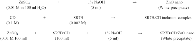
Sudan Red-7B/Cyclodextrin doped ZnO nanocomposites are synthesized and analyzed by various spectral and microscopic methods. The doping effect of SR7B/CD on ZnO nano investigated by UV-visible, fluorescence, FTIR, DTA, XRD, FE-SEM and TEM methods. The effect of different polarities of the solvents, α-cyclodextrin (α-CD) and β-cyclodextrin (β-CD), on MV was studied by various spectral methods. The inclusion behaviour of SR7B on both CDs was determined by PM3 method. The solvent and CD studies show that the azo-imino tautomer is present in the SR7B molecule and that, depending upon the polarity of the solvents, absorbance and emission intensities of the azo-imino tautomer is varied. With increasing CD concentrations, the shorter wavelength emission intensity of the SR7B regularly increased while the longer wavelength emission intensity decreased. The horizontal bond length of SR7B is longer than the CD cavities; hence, this molecule is partially encapsulated in the CD cavity. HOMO-LUMO gap for MV/β-CD inclusion complex was more negative, which supports that this complex is more stable than MV/α-CD inclusion complex. Red or blue shifted absorption and fluorescence maxima were seen in SR7B/CD/ZnO nanocomposites than SR7B/CD inclusion complex. Nanoparticle size was measured by TEM-EDS and X-RD methods. TEM image showed that nanosheets are formed in SR7B/CD/ZnO.
| Published in | Science Journal of Chemistry (Volume 13, Issue 3) |
| DOI | 10.11648/j.sjc.20251303.13 |
| Page(s) | 65-75 |
| Creative Commons |
This is an Open Access article, distributed under the terms of the Creative Commons Attribution 4.0 International License (http://creativecommons.org/licenses/by/4.0/), which permits unrestricted use, distribution and reproduction in any medium or format, provided the original work is properly cited. |
| Copyright |
Copyright © The Author(s), 2025. Published by Science Publishing Group |
Sudan Red-7B, Tautomer, Zinc Oxide Nano, Cyclodextrin, Inclusion Complex
Solvents | abs | log | flu |
|---|---|---|---|
Cyclohexane | 521 | 3.27 | 591 |
367 | 3.17 | 410 | |
247 | 3.46 | ||
1, 4-Dioxane | 534 | 3.48 | 591 |
369 | 3.34 | 412 | |
248 | 3.64 | ||
Ethyl acetate | 534 | 3.52 | 597 |
365 | 3.39 | 414 | |
250 | 3.61 | ||
Acetonitrile | 534 | 3.20 | 595 |
364 | 3.05 | 414 | |
247 | 3.33 | ||
2-Propanol | 533 | 3.47 | 414 |
366 | 3.34 | 598 | |
246 | 3.63 | ||
Ethanol | 536 | 3.12 | 593 |
366 | 2.98 | 414 | |
246 | 3.28 | ||
Water | 565 | 2.88 | 584 |
368 | 3.13 | 364 | |
244 | 3.32 | ||
224 | 3.25 | ||
α-CD -0.01 M | 562 | 3.18 | 584 |
365 | 3.39 | 362 | |
242 | 3.57 | ||
222 | 3.57 | ||
β-CD 0.01 M | 563 | 3.17 | 584 |
366 | 3.35 | 363 | |
243 | 3.53 | ||
223 | 3.53 | ||
α-CD K (1: 1) x105 M-1 | 54 | - | 139 |
β-CD K (1: 1) x105 M-1 | 144 | - | 176 |
α-CD G (kcalmol-1) | -11.0 | - | -13.4 |
β-CD G (kcalmol-1) | -13.5 | - | -14.0 |
Excitation wavelength (nm) | - | - | 320 |
Properties | SR7B | α-CD | β-CD | SR7B/α-CD | SR7B/β-CD |
|---|---|---|---|---|---|
EHOMO (eV) | -8.29 | -10.37 | -10.35 | -8.42 | -8.44 |
ELUMO (eV) | -1.02 | 1.26 | 1.23 | -1.22 | -1.32 |
EHOMO – ELUMO (eV) | 7.26 | -11.63 | -11.58 | 7.19 | 7.12 |
Dipole (D) | 1.46 | 11.34 | 12.29 | 10.96 | 10.56 |
E (kcal mol-1) | -170.91 | -1247.62 | -1457.63 | -1083.08 | -1295.89 |
ΔE (kcal mol-1) | - | - | - | -6.37 | -9.17 |
G (kcal mol-1) | -266.73 | -676.37 | -789.52 | -945.25 | -1002.41 |
ΔG (kcal mol-1) | - | - | - | -2.15 | -53.84 |
H (kcal mol-1) | -210.97 | -570.84 | -667.55 | -796.77 | -852.93 |
ΔH (kcal mol-1) | - | - | - | -14.96 | -25.59 |
S (kcal/mol-Kelvin) | 0.187 | 0.353 | 0.409 | 0.498 | 0.561 |
ΔS (kcal/mol-Kelvin) | - | - | - | 0.042 | 0.035 |
ZPE | 250.16 | 635.09 | 740.56 | 886.52 | 941.11 |
FTIR | Fourier Transform Infrared Spectroscopy |
DTA | Differential Thermal Analysis |
XRD | X-ray Diffraction |
SEM | Scanning Electron Microscopy |
TEM | Transmission Electron Microscopy |
HOMO | Highest Occupied Molecular Orbital |
LUMO | Lowest Unoccupied Molecular Orbital |
SR7B | Sudan Red 7B |
ZnO NPs | Zinc Oxide Nanoparticles |
α-CD | Alpha Cyclodextrin |
β-CD | Beta Cyclodextrin |
PM3 | Parametric Method 3 |
ΔE | Internal Energy Change |
ΔH | Enthalpy Change |
ΔG | Free Energy Change |
ΔS | Entropy Change |
| [1] | Zince, T., Bindenwald, M. On the Action of Phosphoryl Bromide on Aromatic Amines. Chem Ber 1884, 17, 3026-33. |
| [2] | Richard Kuhn, Friedrich Bär, On Chinophthalone, Annalen 1935, 516, 143-161, |
| [3] | Frierz-David, H. E., Blangey, L., Streif, H. On the Knowledge of p-Hydroxy Azo Dye, Helvetica Chimica Acta 1946, 29, 1718-64. |
| [4] | Kishimoto, S., Kitahara S., Manabe O., Hiyama H. Tautomerism and Dissociation of 4-Arylazo-1-Naphthols in Various Solvents, J. Org Chem 1978, 43, 3882-86; |
| [5] | Antonov, L., Stoyanov, S., Stoyanov, T. Quantitative analysis of tautomeric equilibrium in 1-phenylazo-4-naphthols—a new approach, Dyes and Pigments, 1994, 26, 149-158. |
| [6] | Antonov, L., Stoyanov, S. 1-Naphthol. Dyes and Pigments, Dyes and Pigments, 1995, 28, 31-39. |
| [7] | Antonov, L., Fabian, W. M. F., Nedeltcheva, D., Kamounah, F. S. Tautomerism of 2-hydroxy naphthaldehyde Schiff bases. J. Chem. Soc., Perkin Trans. 2, 2000, 1173–1179. |
| [8] | Antonov, L., Stoyanov, S., Stoyanova, T. Tautomeric equilibrium in 1-phenylazo-2-naphthol—A quantitative study, Dyes and Pigments 1995, 27, 133-142. |
| [9] | Antonova, L., Kawauchib, S., Satohb, M., Komiyama, J. Ab initio modeling of the solvent influence on the azo-hydrazone tautomerism, Dyes and Pigments 1999, 40, 163-170. |
| [10] | Joshi, H., Kamounah, F. S., van der Zwan, G., Gooijer, C., Antonov, L. Temperature dependent absorption spectroscopy of some tautomeric azo dyes and Schiff bases. J Chem Soc Perkin Trans 2: 2001, 2303-08. |
| [11] | Hem Joshi, Fadhil S. Kamounah, Cees Gooijera, Gert van der Zwan, Liudmi Antonov, Excited state intramolecular proton transfer in some tautomeric azo dyes and Schiff bases containing an intramolecular hydrogen bond. J. Photochem. and Photobiol. A: Chem. 2002, 152, 183–191. |
| [12] | Fadhil S. Kamounah, Liudmi Antonov, Vesselin Petrov, Gert van der Zwan, An integrated approach to the study of the tautomerism of 4-((Phenylimino) methyl) naphthalene-1-ol, J. Phys. Org. Chem. 2007, 20, 313–320. |
| [13] | Zollinger, H. Color Chemistry: Syntheses, Properties and Applications of Organic Dyes and Pigments. VCH, Weinheim 1991. |
| [14] | Smoluch, M., Joshi, H., Gerssen, A., Gooijer, C. van der Zwan, G., Fast excited-state intramolecular proton transfer and sub nanosecond dynamic Stokes shift of time-resolved fluorescence spectra of the 5-methoxysalicylic acid/diethyl ether complex. J. Phys. Chem. 2005, 109, 535–541. |
| [15] | Alarcón, S. H., Olivieri, A. C., Labadie, G. R., Cravero, R. M., Gonzales-Sierra, M. Tautomerism of representative aromatic—hydroxy carbaldehyde anils as studied by spectroscopic methods and AM1 calculations. Synthesis of 10-hydroxyphenanthrene-9-carbaldehyde. Tetrahedron. 1995, 51, 4619–4626. |
| [16] | Antony Muthu Prabhu, A., Venkatesh, G., Sankaranarayanan, R. K., Rajendiran, N. Azonium-ammonium tautomerism and inclusion complexation of 4-amino-2’,3-dimethyl azobenzene. Indian J. Chem. 2010, 49A, 407–417. |
| [17] | Venkatesh, G., Sankaranarayanan, R. K., Antony Muthu Prabhu, A., Rajendiran, N. Azonium-Ammonium Tautomerism and Inclusion Complexation of 1-(2, 4-diamino phenylazo) naphthalene and 4-amino azobenzene. J. Fluores. 2011, 21, 1485-1497. |
| [18] | Prema Kumari, J., Antony Muthu Prabhu, A., Venkatesh, G., Subramanian, V. K., Rajendiran, N. Effect of solvents and pH on β-CD Inclusion complexation of 2, 4-dihydroxy azobenzene and 4-hydroxy azobenzene. J. Solution Chem, 2011, 40, 327–347. |
| [19] | Venkatesh, G., Saravanan, J., Rajendiran, N. Cyclodextrin covered organic micro rod and micro sheet derived from supramolecular self-assembly of 2, 4-dihydroxy azobenzene and 4-hydroxy azobenzene inclusion complexes. Bull. Chem. Soc. Jpn. 2014, 87, 283-293. |
| [20] | Rajendiran, N., Venkatesh, G., Saravanan, J. Encapsulation of thiazoresorcinol and thiazocresol dye within α- and β-CD cavities: Spectral and molecular modeling studies. J. Mol. Struc. 2014, 1072, 242-252. |
| [21] | Antony Muthu Prabhu, A., Venkatesh, G. Rajendiran, N. Azo-Hydrazo tautomerism in 1-phenyazo-2-naphthol dyes in various solvents, pH and β-CD. J. Fluorescence, 2010, 20, 961–972. |
| [22] | Rajendiran, N., Sankaranarayanan, R. K. Azo dye/Cyclodextrin: New findings of identical nanorods through 2: 2 inclusion complexes. Carbohydrate Polymers, 2014, 106, 422-431. |
| [23] | Supraja, N., Prasad, T. N. V. K. V., Giridhara Krishna, T. David, E. Synthesis, characterization, and evaluation of the antimicrobial efficacy of Boswellia ovalifoliolata stem bark-extract-mediated zinc oxide nanoparticles. Appl. Nanosci. 2016, 6, 581–590, |
| [24] | Niranjan Bala, S. Saha, M. Chakraborty, M. Maiti, S. Das, R. Basub, P. Nandy, Green synthesis of zinc oxide nanoparticles using Hibiscus subdariffa leaf extract: effect of temperature on synthesis, anti-bacterial activity and anti-diabetic activity. RSC Adv., 2015, 5, 4993–5003. |
| [25] | Xiong, H. M. ZnO Nanoparticles Applied to Bioimaging and Drug Delivery, Adv. Mater., 2013, 25(37), 5329–5335. |
| [26] | Sharmaa, D., Rajputa, J., Kaitha, B. S., Kaurb, M. Synthesis of ZnO nanoparticles and study of their antibacterial and antifungal properties. Thin Solid Films, 2010, 519(3), 1224-1229. |
| [27] | Nair, S. Role of size scale of ZnO nanoparticles and microparticles on toxicity toward bacteria and osteoblast cancer cells. J. Mater. Sci.: Mater. Med., 2009, 20 (Suppl. 1), S 235–S241. |
| [28] | Kirthi, A. V., Rahuman, A. A., Rajakumar, G., Marimuthu, S. Acaricidal, pediculocidal and larvicidal activity of synthesized ZnO nanoparticles using wet chemical route against blood feeding parasites. Parasitol. Res., 2011, 109, 461–472. |
| [29] | Alkaladi, A., Abdelazim, A. M., Afifi, M. Antidiabetic Activity of Zinc Oxide and Silver Nanoparticles on Streptozotocin-Induced Diabetic Rats. Int. J. Mol. Sci., 2014, 15, 2015–2023. |
| [30] | Dagdeviren, C., Hwang, S. W., Su, Y., Kim, S. Transient, Biocompatible Electronics and Energy Harvesters Based on ZnO. Small, 2013, 9(20), 3398–3404. |
| [31] | Zhang, Yuanyuan, Leu, Yu-Rui, Aitken J. Robert Riediker Michael, Inventory of Engineered Nanoparticle-Containing Consumer Products Available in the Singapore Retail Market and Likelihood of Release into the Aquatic Environment. International J Environmental Research and Public Health. 2015, 12, 8717–8743. |
| [32] | Wang, L., Kang, Y., Liu, X., Zhang, S. ZnO nanorod gas sensor for ethanol detection. Sens. Actuators, B, 2012, 162(1), 237–243. |
| [33] | Cross, S. E., Innes, B., Roberts, M. S., Tsuzuki, T. Human skin penetration of sunscreen nanoparticles: in vitro assessment of a novel micronized zinc oxide formulation, Skin Pharmacol. Physiol., 2007, 20(3), 148–154. |
| [34] | Zhou, J., Xu, N., Wang, Z. L. Dissolving behavior and stability of ZnO wires in biofluids: a study on biodegradability and biocompatibility of ZnO nanostructures. Adv. Mater., 2006, 18(18), 2432–2435. |
| [35] | Rasmussen, J. W., Martinez, E., Louka, P., Wingett, D. G. Zinc oxide nanoparticles for selective destruction of tumor cells and potential for drug delivery applications. Expert Opin. Drug Delivery, 2010, 7(9), 1063–1077. |
| [36] | Siva Kumar, Surabhi, Putcha Venkateswarlu, Vanka Ranga Rao, Gollapalli Nageswara Rao, Synthesis, characterization and optical properties of zinc oxide nanoparticles. International Nano Letters, 2013, 3, 30-35. |
| [37] | Sukesh Kashiram Tumram, Rajdip Bandyopadhyaya, Zinc oxide nanostructures: Experiments probing their transformation to nanorods. Materials Science and Engineering: B, 2023, 296, 116569- 74. |
| [38] | Ramasamy, P., Mani, A., Sneha, B., Nivetha, E., Venkatesan, M., Rajendiran, N. Azo-hydrazo tautomerism in Sudan Red-B and Cyclodextrin/Sudan Red-B doped ZnO nanomaterials. J Molecular Structure, 2025; 1329: 141423-32. |
| [39] | Mani, A., Ramasamy, P., Antony Muthu Prabhu, A., Rajendiran, N. Investigation of Ag and Ag/Co bimetallic nanoparticles with naproxen-cyclodextrin inclusion complex. J. Molecular Structure 2023; 1284: 135301-10. |
| [40] | Mani, A., Venkatesh, G., Senthilraja, P., Rajendiran, N. Synthesis and Characterisation of Ag-Co-Venlafaxine-Cyclodextrin Nanorods. European J Advanced Chemistry Research, 2024; 5: 9-16. |
| [41] | Mani, A., Ramasamy, P., Antony Muthu Prabhu, A., Senthilraja, P., Rajendiran, N. Synthesis and Analysis of Ag/Olanzapine /Cyclodextrin and Ag/Co/Olanzapine /Cyclodextrin Inclusion Complex Nanorods. Physics and Chemistry of Liquids, 2024; 62: 196-209. |
| [42] | Mani, A., Ramasamy, P., Antony Muthu Prabhu, A., Senthilraja, P., Rajendiran, N. Synthesis and Characterisation of Ag/Co/Chloroquine/Cyclodextrin Inclusion Complex Nanomaterials. J Sol-Gel Science and Technology, 2025. |
| [43] | Prema Kumari, J., Antony Muthu Prabhu, A., Venkatesh, G., Subramanian, V. K., Rajendiran, N. Spectral characteristics of sulfadiazine, sulfisomidine: Effect of solvents, pH and β-CD. Physics and Chemistry of Liquids, 2011, 49, 108–132. |
| [44] | Jude Jenita, M., Antony Muthu Prabhu, A., Rajendiran, N. Theoretical study of inclusion complexation of tricyclic antidepressant drugs with β-CD. Indian J. Chemistry A, 2012, 51 A, 1686-1694. |
| [45] | Rajendiran, N., Saravanan, J. Inclusion complexation of sulfa pyridine with α- and β-CDs: Spectral and molecular modeling study. J. Molecular Structure, 2013, 1054-1055 215–222. |
| [46] | Antony Muthu Prabhu, A., Rajendiran, N., Encapsulation of labetalol, and pseudoephedrine in β-CD cavity: Spectral and molecular modeling studies. J. Fluorescence, 2012, 22, 1461-1474. |
| [47] | Rajendiran, N., Sankaranarayanan, R. K., Saravanan, J. A study of supramolecular host–guest interaction of dothiepin and doxepin drugs with cyclodextrin macrocycles. J Molecular Structure, 2014, 106, 7252-260. |
| [48] | Rajendiran, N., Venkatesh, G., Saravanan, J. Supramolecular aggregates formed by sulfadiazine and sulfisomidine inclusion complexes with α- and β-cyclodextrin. Spectrochimica Acta, A, 2014, 129, 157-162, |
| [49] | Rajendiran, N., Venkatesh, G., Mohandoss, T. Fabrication of 2 D nano sheet through self assembly behavior of sulfamethoxy pyridazine inclusion complex with α- and β-cyclodextrins. Spectrochim Acta A, 2014, 123, 158-166, |
| [50] | Rajendiran, N., Sankaranarayanan, R. K., Venkatesh, G. Excimer emission in inclusion complexes of dibenzofuran and 5-dibenzosuberenone with α- and β-cyclodextrins. Bull Chem Soc Japan, 2014, 87, 797-808. |
APA Style
Ramasamy, P., Rajendiran, N., Mani, A., Venkatesh, G., Prabhu, A. A. M. (2025). Azo-Imino Tautomerism in Sudan Red 7B/Cyclodextrin Coated ZnO Nanocomposites: Evidence by Spectral and Microscopic Perspectives. Science Journal of Chemistry, 13(3), 65-75. https://doi.org/10.11648/j.sjc.20251303.13
ACS Style
Ramasamy, P.; Rajendiran, N.; Mani, A.; Venkatesh, G.; Prabhu, A. A. M. Azo-Imino Tautomerism in Sudan Red 7B/Cyclodextrin Coated ZnO Nanocomposites: Evidence by Spectral and Microscopic Perspectives. Sci. J. Chem. 2025, 13(3), 65-75. doi: 10.11648/j.sjc.20251303.13
@article{10.11648/j.sjc.20251303.13,
  author = {Palanichamy Ramasamy and Narayanasamy Rajendiran and Ayyadurai Mani and Govindaraj Venkatesh and Albert Antony Muthu Prabhu},
  title = {Azo-Imino Tautomerism in Sudan Red 7B/Cyclodextrin Coated ZnO Nanocomposites: Evidence by Spectral and Microscopic Perspectives
},
  journal = {Science Journal of Chemistry},
  volume = {13},
  number = {3},
  pages = {65-75},
  doi = {10.11648/j.sjc.20251303.13},
  url = {https://doi.org/10.11648/j.sjc.20251303.13},
  eprint = {https://article.sciencepublishinggroup.com/pdf/10.11648.j.sjc.20251303.13},
  abstract = {Sudan Red-7B/Cyclodextrin doped ZnO nanocomposites are synthesized and analyzed by various spectral and microscopic methods. The doping effect of SR7B/CD on ZnO nano investigated by UV-visible, fluorescence, FTIR, DTA, XRD, FE-SEM and TEM methods. The effect of different polarities of the solvents, α-cyclodextrin (α-CD) and β-cyclodextrin (β-CD), on MV was studied by various spectral methods. The inclusion behaviour of SR7B on both CDs was determined by PM3 method. The solvent and CD studies show that the azo-imino tautomer is present in the SR7B molecule and that, depending upon the polarity of the solvents, absorbance and emission intensities of the azo-imino tautomer is varied. With increasing CD concentrations, the shorter wavelength emission intensity of the SR7B regularly increased while the longer wavelength emission intensity decreased. The horizontal bond length of SR7B is longer than the CD cavities; hence, this molecule is partially encapsulated in the CD cavity. HOMO-LUMO gap for MV/β-CD inclusion complex was more negative, which supports that this complex is more stable than MV/α-CD inclusion complex. Red or blue shifted absorption and fluorescence maxima were seen in SR7B/CD/ZnO nanocomposites than SR7B/CD inclusion complex. Nanoparticle size was measured by TEM-EDS and X-RD methods. TEM image showed that nanosheets are formed in SR7B/CD/ZnO.
},
 year = {2025}
}
TY  - JOUR
T1  - Azo-Imino Tautomerism in Sudan Red 7B/Cyclodextrin Coated ZnO Nanocomposites: Evidence by Spectral and Microscopic Perspectives

AU  - Palanichamy Ramasamy
AU  - Narayanasamy Rajendiran
AU  - Ayyadurai Mani
AU  - Govindaraj Venkatesh
AU  - Albert Antony Muthu Prabhu
Y1  - 2025/06/23
PY  - 2025
N1  - https://doi.org/10.11648/j.sjc.20251303.13
DO  - 10.11648/j.sjc.20251303.13
T2  - Science Journal of Chemistry
JF  - Science Journal of Chemistry
JO  - Science Journal of Chemistry
SP  - 65
EP  - 75
PB  - Science Publishing Group
SN  - 2330-099X
UR  - https://doi.org/10.11648/j.sjc.20251303.13
AB  - Sudan Red-7B/Cyclodextrin doped ZnO nanocomposites are synthesized and analyzed by various spectral and microscopic methods. The doping effect of SR7B/CD on ZnO nano investigated by UV-visible, fluorescence, FTIR, DTA, XRD, FE-SEM and TEM methods. The effect of different polarities of the solvents, α-cyclodextrin (α-CD) and β-cyclodextrin (β-CD), on MV was studied by various spectral methods. The inclusion behaviour of SR7B on both CDs was determined by PM3 method. The solvent and CD studies show that the azo-imino tautomer is present in the SR7B molecule and that, depending upon the polarity of the solvents, absorbance and emission intensities of the azo-imino tautomer is varied. With increasing CD concentrations, the shorter wavelength emission intensity of the SR7B regularly increased while the longer wavelength emission intensity decreased. The horizontal bond length of SR7B is longer than the CD cavities; hence, this molecule is partially encapsulated in the CD cavity. HOMO-LUMO gap for MV/β-CD inclusion complex was more negative, which supports that this complex is more stable than MV/α-CD inclusion complex. Red or blue shifted absorption and fluorescence maxima were seen in SR7B/CD/ZnO nanocomposites than SR7B/CD inclusion complex. Nanoparticle size was measured by TEM-EDS and X-RD methods. TEM image showed that nanosheets are formed in SR7B/CD/ZnO.

VL  - 13
IS  - 3
ER  -
Department of Chemistry, Annamalai University, Annamalai Nagar, Tamilnadu, India
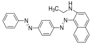
Figure 1. Chemical structure of sudan red-7B [SR7B].
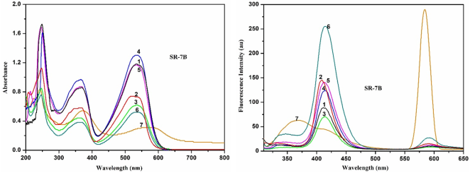
Figure 2. Absorption and fluorescence spectra of SR7B in different solvents at 303 K: 1) cyclohexane 2) 1, 4-dioxane 3) ethyl acetate 4) acetonitrile 5) 2-propanol 6) ethanol 7) water.
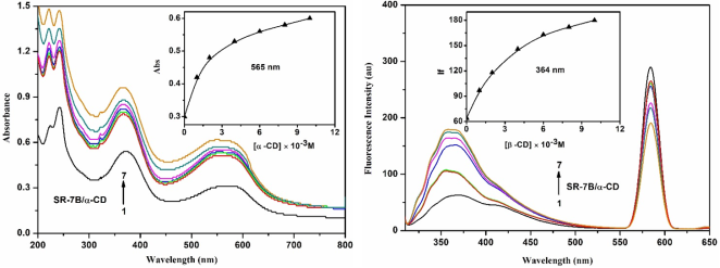
Figure 3. Absorption and fluorescence spectra of SR7B in different α-CD concentrations (M): (1) 0, (2) 0.001, (3) 0.002, (4) 0.004, (5) 0.006, (6) 0.008, (7) 0.01. Insert figure: absorbance/IF vs [α-CD].
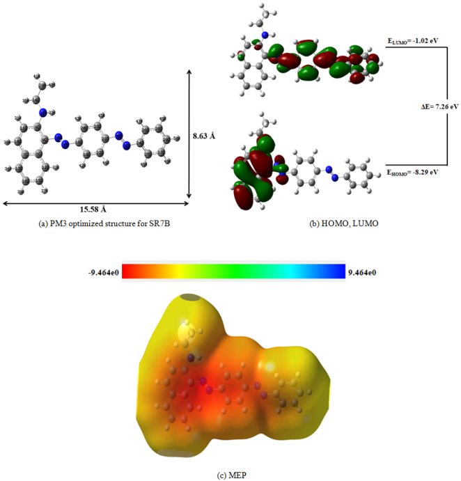
Figure 4. PM3 optimized structures of (a) SR7B, (b) HOMO, LUMO and (c) MEP of SR7B. The blue color indicates for nitrogen atom, while in HOMO-LUMO, the green and red colors denote negative and positive phases of the molecules.
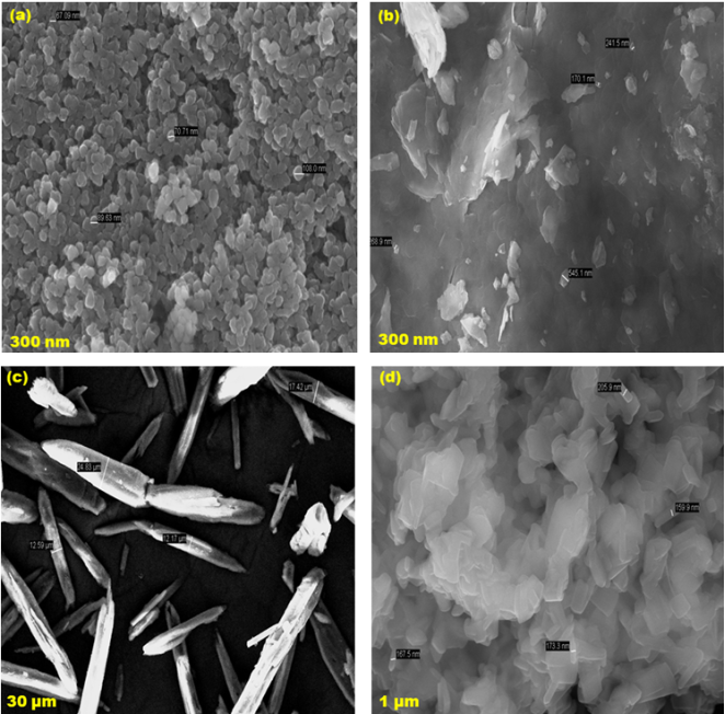
Figure 5. FE-SEM images for (a) ZnO, (b) ZnO/β-CD, (c) SR7B, (d) ZnO/SR7B/β-CD inclusion complexes.
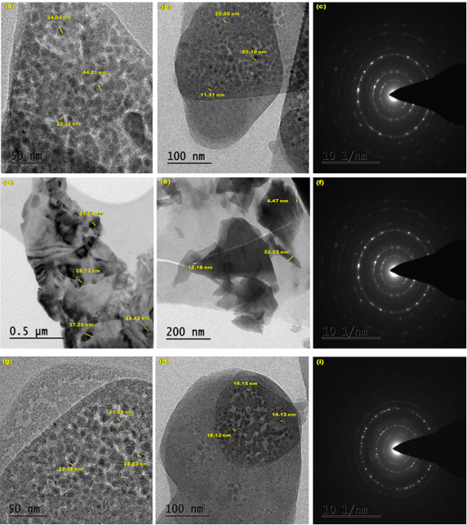
Figure 6. HR-TEM images for (a-c) ZnO, (d-f) ZnO/β-CD, (g-i) ZnO/SR7B/β-CD.
Information

