Introduction. Spinal arachnoid cysts (SAC) are uncommon lesions that result from a small defect in the arachnoid resulting in an intradural or extradural arachnoid hernia. Their diagnosis is misleading. Surgery is required when SAC is symptomatic. The modalities of this surgery are still the subject of controversy. We report an unusual case of intradural lumbar SAC. Observation. A 43-year-old man consulted for a right L5 lumbosciatica evolving for at least 7 years. Rheumatological examination noted lumbar spinal syndrome and radicular syndrome. The neurological examination was normal. Lumbar CT scan had shown discrete protrusive disc disease. Medical treatment had been prescribed. In view of the persistence of pain, magnetic resonance imaging (MRI) was prescribed. This was performed 6 months later when the patient had experienced a worsening type of cauda equina syndrome. This MRI had objectified signs in favor of an intradural lumbar SAC. The patient was transferred to neurosurgery. Cyst excision was performed and histology had confirmed the arachnoid nature of the cyst wall. The postoperative follow-up was simple and the patient had to recover completely from his neurological deficits. Conclusion. The diagnosis of SAC can be misleading and late in the stage of significant neurological disorders. Do not hesitate to request an MRI for a rebellious lumbosciatica with a normal CT scan in a patient without neurological deficits.
This is an Open Access article, distributed under the terms of the Creative Commons Attribution 4.0 International License (http://creativecommons.org/licenses/by/4.0/), which permits unrestricted use, distribution and reproduction in any medium or format, provided the original work is properly cited.
Spinal arachnoid cysts (SAC) are uncommon lesions in the spinal canal. They result from a small defect of the arachnoid resulting in an accumulation of cerebrospinal fluid (CSF) leading to a hernia of the arachnoid membrane intradural or extradural in case of a coexisting dural defect
[1]
Eroglu U, Bozkurt M, Kahilogullari G, Dogan I, Ozgural O, Shah KJ and al. Surgical Management of Spinal Arachnoid Cysts in Adults. World Neurosurgery, 2019; 122: e1146-e1152.
. In descending order of frequency, they occur in the thoracic, lumbar and cervical spine
[2]
Fam MD., Woodroffe RW., Helland L, Noeller J, Dahdaleh NS., Menezes A H and Hitchon PW. Spinal arachnoid cysts in adults: diagnosis and management. A single-center experience. Journal of Neurosurgery: Spine, 1-9.
Sadek A-R and Nader-Sepahi A. Spinal Arachnoid Cysts: Presentation, management and pathophysiology. Clinical Neurology and Neurosurgery, 2019; 180: 87-96.
The most commonly used classification separated cysts into 3 broad categories based on their location. Type I cysts are extradural arachnoid cysts (EAC) without nerve root damage. Type I cysts are further subclassified as EAC (type Ia) and sacral meningocele (type Ib). Type II cysts are EAC with nerve root involvement. Finally, type III cysts are intradural arachnoid cysts (IAC)
[5]
Nabors MW, Pait TG, Byrd EB, Karim NO, Davis DO, Kobrine AI, et al. Updated assessment and current classification of spinal meningeal cysts. J Neurosurg. 1988; 68: 366-377.
[5]
. Intramedullary cases have also been described
[5]
Nabors MW, Pait TG, Byrd EB, Karim NO, Davis DO, Kobrine AI, et al. Updated assessment and current classification of spinal meningeal cysts. J Neurosurg. 1988; 68: 366-377.
[5]
. The origin of the SAC is not well known. They can be congenital or secondary to various conditions. Thus, a more recent classification of spinal meningeal pathologies agrees that SACs are basically intradural lesions that are either of primary (i.e., idiopathic) origin or secondary to inflammatory reactions following trauma, infection, hemorrhage or surgery
[6]
Klekamp J. A New classification for pathologies of spinal meninges-part 2: primaryand secondary intradural arachnoid cysts, Neurosurgery 81 (2) (2017) 217-229.
[6]
.
Magnetic resonance imaging (MRI) is the test of choice for the diagnosis of these SACs which are generally asymptomatic but can have variable clinical manifestations which make the diagnosis err and make it late. Surgery is required when SACs are symptomatic. The modalities of this surgery are still the subject of controversy
[7]
Kumar A. Arachnoid Cysts: Clinical and Surgical Management. Arachnoid Cysts Elsevier, 2018; 271-283.
We report an unusual case of intradural lumbar SAC whose diagnosis was misleading that we managed effectively.
2. Result
Forty-three-year-old man was admitted in rheumatology for a right L5 lumbosciatica. This pain had been evolving for at least 7 years and had been managed by different doctors (general practitioners, rheumatologists, neurosurgeons, physical medicine and rehabilitation doctors) without success. The patient reported no medical history. At admission, the patient rated her pain on the Visual Analog Scale (VAS) at 7/10.
On physical examination, standing and walking were possible but there was lameness when walking due to pain. Lumbar spinal syndrome (made of pain on palpation of the lumbar spinous processes at L5, contracture of the paravertebral muscles) and radicular syndrome (made of a bell’s sign in L5S1 right, a sign of Lasegue positive at 50 degrees) were noted. There were no neurological deficits.
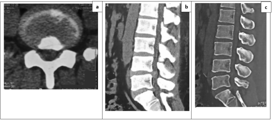
The non-injected computed tomography (CT) scan of the lumbar spine performed 7 years ago at the beginning of lumbosciatica had objectified discrete protrusive disc disease (Figure 1). We concluded that he had common lumbosciatica and prescribed methyl prednisolone combined with paracetamol, tramadol and a muscle relaxant to be taken orally. But a week later the patient reported the persistent see the worsening of the pain despite this treatment. His physical examination was superimposed on that of his admission. So, the tramadol was replaced by morphine and a lumbar MRI were ordered. The patient returned six months later with this MRI for financial reasons, according to him. By this time, his clinical condition had worsened. He explained that morphine did not improve his pain satisfactorily. He therefore went to see traditional medicine without success. Thus, he performed the MRI in front of the signs of aggravation. The lumbosciatica had become bilateral (predominant in the right lower limb), permanent, insomniating and very disabling. He also reported sphincter disorders such as stool and urine retention that motivated the placement of an indwelling urinary catheter. The physical examination noted almost impossible walking and motor deficit L5 and S1 rated at 2/5 on the right and 4/5 on the left.
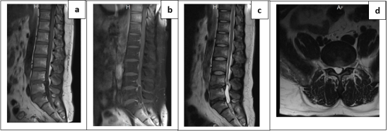
Lumbar spine MRI revealed a cystic lesion with the same signal as CSF. This lesion was hypo signal T1, hypersignal T2, with a thin wall not enhanced by the injection of contrast sitting in the spinal canal in the right posterior part of the L5 body slightly rising on L4. This lesion was more in favor of an arachnoid cyst but cystic ependymoma of the cauda equina had also been suggested (Figure 2).
The diagnosis of an intradural lumbar SAC causing cauda equina syndrome was retained and the patient was therefore transferred in neurosurgery.
He underwent surgery three days after his transfer in neurosurgery. An L5 laminectomy and a lower L4 hemilaminectomy were performed. At the time of the dural opening, the cerebrospinal fluid (CSF) squirted at the height of the cystic lesion. After complete emptying of the cyst the wall which was intradural and which resembled arachnoid was resected and transmitted in pathological anatomy. The intraoperative diagnosis of intradural SAC was retained.
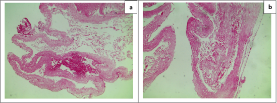
The pathological anatomy had concluded to an arachnoid cyst in front of the microscopic aspect of a "fringed" cystic wall achieving "pseudo villous" aspects. The coating of these fringes was regular without dysplastic lesions. These were usually flattened, sometimes oval cells with optically empty cytoplasm. The wall was loose, avascular fibrous and reworked by a discrete lymphocyte-like inflammatory infiltrate. There were no suspicious elements of malignancy. (Figure 3).
The postoperative follow-up was simple. The patient was allowed to be discharged three days after surgery. The removal of the urinary catheter was possible one week after surgery. One month after surgery the patient had no complaints and his neurological examination was normal. After a 2-year follow-up, the clinical examination remained normal.
Sagittal sections weighted sequence T1 (a), T1 with injection of gadolinium (b), T2 (c), and axial section T2 (d) showing a lesion in hypo signal T1, hypersignal T2, with a thin wall not enhanced by the contrast sitting in the spinal canal in the right posterior part of the L5 body rising slightly on L4. This lesion was more in favor of an arachnoid cyst but a cystic ependymoma of cauda equina had also been suggested.
Figure 3. Histological images of the operating specimen (cyst wall).
Magnification Gx40 (a) and Gx100 (b) showing the aspect of a "fringed" cystic wall achieving "pseudo villous" aspects. The coating of these fringes was regular without dysplastic lesions. These were usually flattened, sometimes oval cells with optically empty cytoplasm. The wall was loose, avascular fibrous and reworked by a discrete lymphocyte-like inflammatory infiltrate. There were no suspicious elements of malignancy.
3. Discussion
Almost all studies note that CAS is a relatively rare condition that accounts for 1% to 3% of all mass lesions in the spinal canal. The studies involved either a single case
[8]
Nath PC., Mishra SS., Deo RC and Satapathy MC. Intradural Spinal Arachnoid Cyst: A Long-Term Postlaminectomy Complication: A Case Report and Review of the Literature. World Neurosurgery, 2016; 85, 367. e1-367. e4.
or a small number of cases over a long period of time (11 cases in years
[9]
Kanwaljeet G, Sachin AB, Shashank SK and Bhawani SS. Spinal arachnoid cysts - our experience and review of literature. British Journal of Neurosurgery, 2017; 31 (2): 172-178.
Eroglu U, Bozkurt M, Kahilogullari G, Dogan I, Ozgural O, Shah KJ and al. Surgical Management of Spinal Arachnoid Cysts in Adults. World Neurosurgery, 2019; 122: e1146-e1152.
Sadek A-R and Nader-Sepahi A. Spinal Arachnoid Cysts: Presentation, management and pathophysiology. Clinical Neurology and Neurosurgery, 2019; 180: 87-96.
Fam MD., Woodroffe RW., Helland L, Noeller J, Dahdaleh NS., Menezes A H and Hitchon PW. Spinal arachnoid cysts in adults: diagnosis and management. A single-center experience. Journal of Neurosurgery: Spine, 1-9.
Nath PC., Mishra SS., Deo RC and Satapathy MC. Intradural Spinal Arachnoid Cyst: A Long-Term Postlaminectomy Complication: A Case Report and Review of the Literature. World Neurosurgery, 2016; 85, 367. e1-367. e4.
, intradural arachnoid cysts (IAC) are even less common than extradural arachnoid cysts (EAC); for other authors
[1]
Eroglu U, Bozkurt M, Kahilogullari G, Dogan I, Ozgural O, Shah KJ and al. Surgical Management of Spinal Arachnoid Cysts in Adults. World Neurosurgery, 2019; 122: e1146-e1152.
Eroglu U, Bozkurt M, Kahilogullari G, Dogan I, Ozgural O, Shah KJ and al. Surgical Management of Spinal Arachnoid Cysts in Adults. World Neurosurgery, 2019; 122: e1146-e1152.
Young adult males were the most concerned. Authors have reported the case of a 45-year-old male subject
[8]
Nath PC., Mishra SS., Deo RC and Satapathy MC. Intradural Spinal Arachnoid Cyst: A Long-Term Postlaminectomy Complication: A Case Report and Review of the Literature. World Neurosurgery, 2016; 85, 367. e1-367. e4.
. Elsewhere, the average age was 32.9 ± 20.8 years
[9]
Kanwaljeet G, Sachin AB, Shashank SK and Bhawani SS. Spinal arachnoid cysts - our experience and review of literature. British Journal of Neurosurgery, 2017; 31 (2): 172-178.
Fam MD., Woodroffe RW., Helland L, Noeller J, Dahdaleh NS., Menezes A H and Hitchon PW. Spinal arachnoid cysts in adults: diagnosis and management. A single-center experience. Journal of Neurosurgery: Spine, 1-9.
Cuoco JA, Muthukumar S, Rogers CM, Entwistle JJ, Patel VM, Olasunkanmi AL, et al. Spinal Intradural Arachnoid Cysts in Adults: An Institutional Experience and Literature Review. Neurosurgery. 2023; 92(3): 450-463.
Fam MD., Woodroffe RW., Helland L, Noeller J, Dahdaleh NS., Menezes A H and Hitchon PW. Spinal arachnoid cysts in adults: diagnosis and management. A single-center experience. Journal of Neurosurgery: Spine, 1-9.
Sadek A-R and Nader-Sepahi A. Spinal Arachnoid Cysts: Presentation, management and pathophysiology. Clinical Neurology and Neurosurgery, 2019; 180: 87-96.
The exact origin of SAC is controversial and several theories exist. They are often attributed to birth defects. Acquired SAC may be due to arachnoid adhesions that develop as a result of inflammation, which may result from infection (meningitis), hemorrhage, or an iatrogenic cause such as injection of contrast medium or anesthetic or intraoperative fibrin glue contaminants. Some acquired cysts can be caused by trauma related to a lumbar puncture, anesthetic or surgical procedures. Other cysts are idiopathic
[7]
Kumar A. Arachnoid Cysts: Clinical and Surgical Management. Arachnoid Cysts Elsevier, 2018; 271-283.
The compressive and symptomatic SAC is exceptional so we think about it very little. Also, the symptomatology is nonspecific. All of this can cause the diagnosis to err for months or even years and allow neurological disorders to take hold
[7]
Kumar A. Arachnoid Cysts: Clinical and Surgical Management. Arachnoid Cysts Elsevier, 2018; 271-283.
. In the literature pain is the most common symptom. It is inaugural and neurological disorders occur slowly and gradually over several months or even years
[1]
Eroglu U, Bozkurt M, Kahilogullari G, Dogan I, Ozgural O, Shah KJ and al. Surgical Management of Spinal Arachnoid Cysts in Adults. World Neurosurgery, 2019; 122: e1146-e1152.
Sadek A-R and Nader-Sepahi A. Spinal Arachnoid Cysts: Presentation, management and pathophysiology. Clinical Neurology and Neurosurgery, 2019; 180: 87-96.
Nath PC., Mishra SS., Deo RC and Satapathy MC. Intradural Spinal Arachnoid Cyst: A Long-Term Postlaminectomy Complication: A Case Report and Review of the Literature. World Neurosurgery, 2016; 85, 367. e1-367. e4.
Kanwaljeet G, Sachin AB, Shashank SK and Bhawani SS. Spinal arachnoid cysts - our experience and review of literature. British Journal of Neurosurgery, 2017; 31 (2): 172-178.
. This is why do not hesitate to ask for appropriate imaging in the face of pain that persists or worsens despite well-conducted medical treatment for several weeks.
The CT scan may not allow to objectify the SAC as was the case in our observation especially that the cyst was located at the root of the cauda equina. MRI is the best exam. It makes it possible to highlight SAC as a cystic lesion having the same signal as CSF with a thin wall not enhanced by the injection of contrast
[2]
Fam MD., Woodroffe RW., Helland L, Noeller J, Dahdaleh NS., Menezes A H and Hitchon PW. Spinal arachnoid cysts in adults: diagnosis and management. A single-center experience. Journal of Neurosurgery: Spine, 1-9.
Kanwaljeet G, Sachin AB, Shashank SK and Bhawani SS. Spinal arachnoid cysts - our experience and review of literature. British Journal of Neurosurgery, 2017; 31 (2): 172-178.
. The diffusion sequence helps differentiate an epidermoid cyst or cystic ependymoma from a SAC. It can also help differentiate a cyst from an abscess. Flow MRI can assess the presence of communication between the cyst and the subarachnoid space
[7]
Kumar A. Arachnoid Cysts: Clinical and Surgical Management. Arachnoid Cysts Elsevier, 2018; 271-283.
. Myelography CT can achieve the same results as MRI by highlighting SAC and its possible communication with the subarachnoid space
[2]
Fam MD., Woodroffe RW., Helland L, Noeller J, Dahdaleh NS., Menezes A H and Hitchon PW. Spinal arachnoid cysts in adults: diagnosis and management. A single-center experience. Journal of Neurosurgery: Spine, 1-9.
Kanwaljeet G, Sachin AB, Shashank SK and Bhawani SS. Spinal arachnoid cysts - our experience and review of literature. British Journal of Neurosurgery, 2017; 31 (2): 172-178.
. This examination is no longer carried out in our country because of its invasive nature. However, MRI poses the problem of its geographical and financial accessibility in our country. It is therefore not a common prescription in front of a non-deficient lumbosciatica as was the case in the patient of our observation at the beginning. Thus, it was 7 years after having performed a CT scan that the diagnosis of arachnoid cyst was evoked on MRI. This at a very advanced stage where the patient had significant neurological deficits.
The usual management of symptomatic SAC is excision of the cyst with closure of the dural defect in extradural cysts, while in the case of intradural cysts, especially those located before the cord, fenestration of the cyst is usually performed
[1]
Eroglu U, Bozkurt M, Kahilogullari G, Dogan I, Ozgural O, Shah KJ and al. Surgical Management of Spinal Arachnoid Cysts in Adults. World Neurosurgery, 2019; 122: e1146-e1152.
Kanwaljeet G, Sachin AB, Shashank SK and Bhawani SS. Spinal arachnoid cysts - our experience and review of literature. British Journal of Neurosurgery, 2017; 31 (2): 172-178.
. The total removal of the SAC wall remains controversial
[1]
Eroglu U, Bozkurt M, Kahilogullari G, Dogan I, Ozgural O, Shah KJ and al. Surgical Management of Spinal Arachnoid Cysts in Adults. World Neurosurgery, 2019; 122: e1146-e1152.
The follow-up of this surgery was most often favorable [1-3.9]. Thus, authors specify that postoperative MRI showed complete resolution of SAC in 14 of 16 patients
[2]
Fam MD., Woodroffe RW., Helland L, Noeller J, Dahdaleh NS., Menezes A H and Hitchon PW. Spinal arachnoid cysts in adults: diagnosis and management. A single-center experience. Journal of Neurosurgery: Spine, 1-9.
. For still others, 6 months after surgery, all patients had experienced improvement
[3]
Sadek A-R and Nader-Sepahi A. Spinal Arachnoid Cysts: Presentation, management and pathophysiology. Clinical Neurology and Neurosurgery, 2019; 180: 87-96.
Recent data from the literature confirm the rarity of spinal arachnoid cysts and highlight the diagnostic difficulty of these lesions. In a literature review associated with an institutional series of 29 patients, Cuoco et al. showed that intradural arachnoid cysts in adults remain uncommon conditions, the diagnosis of which is often delayed due to nonspecific symptoms dominated by spinal and radicular pain. The authors emphasize the importance of MRI in the early identification of these lesions and in therapeutic planning
[11]
Cuoco JA, Muthukumar S, Rogers CM, Entwistle JJ, Patel VM, Olasunkanmi AL, et al. Spinal Intradural Arachnoid Cysts in Adults: An Institutional Experience and Literature Review. Neurosurgery. 2023; 92(3): 450-463.
Kalsi et al., in a systematic review published in 2022, report that the majority of patients present with a chronic course with progressive worsening of neurological symptoms before diagnosis. This observation is comparable to that of our patient, in whom the symptoms evolved for several years before the cyst was identified by MRI
[12]
Kalsi P, Hejrati N, Charalampidis A, Wu PH, Schneider M, Wilson JRF, et al. Spinal arachnoid cysts: A case series and systematic review of the literature. Brain Spine. 2022; 2: 100904.
Regarding the etiopathogenesis, Wang et al. A systematic review of symptomatic secondary arachnoid cysts showed that inflammatory processes, trauma, spinal surgery, and subarachnoid hemorrhage are the main acquired factors contributing to their development. However, a significant proportion of cases remain idiopathic, as in our observation
[13]
Wang YB, Wang DH, Deng SL. Symptomatic secondary spinal arachnoid cysts: a systematic review. Spine J. 2023; 23(8): 1199-1211.
MRI remains the gold standard examination. Conventional sequences generally allow differentiation of the cyst from the adjacent neural parenchyma, while diffusion-weighted imaging and CSF flow analysis techniques improve the differential diagnosis with tumoral or infectious cystic lesions
[11]
Cuoco JA, Muthukumar S, Rogers CM, Entwistle JJ, Patel VM, Olasunkanmi AL, et al. Spinal Intradural Arachnoid Cysts in Adults: An Institutional Experience and Literature Review. Neurosurgery. 2023; 92(3): 450-463.
Kalsi P, Hejrati N, Charalampidis A, Wu PH, Schneider M, Wilson JRF, et al. Spinal arachnoid cysts: A case series and systematic review of the literature. Brain Spine. 2022; 2: 100904.
From a therapeutic standpoint, recent publications confirm that surgery remains indicated in symptomatic patients or those presenting with neurological deterioration. Complete excision, when technically feasible, or failing that, fenestration of the cyst with restoration of CSF circulation, yields good functional results in the majority of cases
[11]
Cuoco JA, Muthukumar S, Rogers CM, Entwistle JJ, Patel VM, Olasunkanmi AL, et al. Spinal Intradural Arachnoid Cysts in Adults: An Institutional Experience and Literature Review. Neurosurgery. 2023; 92(3): 450-463.
Kalsi P, Hejrati N, Charalampidis A, Wu PH, Schneider M, Wilson JRF, et al. Spinal arachnoid cysts: A case series and systematic review of the literature. Brain Spine. 2022; 2: 100904.
El-Hajj VG, Edström E, Elmi-Terander A, Fletcher-Sandersjöö A, et al. An unusual cause of chronic neuropathic pain: report of a case of multiple intradural spinal arachnoid cysts and review of the literature. Acta Neurochir (Wien). 2023; 165: 2699-2705.
Finally, the postoperative results reported in contemporary series are generally favorable, with significant improvement in pain and neurological deficits. These data are consistent with the outcome observed in our patient, who experienced complete neurological recovery after surgical excision of the cyst and has shown no clinical recurrence after two years of follow-up
[15]
Messerer R, Aldugman M, Morgado A, Barrey C. Spinal extradural arachnoid cysts: a rare entity and review of the literature. Eur Spine J. 2024; 33(3): 872-880.
Khan MSI, Ahmed N, Barua KK, Chaurasia B, Vats A, Goel A. Pathogenesis, management strategies, and outcome of non-communicating extradural spinal arachnoid cysts: a systematic review. Br J Neurosurg. 2023; 37(2): 213-219.
The compressive and symptomatic SAC is exceptional so we think about it very little. Its symptoms are non-specific and develop slowly and gradually that can lead to significant neurological deficits. CT was of no contribution to the diagnosis, especially since the cyst was located at the roots of the cauda equina.
Therapeutically, the removal of the lesion had been carried out without any difficulty. The post-operative outcome was good. This clinical case edifies us on the fact that we should not hesitate to prescribe an MRI in front of a lumbosciatica rebellious to any drug treatment after several weeks even if it is non-deficient.
5. Recommendations
In light of this clinical observation and the literature, the following recommendations can be made:
In any case of chronic lumbosciatica that is resistant to well-conducted medical treatment and has an atypical course, further investigation with MRI should be considered, even when CT scans reveal only minimal disc pathology.
The presence of alarm signs such as progressive worsening of pain, the onset of motor deficits, sensory disturbances, or sphincter dysfunction should prompt the urgent performance of a spinal MRI.
Spinal arachnoid cysts should be included in the differential diagnosis of unexplained chronic radicular syndromes, particularly when the results of conventional imaging are inconsistent with the clinical symptoms.
MRI is the gold standard for the diagnosis of spinal arachnoid cysts and for preoperative treatment planning.
A multidisciplinary collaboration involving rheumatologists, neurosurgeons, radiologists, and pathologists is recommended to optimize the diagnostic and therapeutic management of these rare lesions.
In symptomatic patients or those experiencing neurological deterioration, early surgical intervention should be offered to prevent the development of irreversible neurological deficits.
A histopathological examination of the resected cyst wall should be systematically performed to confirm the diagnosis and rule out a tumor.
Postoperative clinical and radiological follow-up is recommended to detect any recurrence and assess long-term neurological recovery.
In resource-limited countries, strategies aimed at improving financial and geographic access to MRI could help reduce diagnostic delays observed in patients with spinal arachnoid cysts.
Acknowledgments
Our thanks go to:
Sir Ousmane Nere Director General of Yalgado Ouedraogo University Hospital Center.
Sir Saydou Nombre Director General of Bogodogo University Hospital Center.
Teacher Denlewende Sylvain Zabsonrehead of neurosurgery department of Yalgado Ouedraogo University Hospital in Ouagadougou, my colleagues in the neurosurgery department (Yacouba Haro, Inoussa Zoungrana, Abdoulaye Sanou).
Author Contributions
Henri Lankoande: Conceptualization, Resources, Investigation, Writing – original draft
Authors declare any perceived conflict of interest for each author related to the manuscript or its subject matter.
Appendix
Lessons Learned from the Case
This clinical case highlights several important aspects of the management of intradural spinal arachnoid cysts.
First, the initial symptoms can be misleading and mimic common lumbosacral radiculopathy. In our observation, the presence of a subtle disc protrusion on CT scan led to a diagnosis of degenerative spinal disease, thus delaying the identification of the true cause of the symptoms.
Second, the progressive evolution of symptoms over several years underscores the insidious nature of this condition. Secondary worsening with the development of cauda equina syndrome ultimately led to an MRI, which confirmed the diagnosis.
Third, this case illustrates the major diagnostic value of MRI in the investigation of atypical or treatment-resistant lumbosacral radiculopathy, particularly when previous investigations fail to explain the severity of the symptoms. Finally, the favorable postoperative course with complete neurological recovery confirms the effectiveness of surgical treatment when indicated, even in the presence of severe neurological deficits.
References
[1]
Eroglu U, Bozkurt M, Kahilogullari G, Dogan I, Ozgural O, Shah KJ and al. Surgical Management of Spinal Arachnoid Cysts in Adults. World Neurosurgery, 2019; 122: e1146-e1152.
Fam MD., Woodroffe RW., Helland L, Noeller J, Dahdaleh NS., Menezes A H and Hitchon PW. Spinal arachnoid cysts in adults: diagnosis and management. A single-center experience. Journal of Neurosurgery: Spine, 1-9.
Sadek A-R and Nader-Sepahi A. Spinal Arachnoid Cysts: Presentation, management and pathophysiology. Clinical Neurology and Neurosurgery, 2019; 180: 87-96.
Nabors MW, Pait TG, Byrd EB, Karim NO, Davis DO, Kobrine AI, et al. Updated assessment and current classification of spinal meningeal cysts. J Neurosurg. 1988; 68: 366-377.
[6]
Klekamp J. A New classification for pathologies of spinal meninges-part 2: primaryand secondary intradural arachnoid cysts, Neurosurgery 81 (2) (2017) 217-229.
[7]
Kumar A. Arachnoid Cysts: Clinical and Surgical Management. Arachnoid Cysts Elsevier, 2018; 271-283.
Nath PC., Mishra SS., Deo RC and Satapathy MC. Intradural Spinal Arachnoid Cyst: A Long-Term Postlaminectomy Complication: A Case Report and Review of the Literature. World Neurosurgery, 2016; 85, 367. e1-367. e4.
Kanwaljeet G, Sachin AB, Shashank SK and Bhawani SS. Spinal arachnoid cysts - our experience and review of literature. British Journal of Neurosurgery, 2017; 31 (2): 172-178.
Cuoco JA, Muthukumar S, Rogers CM, Entwistle JJ, Patel VM, Olasunkanmi AL, et al. Spinal Intradural Arachnoid Cysts in Adults: An Institutional Experience and Literature Review. Neurosurgery. 2023; 92(3): 450-463.
Kalsi P, Hejrati N, Charalampidis A, Wu PH, Schneider M, Wilson JRF, et al. Spinal arachnoid cysts: A case series and systematic review of the literature. Brain Spine. 2022; 2: 100904.
El-Hajj VG, Edström E, Elmi-Terander A, Fletcher-Sandersjöö A, et al. An unusual cause of chronic neuropathic pain: report of a case of multiple intradural spinal arachnoid cysts and review of the literature. Acta Neurochir (Wien). 2023; 165: 2699-2705.
Messerer R, Aldugman M, Morgado A, Barrey C. Spinal extradural arachnoid cysts: a rare entity and review of the literature. Eur Spine J. 2024; 33(3): 872-880.
Khan MSI, Ahmed N, Barua KK, Chaurasia B, Vats A, Goel A. Pathogenesis, management strategies, and outcome of non-communicating extradural spinal arachnoid cysts: a systematic review. Br J Neurosurg. 2023; 37(2): 213-219.
Lankoande, H., Tiendrebeogo/Zabsonre, W. J. S., Zabsonre, D. S., Haro, Y., Ouattara, S., et al. (2026). Lumbosciatica Indicative of an Intradural Lumbar Spinal Arachnoid Cyst. American Journal of Health Research, 14(4), 198-203. https://doi.org/10.11648/j.ajhr.20261404.13
Lankoande, H.; Tiendrebeogo/Zabsonre, W. J. S.; Zabsonre, D. S.; Haro, Y.; Ouattara, S., et al. Lumbosciatica Indicative of an Intradural Lumbar Spinal Arachnoid Cyst. Am. J. Health Res.2026, 14(4), 198-203. doi: 10.11648/j.ajhr.20261404.13
Lankoande H, Tiendrebeogo/Zabsonre WJS, Zabsonre DS, Haro Y, Ouattara S, et al. Lumbosciatica Indicative of an Intradural Lumbar Spinal Arachnoid Cyst. Am J Health Res. 2026;14(4):198-203. doi: 10.11648/j.ajhr.20261404.13
@article{10.11648/j.ajhr.20261404.13,
  author = {Henri Lankoande and Wendlassida Joelle Stephanie Tiendrebeogo/Zabsonre and Denlewende Sylvain Zabsonre and Yakouba Haro and Souleymane Ouattara and Fulgence Kabore and Abdoulaye Sanou and Inoussa Zoungrana and Dieu-Donne Ouedraogo},
  title = {Lumbosciatica Indicative of an Intradural Lumbar Spinal Arachnoid Cyst},
  journal = {American Journal of Health Research},
  volume = {14},
  number = {4},
  pages = {198-203},
  doi = {10.11648/j.ajhr.20261404.13},
  url = {https://doi.org/10.11648/j.ajhr.20261404.13},
  eprint = {https://article.sciencepublishinggroup.com/pdf/10.11648.j.ajhr.20261404.13},
  abstract = {Introduction. Spinal arachnoid cysts (SAC) are uncommon lesions that result from a small defect in the arachnoid resulting in an intradural or extradural arachnoid hernia. Their diagnosis is misleading. Surgery is required when SAC is symptomatic. The modalities of this surgery are still the subject of controversy. We report an unusual case of intradural lumbar SAC. Observation. A 43-year-old man consulted for a right L5 lumbosciatica evolving for at least 7 years. Rheumatological examination noted lumbar spinal syndrome and radicular syndrome. The neurological examination was normal. Lumbar CT scan had shown discrete protrusive disc disease. Medical treatment had been prescribed. In view of the persistence of pain, magnetic resonance imaging (MRI) was prescribed. This was performed 6 months later when the patient had experienced a worsening type of cauda equina syndrome. This MRI had objectified signs in favor of an intradural lumbar SAC. The patient was transferred to neurosurgery. Cyst excision was performed and histology had confirmed the arachnoid nature of the cyst wall. The postoperative follow-up was simple and the patient had to recover completely from his neurological deficits. Conclusion. The diagnosis of SAC can be misleading and late in the stage of significant neurological disorders. Do not hesitate to request an MRI for a rebellious lumbosciatica with a normal CT scan in a patient without neurological deficits.},
 year = {2026}
}
TY  - JOUR
T1  - Lumbosciatica Indicative of an Intradural Lumbar Spinal Arachnoid Cyst
AU  - Henri Lankoande
AU  - Wendlassida Joelle Stephanie Tiendrebeogo/Zabsonre
AU  - Denlewende Sylvain Zabsonre
AU  - Yakouba Haro
AU  - Souleymane Ouattara
AU  - Fulgence Kabore
AU  - Abdoulaye Sanou
AU  - Inoussa Zoungrana
AU  - Dieu-Donne Ouedraogo
Y1  - 2026/07/27
PY  - 2026
N1  - https://doi.org/10.11648/j.ajhr.20261404.13
DO  - 10.11648/j.ajhr.20261404.13
T2  - American Journal of Health Research
JF  - American Journal of Health Research
JO  - American Journal of Health Research
SP  - 198
EP  - 203
PB  - Science Publishing Group
SN  - 2330-8796
UR  - https://doi.org/10.11648/j.ajhr.20261404.13
AB  - Introduction. Spinal arachnoid cysts (SAC) are uncommon lesions that result from a small defect in the arachnoid resulting in an intradural or extradural arachnoid hernia. Their diagnosis is misleading. Surgery is required when SAC is symptomatic. The modalities of this surgery are still the subject of controversy. We report an unusual case of intradural lumbar SAC. Observation. A 43-year-old man consulted for a right L5 lumbosciatica evolving for at least 7 years. Rheumatological examination noted lumbar spinal syndrome and radicular syndrome. The neurological examination was normal. Lumbar CT scan had shown discrete protrusive disc disease. Medical treatment had been prescribed. In view of the persistence of pain, magnetic resonance imaging (MRI) was prescribed. This was performed 6 months later when the patient had experienced a worsening type of cauda equina syndrome. This MRI had objectified signs in favor of an intradural lumbar SAC. The patient was transferred to neurosurgery. Cyst excision was performed and histology had confirmed the arachnoid nature of the cyst wall. The postoperative follow-up was simple and the patient had to recover completely from his neurological deficits. Conclusion. The diagnosis of SAC can be misleading and late in the stage of significant neurological disorders. Do not hesitate to request an MRI for a rebellious lumbosciatica with a normal CT scan in a patient without neurological deficits.
VL  - 14
IS  - 4
ER  -
Lankoande, H., Tiendrebeogo/Zabsonre, W. J. S., Zabsonre, D. S., Haro, Y., Ouattara, S., et al. (2026). Lumbosciatica Indicative of an Intradural Lumbar Spinal Arachnoid Cyst. American Journal of Health Research, 14(4), 198-203. https://doi.org/10.11648/j.ajhr.20261404.13
Lankoande, H.; Tiendrebeogo/Zabsonre, W. J. S.; Zabsonre, D. S.; Haro, Y.; Ouattara, S., et al. Lumbosciatica Indicative of an Intradural Lumbar Spinal Arachnoid Cyst. Am. J. Health Res.2026, 14(4), 198-203. doi: 10.11648/j.ajhr.20261404.13
Lankoande H, Tiendrebeogo/Zabsonre WJS, Zabsonre DS, Haro Y, Ouattara S, et al. Lumbosciatica Indicative of an Intradural Lumbar Spinal Arachnoid Cyst. Am J Health Res. 2026;14(4):198-203. doi: 10.11648/j.ajhr.20261404.13
@article{10.11648/j.ajhr.20261404.13,
  author = {Henri Lankoande and Wendlassida Joelle Stephanie Tiendrebeogo/Zabsonre and Denlewende Sylvain Zabsonre and Yakouba Haro and Souleymane Ouattara and Fulgence Kabore and Abdoulaye Sanou and Inoussa Zoungrana and Dieu-Donne Ouedraogo},
  title = {Lumbosciatica Indicative of an Intradural Lumbar Spinal Arachnoid Cyst},
  journal = {American Journal of Health Research},
  volume = {14},
  number = {4},
  pages = {198-203},
  doi = {10.11648/j.ajhr.20261404.13},
  url = {https://doi.org/10.11648/j.ajhr.20261404.13},
  eprint = {https://article.sciencepublishinggroup.com/pdf/10.11648.j.ajhr.20261404.13},
  abstract = {Introduction. Spinal arachnoid cysts (SAC) are uncommon lesions that result from a small defect in the arachnoid resulting in an intradural or extradural arachnoid hernia. Their diagnosis is misleading. Surgery is required when SAC is symptomatic. The modalities of this surgery are still the subject of controversy. We report an unusual case of intradural lumbar SAC. Observation. A 43-year-old man consulted for a right L5 lumbosciatica evolving for at least 7 years. Rheumatological examination noted lumbar spinal syndrome and radicular syndrome. The neurological examination was normal. Lumbar CT scan had shown discrete protrusive disc disease. Medical treatment had been prescribed. In view of the persistence of pain, magnetic resonance imaging (MRI) was prescribed. This was performed 6 months later when the patient had experienced a worsening type of cauda equina syndrome. This MRI had objectified signs in favor of an intradural lumbar SAC. The patient was transferred to neurosurgery. Cyst excision was performed and histology had confirmed the arachnoid nature of the cyst wall. The postoperative follow-up was simple and the patient had to recover completely from his neurological deficits. Conclusion. The diagnosis of SAC can be misleading and late in the stage of significant neurological disorders. Do not hesitate to request an MRI for a rebellious lumbosciatica with a normal CT scan in a patient without neurological deficits.},
 year = {2026}
}
TY  - JOUR
T1  - Lumbosciatica Indicative of an Intradural Lumbar Spinal Arachnoid Cyst
AU  - Henri Lankoande
AU  - Wendlassida Joelle Stephanie Tiendrebeogo/Zabsonre
AU  - Denlewende Sylvain Zabsonre
AU  - Yakouba Haro
AU  - Souleymane Ouattara
AU  - Fulgence Kabore
AU  - Abdoulaye Sanou
AU  - Inoussa Zoungrana
AU  - Dieu-Donne Ouedraogo
Y1  - 2026/07/27
PY  - 2026
N1  - https://doi.org/10.11648/j.ajhr.20261404.13
DO  - 10.11648/j.ajhr.20261404.13
T2  - American Journal of Health Research
JF  - American Journal of Health Research
JO  - American Journal of Health Research
SP  - 198
EP  - 203
PB  - Science Publishing Group
SN  - 2330-8796
UR  - https://doi.org/10.11648/j.ajhr.20261404.13
AB  - Introduction. Spinal arachnoid cysts (SAC) are uncommon lesions that result from a small defect in the arachnoid resulting in an intradural or extradural arachnoid hernia. Their diagnosis is misleading. Surgery is required when SAC is symptomatic. The modalities of this surgery are still the subject of controversy. We report an unusual case of intradural lumbar SAC. Observation. A 43-year-old man consulted for a right L5 lumbosciatica evolving for at least 7 years. Rheumatological examination noted lumbar spinal syndrome and radicular syndrome. The neurological examination was normal. Lumbar CT scan had shown discrete protrusive disc disease. Medical treatment had been prescribed. In view of the persistence of pain, magnetic resonance imaging (MRI) was prescribed. This was performed 6 months later when the patient had experienced a worsening type of cauda equina syndrome. This MRI had objectified signs in favor of an intradural lumbar SAC. The patient was transferred to neurosurgery. Cyst excision was performed and histology had confirmed the arachnoid nature of the cyst wall. The postoperative follow-up was simple and the patient had to recover completely from his neurological deficits. Conclusion. The diagnosis of SAC can be misleading and late in the stage of significant neurological disorders. Do not hesitate to request an MRI for a rebellious lumbosciatica with a normal CT scan in a patient without neurological deficits.
VL  - 14
IS  - 4
ER  -

 ,
Wendlassida Joelle Stephanie Tiendrebeogo/Zabsonre,
Denlewende Sylvain Zabsonre,
Yakouba Haro,
Souleymane Ouattara,
Fulgence Kabore,
Abdoulaye Sanou,
Inoussa Zoungrana,
Dieu-Donne Ouedraogo
,
Wendlassida Joelle Stephanie Tiendrebeogo/Zabsonre,
Denlewende Sylvain Zabsonre,
Yakouba Haro,
Souleymane Ouattara,
Fulgence Kabore,
Abdoulaye Sanou,
Inoussa Zoungrana,
Dieu-Donne Ouedraogo



 Share This Article
 Twitter
 Linked In
 Facebook
 Copy
Copy Download