Introduction. Giant intramuscular lipoma is a rare benign tumor. Because of its deep location, it may cause nerve compression and raise concern for an atypical lipomatous tumor. We report a giant lipoma of the soleus muscle complicated by tibial nerve neuralgia after traditional massage and briefly describe the clinical, imaging, surgical and histological findings. Clinical examination, ultrasonography, operative findings and histology were reviewed in this single case observation. Case presentation. A 58-year-old female farmer with no medical history presented with a left calf swelling that had slowly enlarged over five years. After one month of traditional massage consisting of repeated deep manual pressure and kneading applied locally to the calf, she developed neurogenic pain radiating to the foot, associated with plantar paresthesia and a positive Tinel sign. Ultrasonography showed a heterogeneous calcified mass measuring 15 × 7 × 4 cm within the soleus muscle. A marginal en bloc resection was performed with release of the posterior tibial neurovascular bundle and neurolysis. Histology confirmed a benign lipoma. Conclusion. Traditional massage may have hypothetically decompensated chronic tibial nerve compression related to the lipoma. Complete excision with neurolysis led to rapid neurological improvement, with no clinical recurrence at six months; longer follow-up remains recommended.
This is an Open Access article, distributed under the terms of the Creative Commons Attribution 4.0 International License (http://creativecommons.org/licenses/by/4.0/), which permits unrestricted use, distribution and reproduction in any medium or format, provided the original work is properly cited.
Giant Lipoma, Intramuscular Lipoma, Calf, Soleus Muscle, Tibial Nerve, Traditional Massage, Nerve Compression,
Case Report
1. Introduction
Lipomas are the most common benign soft-tissue tumors. The term “giant” is usually applied when the lesion exceeds 10 cm in its largest diameter or weighs more than 1,000 g, a situation in which atypical lipomatous tumor or well-differentiated liposarcoma must be ruled out
[1]
Sanchez MR, Golomb FM, Moy JA, Potozkin JR. Giant lipoma: case report and review of the literature. J Am Acad Dermatol. 1993; 28(2 Pt 1): 266-268.
Rydholm A, Berg NO. Size, site and clinical incidence of lipoma. Factors in the differential diagnosis of lipoma and sarcoma. Acta Orthop Scand. 1983; 54(6): 929-934.
Achar S, Yamanaka J, Oberstar J. Soft Tissue Masses: Evaluation and Treatment. Am Fam Physician. 2022; 105(6): 602-612.
[1-3]
.
Intramuscular lipomas are rare and may remain silent for a long time. In the calf, growth near the tibial neurovascular bundle may cause neuropathic pain, plantar paresthesia and a positive Tinel sign through irritation or compression of the tibial nerve
[4]
McTighe S, Chernev I. Intramuscular lipoma: a review of the literature. Orthop Rev (Pavia). 2014; 6(4): 5618.
Badr IT, Hassan S, Fotoh DS, Moawad MM. Extrinsic compression neuropathy of the tibial nerve secondary to accessory soleus muscle in a young teenager. J Clin Orthop Trauma. 2019; 11(2): 302-306.
Deep traditional massage may hypothetically act as a decompensating factor in a pre-existing bulky lesion, through mechanical irritation, edema or microtrauma. We report a giant intramuscular lipoma of the soleus muscle presenting as tibial nerve neuralgia after traditional massage
[6]
Yin P, Gao N, Wu J, Litscher G, Xu S. Adverse events of massage therapy in pain-related conditions: a systematic review. Evid Based Complement Alternat Med. 2014; 2014: 480956.
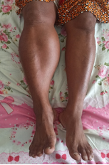
A 58-year-old female farmer, with no known medical or surgical history and no comorbidities, was seen for a large swelling of the left calf that had progressively enlarged over five years, with no history of initial trauma. Because of a feeling of fatigue in the left lower limb, she had undergone local traditional massage consisting of repeated deep manual pressure and kneading applied locally to the calf for about one month. After these massages, she developed intense neurogenic pain in the left calf, radiating to the foot, worsened by walking and prolonged standing, and associated with paresthesia of the sole and toes. Clinical examination revealed a large left calf mass that was firm, tender and mobile, with no local inflammatory skin changes or distal vascular compromise. Peripheral pulses were present. Neurological examination found paresthesia in the sensory territory of the tibial nerve, with a positive Tinel sign, suggesting neuralgia due to irritation or compression of the posterior tibial nerve.
Ultrasonography showed a heterogeneous hypoechoic mass with internal calcifications, measuring 15 × 7 × 4 cm, arising from the soleus muscle. Given the depth, giant size and heterogeneous appearance of the lesion, excision with pathological analysis was indicated to rule out an atypical lipomatous tumor.
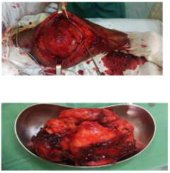
Marginal resection was performed under spinal anesthesia, with the patient in the prone position, through a posterior approach. After release of the posterior tibial vascular bundle and neurolysis of the tibial nerve, the tumor was removed en bloc with a macroscopically healthy margin. Pathological examination confirmed a benign lipoma. The postoperative course was uneventful, with marked regression of neurological signs from the first postoperative week. No clinical recurrence was observed after six months of follow-up.
. Large size, deep location and progressive growth require a structured diagnostic approach, because deep lipomas may mimic atypical lipomatous tumors
[2]
Rydholm A, Berg NO. Size, site and clinical incidence of lipoma. Factors in the differential diagnosis of lipoma and sarcoma. Acta Orthop Scand. 1983; 54(6): 929-934.
. In our case, the five-year history, mobility, absence of systemic signs and benign histology supported the diagnosis of lipoma, whereas the heterogeneous ultrasound appearance and calcifications justified pathological confirmation.
Ultrasonography helped determine the intramuscular location and size of the mass. However, magnetic resonance imaging (MRI) remains the reference examination for deep lipomatous tumors, as it assesses neurovascular relationships and looks for signs of atypia such as thick septa, non-fatty components, nodules or contrast enhancement
[8]
Kransdorf MJ, Bancroft LW, Peterson JJ, Murphey MD, Foster WC, Temple HT. Imaging of fatty tumors: distinction of lipoma and well-differentiated liposarcoma. Radiology. 2002; 224(1): 99-104.
Sudjai N, Siriwanarangsun P, Lektrakul N, Saiviroonporn P, Maungsomboon S, Phimolsarnti R, et al. Tumor-to-bone distance and radiomic features on MRI distinguish intramuscular lipomas from well-differentiated liposarcomas. J Orthop Surg Res. 2023; 18: 255.
. The absence of preoperative MRI is a limitation of this observation, but the tumor size and compressive symptoms made surgery appropriate.
The neurological symptoms can be explained by the close relationship between the tibial nerve and the posterior compartment of the leg. A mass arising from the soleus muscle may cause extrinsic compression or mechanical irritation of the nerve, resulting in plantar paresthesia, radiation to the toes and a positive Tinel sign
[5]
Badr IT, Hassan S, Fotoh DS, Moawad MM. Extrinsic compression neuropathy of the tibial nerve secondary to accessory soleus muscle in a young teenager. J Clin Orthop Trauma. 2019; 11(2): 302-306.
. The rapid improvement after excision and neurolysis supports a probably reversible compressive neuropathy.
Traditional massage cannot be considered the cause of the lipoma, which had been evolving for years. However, the chronology suggests a triggering or aggravating role in the painful decompensation. Hypothetically, repeated microtrauma, local edema, nerve irritation, inflammation or a transient increase in pressure within a compartment already occupied by the tumor may be involved. Traumatic complications related to massage have been reported, although they remain rare
[6]
Yin P, Gao N, Wu J, Litscher G, Xu S. Adverse events of massage therapy in pain-related conditions: a systematic review. Evid Based Complement Alternat Med. 2014; 2014: 480956.
. Her farming activity, involving prolonged standing and repeated lower-limb load, may have contributed to earlier awareness of calf fatigue, but cannot be considered a proven etiological factor.
Treatment of symptomatic intramuscular lipomas is based on complete excision, most often marginal, while preserving the neurovascular structures. In our observation, en bloc excision combined with release of the posterior tibial vascular bundle and neurolysis allowed rapid clinical improvement. The absence of recurrence at six months is favorable, but longer follow-up remains recommended because of the deep and giant nature of the lesion
[11]
Presman B, Jauffred SF, Kornø MR, Petersen MM. Low recurrence rate and risk of distant metastases following marginal surgery of intramuscular lipoma and atypical lipomatous tumors of the extremities and trunk wall. Med Princ Pract. 2020; 29(3): 203-210.
Hosri J, Yammine Y, El Hadi N, Aoun J, Mourad M, Hadi U. Intramuscular Lipoma of the Sternocleidomastoid Muscle: A Rare Entity Revisited. Ear Nose Throat J. 2024.
Giant intramuscular lipoma of the calf is a rare entity that may become symptomatic through compression of the tibial nerve. Traditional massage may hypothetically act as a triggering factor for mainly neurological complications. Marginal en bloc excision combined with neurolysis can allow rapid neurological recovery. Longer follow-up is recommended because of the deep and giant nature of the lesion.
Abbreviations
MRI
Magnetic Resonance Imaging
Author Contributions
Andrimpitia Randrianirina: Conceptualization, Data curation, Investigation, Writing – original draft
Rydholm A, Berg NO. Size, site and clinical incidence of lipoma. Factors in the differential diagnosis of lipoma and sarcoma. Acta Orthop Scand. 1983; 54(6): 929-934.
Badr IT, Hassan S, Fotoh DS, Moawad MM. Extrinsic compression neuropathy of the tibial nerve secondary to accessory soleus muscle in a young teenager. J Clin Orthop Trauma. 2019; 11(2): 302-306.
Yin P, Gao N, Wu J, Litscher G, Xu S. Adverse events of massage therapy in pain-related conditions: a systematic review. Evid Based Complement Alternat Med. 2014; 2014: 480956.
Presman B, Jauffred SF, Kornø MR, Petersen MM. Low recurrence rate and risk of distant metastases following marginal surgery of intramuscular lipoma and atypical lipomatous tumors of the extremities and trunk wall. Med Princ Pract. 2020; 29(3): 203-210.
Sudjai N, Siriwanarangsun P, Lektrakul N, Saiviroonporn P, Maungsomboon S, Phimolsarnti R, et al. Tumor-to-bone distance and radiomic features on MRI distinguish intramuscular lipomas from well-differentiated liposarcomas. J Orthop Surg Res. 2023; 18: 255.
Hosri J, Yammine Y, El Hadi N, Aoun J, Mourad M, Hadi U. Intramuscular Lipoma of the Sternocleidomastoid Muscle: A Rare Entity Revisited. Ear Nose Throat J. 2024.
Randrianirina, A., Ranaivoson, H., Tojomanajara, S., Ralahy, M. F., Razafimahandry, H. J. C., et al. (2026). Giant Compressive Intramuscular Lipoma of the Calf Presenting as Tibial Nerve Neuralgia After Traditional Massage: A Case Report. American Journal of Orthopaedics and Traumatology, 1(1), 36-39. https://doi.org/10.11648/j.ajot.20260101.16
Randrianirina, A.; Ranaivoson, H.; Tojomanajara, S.; Ralahy, M. F.; Razafimahandry, H. J. C., et al. Giant Compressive Intramuscular Lipoma of the Calf Presenting as Tibial Nerve Neuralgia After Traditional Massage: A Case Report. Am. J. Orthop. Traumatol.2026, 1(1), 36-39. doi: 10.11648/j.ajot.20260101.16
Randrianirina A, Ranaivoson H, Tojomanajara S, Ralahy MF, Razafimahandry HJC, et al. Giant Compressive Intramuscular Lipoma of the Calf Presenting as Tibial Nerve Neuralgia After Traditional Massage: A Case Report. Am J Orthop Traumatol. 2026;1(1):36-39. doi: 10.11648/j.ajot.20260101.16
@article{10.11648/j.ajot.20260101.16,
  author = {Andrimpitia Randrianirina and Harinarindra Ranaivoson and Sitrakiniaina Tojomanajara and Malinirina Fanjalalaina Ralahy and Henri Jean Claude Razafimahandry and Gaetan Duval Solofomalala},
  title = {Giant Compressive Intramuscular Lipoma of the Calf Presenting as Tibial Nerve Neuralgia After Traditional Massage: A Case Report},
  journal = {American Journal of Orthopaedics and Traumatology},
  volume = {1},
  number = {1},
  pages = {36-39},
  doi = {10.11648/j.ajot.20260101.16},
  url = {https://doi.org/10.11648/j.ajot.20260101.16},
  eprint = {https://article.sciencepublishinggroup.com/pdf/10.11648.j.ajot.20260101.16},
  abstract = {Introduction. Giant intramuscular lipoma is a rare benign tumor. Because of its deep location, it may cause nerve compression and raise concern for an atypical lipomatous tumor. We report a giant lipoma of the soleus muscle complicated by tibial nerve neuralgia after traditional massage and briefly describe the clinical, imaging, surgical and histological findings. Clinical examination, ultrasonography, operative findings and histology were reviewed in this single case observation. Case presentation. A 58-year-old female farmer with no medical history presented with a left calf swelling that had slowly enlarged over five years. After one month of traditional massage consisting of repeated deep manual pressure and kneading applied locally to the calf, she developed neurogenic pain radiating to the foot, associated with plantar paresthesia and a positive Tinel sign. Ultrasonography showed a heterogeneous calcified mass measuring 15 × 7 × 4 cm within the soleus muscle. A marginal en bloc resection was performed with release of the posterior tibial neurovascular bundle and neurolysis. Histology confirmed a benign lipoma. Conclusion. Traditional massage may have hypothetically decompensated chronic tibial nerve compression related to the lipoma. Complete excision with neurolysis led to rapid neurological improvement, with no clinical recurrence at six months; longer follow-up remains recommended.},
 year = {2026}
}
TY  - JOUR
T1  - Giant Compressive Intramuscular Lipoma of the Calf Presenting as Tibial Nerve Neuralgia After Traditional Massage: A Case Report
AU  - Andrimpitia Randrianirina
AU  - Harinarindra Ranaivoson
AU  - Sitrakiniaina Tojomanajara
AU  - Malinirina Fanjalalaina Ralahy
AU  - Henri Jean Claude Razafimahandry
AU  - Gaetan Duval Solofomalala
Y1  - 2026/05/26
PY  - 2026
N1  - https://doi.org/10.11648/j.ajot.20260101.16
DO  - 10.11648/j.ajot.20260101.16
T2  - American Journal of Orthopaedics and Traumatology
JF  - American Journal of Orthopaedics and Traumatology
JO  - American Journal of Orthopaedics and Traumatology
SP  - 36
EP  - 39
PB  - Science Publishing Group
UR  - https://doi.org/10.11648/j.ajot.20260101.16
AB  - Introduction. Giant intramuscular lipoma is a rare benign tumor. Because of its deep location, it may cause nerve compression and raise concern for an atypical lipomatous tumor. We report a giant lipoma of the soleus muscle complicated by tibial nerve neuralgia after traditional massage and briefly describe the clinical, imaging, surgical and histological findings. Clinical examination, ultrasonography, operative findings and histology were reviewed in this single case observation. Case presentation. A 58-year-old female farmer with no medical history presented with a left calf swelling that had slowly enlarged over five years. After one month of traditional massage consisting of repeated deep manual pressure and kneading applied locally to the calf, she developed neurogenic pain radiating to the foot, associated with plantar paresthesia and a positive Tinel sign. Ultrasonography showed a heterogeneous calcified mass measuring 15 × 7 × 4 cm within the soleus muscle. A marginal en bloc resection was performed with release of the posterior tibial neurovascular bundle and neurolysis. Histology confirmed a benign lipoma. Conclusion. Traditional massage may have hypothetically decompensated chronic tibial nerve compression related to the lipoma. Complete excision with neurolysis led to rapid neurological improvement, with no clinical recurrence at six months; longer follow-up remains recommended.
VL  - 1
IS  - 1
ER  -
Randrianirina, A., Ranaivoson, H., Tojomanajara, S., Ralahy, M. F., Razafimahandry, H. J. C., et al. (2026). Giant Compressive Intramuscular Lipoma of the Calf Presenting as Tibial Nerve Neuralgia After Traditional Massage: A Case Report. American Journal of Orthopaedics and Traumatology, 1(1), 36-39. https://doi.org/10.11648/j.ajot.20260101.16
Randrianirina, A.; Ranaivoson, H.; Tojomanajara, S.; Ralahy, M. F.; Razafimahandry, H. J. C., et al. Giant Compressive Intramuscular Lipoma of the Calf Presenting as Tibial Nerve Neuralgia After Traditional Massage: A Case Report. Am. J. Orthop. Traumatol.2026, 1(1), 36-39. doi: 10.11648/j.ajot.20260101.16
Randrianirina A, Ranaivoson H, Tojomanajara S, Ralahy MF, Razafimahandry HJC, et al. Giant Compressive Intramuscular Lipoma of the Calf Presenting as Tibial Nerve Neuralgia After Traditional Massage: A Case Report. Am J Orthop Traumatol. 2026;1(1):36-39. doi: 10.11648/j.ajot.20260101.16
@article{10.11648/j.ajot.20260101.16,
  author = {Andrimpitia Randrianirina and Harinarindra Ranaivoson and Sitrakiniaina Tojomanajara and Malinirina Fanjalalaina Ralahy and Henri Jean Claude Razafimahandry and Gaetan Duval Solofomalala},
  title = {Giant Compressive Intramuscular Lipoma of the Calf Presenting as Tibial Nerve Neuralgia After Traditional Massage: A Case Report},
  journal = {American Journal of Orthopaedics and Traumatology},
  volume = {1},
  number = {1},
  pages = {36-39},
  doi = {10.11648/j.ajot.20260101.16},
  url = {https://doi.org/10.11648/j.ajot.20260101.16},
  eprint = {https://article.sciencepublishinggroup.com/pdf/10.11648.j.ajot.20260101.16},
  abstract = {Introduction. Giant intramuscular lipoma is a rare benign tumor. Because of its deep location, it may cause nerve compression and raise concern for an atypical lipomatous tumor. We report a giant lipoma of the soleus muscle complicated by tibial nerve neuralgia after traditional massage and briefly describe the clinical, imaging, surgical and histological findings. Clinical examination, ultrasonography, operative findings and histology were reviewed in this single case observation. Case presentation. A 58-year-old female farmer with no medical history presented with a left calf swelling that had slowly enlarged over five years. After one month of traditional massage consisting of repeated deep manual pressure and kneading applied locally to the calf, she developed neurogenic pain radiating to the foot, associated with plantar paresthesia and a positive Tinel sign. Ultrasonography showed a heterogeneous calcified mass measuring 15 × 7 × 4 cm within the soleus muscle. A marginal en bloc resection was performed with release of the posterior tibial neurovascular bundle and neurolysis. Histology confirmed a benign lipoma. Conclusion. Traditional massage may have hypothetically decompensated chronic tibial nerve compression related to the lipoma. Complete excision with neurolysis led to rapid neurological improvement, with no clinical recurrence at six months; longer follow-up remains recommended.},
 year = {2026}
}
TY  - JOUR
T1  - Giant Compressive Intramuscular Lipoma of the Calf Presenting as Tibial Nerve Neuralgia After Traditional Massage: A Case Report
AU  - Andrimpitia Randrianirina
AU  - Harinarindra Ranaivoson
AU  - Sitrakiniaina Tojomanajara
AU  - Malinirina Fanjalalaina Ralahy
AU  - Henri Jean Claude Razafimahandry
AU  - Gaetan Duval Solofomalala
Y1  - 2026/05/26
PY  - 2026
N1  - https://doi.org/10.11648/j.ajot.20260101.16
DO  - 10.11648/j.ajot.20260101.16
T2  - American Journal of Orthopaedics and Traumatology
JF  - American Journal of Orthopaedics and Traumatology
JO  - American Journal of Orthopaedics and Traumatology
SP  - 36
EP  - 39
PB  - Science Publishing Group
UR  - https://doi.org/10.11648/j.ajot.20260101.16
AB  - Introduction. Giant intramuscular lipoma is a rare benign tumor. Because of its deep location, it may cause nerve compression and raise concern for an atypical lipomatous tumor. We report a giant lipoma of the soleus muscle complicated by tibial nerve neuralgia after traditional massage and briefly describe the clinical, imaging, surgical and histological findings. Clinical examination, ultrasonography, operative findings and histology were reviewed in this single case observation. Case presentation. A 58-year-old female farmer with no medical history presented with a left calf swelling that had slowly enlarged over five years. After one month of traditional massage consisting of repeated deep manual pressure and kneading applied locally to the calf, she developed neurogenic pain radiating to the foot, associated with plantar paresthesia and a positive Tinel sign. Ultrasonography showed a heterogeneous calcified mass measuring 15 × 7 × 4 cm within the soleus muscle. A marginal en bloc resection was performed with release of the posterior tibial neurovascular bundle and neurolysis. Histology confirmed a benign lipoma. Conclusion. Traditional massage may have hypothetically decompensated chronic tibial nerve compression related to the lipoma. Complete excision with neurolysis led to rapid neurological improvement, with no clinical recurrence at six months; longer follow-up remains recommended.
VL  - 1
IS  - 1
ER  -





 Share This Article
 Twitter
 Linked In
 Facebook
 Copy
Copy Download