Abstract
Background: Perinatal asphyxia is major cause of neonatal mortality and morbidity. Hypoxic ischaemic brain injury is the most important consequences of perinatal asphyxia which ultimately results in immediate and delayed form of neuronal death. The aim of this study was to find a relationship between glycaemic status and immediate outcomes of perinatal asphyxia. Methods: This prospective study was carried out in Department of Paediatrics, Shaheed Suhrawardy Medical College Hospital, from 16th April 2019 to 15th October 2019. Total 100 term asphyxiated newborn babies with HIE (Stage II and III) admitted within 24 hours were enrolled according to selection criteria, Blood glucose level and other relevant tests were done in all included patients. Results: The mean age of the neonates was 6.31±0.91 hours. Among the patients, 60% were male and 40% were female. Most cases (65%) had normal birth weight, while 35% were low birth weight. Common clinical features included respiratory distress (59%), poor feeding (75%), lethargy (33%), grunting (48%), and petechiae (6%). Moderate encephalopathy (Stage II) was observed in 63% of cases, and severe asphyxia (Stage III) in 37%. Hypoglycaemia was present in 26% of neonates, hyperglycaemia in 3%, and 71% had normal glucose levels. Hypoglycaemia was significantly associated with severe asphyxia, occurring in 45.9% of severe cases compared to 14.2% of moderate cases (p<0.05). Conclusion: There was significant association between glycaemic abnormalities with severity of perinatal asphyxia and immediate outcome of the asphyxiated newborn.
Keywords
Perinatal Asphyxia, Hypoxic-Ischemic Encephalopathy (HIE), Glycaemic Status, Neonatal Outcomes, Hypoglycaemia
1. Introduction
Perinatal Asphyxia (PA)-oxygen deficit at delivery-can lead to severe hypохіс ischaemic organ damage in newborns followed by a fatal outcome or severe lifelong pathologies
| [1] | Golubnitschaja O, Yeghiazaryan K, Cebioglu M, Morelli M, Marschitz M. Birth asphyxia as the major complication in newborns: moving towards improved individual outcomes by prediction, targeted prevention and tailored medical care. EPМА J. 2011; 2(2): 197-210. |
[1]
. The severe insults often cause neurodevelopmental diseases, mental retardation and epilepsies
| [2] | MacLennan A. A template for defining a causal relation between acute intrapartum events and cerebral palsy: international consensus statement. BMJ. 1999; 319: 1054-9. |
[2]
. The mild insults lead to so-called "minimal brain- damage disorders such as attention deficits and hyperactivity, but can also be associated with the development of schizophrenia and life-long functional psychotic syndromes
| [1] | Golubnitschaja O, Yeghiazaryan K, Cebioglu M, Morelli M, Marschitz M. Birth asphyxia as the major complication in newborns: moving towards improved individual outcomes by prediction, targeted prevention and tailored medical care. EPМА J. 2011; 2(2): 197-210. |
[1]
. Different clinical parameters have been used to both diagnose and predict the prognosis for PA, including non-reassuring foetal heart rate patterns, prolonged labour, meconium-stained fluid, low 1-minute Apgar score, and mild to severe acidemía, defined as arterial blood pH less than 7 or base excess greater than 12 mmol/L
| [2] | MacLennan A. A template for defining a causal relation between acute intrapartum events and cerebral palsy: international consensus statement. BMJ. 1999; 319: 1054-9. |
[2]
. Recent study demonstrated that abnormal glycaemic status is an important metabolic condition in the neonate although there is no common consensus with regards to diagnosis, management and follow-up. It is common in preterm, small and large for gestational age and in birth asphyxia
| [3] | Amponsah G, Hagan OCK, E Okai E. Neonatal Hypoglycaemia at Cape Coast Teaching Hospital. J West AfrColl Surg. 2015; 5(2): 100-116. |
[3]
.
The guidelines of the American Academy of Paediatrics (AAP) and the American College of Obstetrics and Gynaecology (ACOG) consider all of the following criteria in diagnosing asphyxia: (i) profound metabolic or mixed acidemia (pH <7.00) in umbilical artery blood sample, if obtained, (ii) persistence of an Apgar score of 0-3 for longer than 5 min, (iii) neonatal neurologic sequelae (e.g., seizures, coma, hypotonia), and (iv) multiple organ involvement (e.g., kidney, lungs, liver, heart, intestines)
| [4] | American College of Obstetrics and Gynecology. Task Force on Neonatal Encephalopathy and Cerebral Palsy. American Academy of Pediatrics. Neonatal Encephalopathy and Cerebral Palsy: Defining the Pathogenesis and Pathophysiology. Edited by Washington, DC, American College of Obstetricians and Gynecologists, 2003. |
[4]
. Clinically, this type of brain injury is called Hypoxic-Ischemic Encephalopathy (HIE). The staging system proposed by Sarnat and Sarnat in 1976 is often useful in classifying the degree of encephalopathy. Mild (stage 1), moderate (stage II), or severe (stage III) HIE is commonly diagnosed using physical examination, which evaluates the level of consciousness, neuromuscular control, tendon and complex reflexes, pupils, heart rate, bronchial and salivary secretions, gastrointestinal motility, presence or absence of myoclonus or seizures, electroencephalography findings, and autonomic functions
| [5] | Sarnat HB, Sarnat MS. Neonatal encephalopathy following fetal distress. A clinical and electroencephalographic study. Arch Neurol. 1976; 33: 696-705. |
[5]
. However, these parameters have no predictive value for long-term neurologic injury after mild to moderate asphyxia
| [6] | Leuthner SR, Das UG. Low Apgar scores and the definition of birth asphyxia. PediatrClin North Am. 2004; 51: 737-45. |
[6]
.
Asphyxia as a risk factor for neonatal hypoglycemia was also reported by other researchers. However, the magnitude of that risk was not indicated in the previous study
| [7] | Mufidati L, Anggraini A, Wibowo T. Asphyxia as a Risk Factor for Neonatal Hypoglycemia. J Nepal PaediatrSoc 2017; 37(2): 111-116. |
[7]
. Hypoglycemia caused by asphyxia can be explained by several mechanisms. During asphyxia, anaerobe glycolysis process results in the production of 2 molecules of adenosine triphosphate (ATP) from each molecule of glucose, instead of 38 ATP which usually were produced during aerobic condition. Anaerobic glycolysis elevates lactic acid production. Severe acidosis, on the other hand, diminishes glycolysis, causes loss of cardiovascular auto regulation, and decreases cardiac function. Furthermore, this process results in local ischemia and limitation in glucose transportation
| [8] | Basu P, Som S, Choudhuri, Das H. Contribution of the blood glucose level in perinatal asphyxia. Eur J Pediatr 2009; 168: 833-38. |
[8]
.
The unexplained catecholamine release and hyper insulinemia will decrease glycogen storage. Newborn with asphyxia demonstrates higher blood insulin levels within first 24 and 72 hours of life, which potentially suppress hepatic glycogenolysis response
| [7] | Mufidati L, Anggraini A, Wibowo T. Asphyxia as a Risk Factor for Neonatal Hypoglycemia. J Nepal PaediatrSoc 2017; 37(2): 111-116. |
[7]
. Besides that, hypoxia of the brain increases glucose consumption. Compared with adult brain, an immature brain of low birth weight infant is more resistant to injury caused by hypoxia. Moreover, combined hypoglycemia and asphyxia results in the failure of compensatory mechanism to save the brain from damage due to persistent hypoxia
| [3] | Amponsah G, Hagan OCK, E Okai E. Neonatal Hypoglycaemia at Cape Coast Teaching Hospital. J West AfrColl Surg. 2015; 5(2): 100-116. |
[3]
.
According to the statistical data collected in years 2000-2002 by the Global Burden of Disease Study, worldwide 56 million deaths occur every year, from that 10.5. million, i.e. 20% represent children aged below 5 years. In this group, the leading cause of death is perinatal complications
| [9] | Lopez AD, Mathers CD. Measuring the global burden of disease and epidemiological transitions: 2002-2030. Ann Trop Med Parasitol. 2006; 100: 481-99. |
[9]
. Current statistical data considering epidemiology of prenatal, perinatal and postnatal pathologies are worldwide have not been systematically analysed; sometimes these data are even controversial as provided for single countries
| [10] | Tomashek KM, Crouse CJ, Iyasu S, Johnson CH, Flowers LM. A comparison of morbidity rates attributable to conditions originating in the perinatal period among newborns discharged from United States hospitals, 1989-90 and 1999-2000. Paediatric Perinat Epidemiol. 2006; 20: 24-34. |
[10]
. One the most reliable issue-related studies performed in the USA has demonstrated the perinatal morbidity comprising 60% of the child death cases giving a general idea of the biggest impact of perinatal complications in childhood and reflecting extensive issue-related problems in corresponding healthcare system as well as massive deficits in knowledge about and/or practical application of targeted prevention and effective treatment of neonatal, perinatal and postnatal pathologies
| [7] | Mufidati L, Anggraini A, Wibowo T. Asphyxia as a Risk Factor for Neonatal Hypoglycemia. J Nepal PaediatrSoc 2017; 37(2): 111-116. |
| [11] | Elamin S, Langhoff-Roos J, Boedker B, Ibrahim SA, Ashmeig AL, Lindmark G. Classification of perinatal death in a developing country. Int J Gynaecol Obstet. 2003: 80: 327-33. |
| [12] | Luo ZC, Liu S, Wilkins R, Kramer MS; Fetal and Infant Health Study Group of the Canadian Perinatal Surveillance System. Risks of stillbirth and early neonatal death by day of week. CMAJ. 2004; 170: 337-41. |
[7, 11, 12]
.
2. Objective
The objective of this study was to find a relationship between glycaemic status and immediate outcomes of perinatal asphyxia.
3. Methodology & Materials
This prospective analytical study was conducted at the Department of Paediatrics, Shaheed Suhrawardy Medical College Hospital, Dhaka, over a six-month period from 16th April 2019 to 15th October 2019. The study included 100 asphyxiated term neonates admitted within the first 24 hours of life, selected using purposive sampling. Neonates were diagnosed with perinatal asphyxia (PNA) based on criteria such as delayed crying for more than one minute or an APGAR score of less than 5 at one minute or less than 7 at five minutes, and were further categorized into Hypoxic-Ischemic Encephalopathy (HIE) Stage II or III. The study excluded pre-term and post-term neonates, those with major congenital abnormalities, very low birth weight, and neonates with conditions like CNS infection, neonatal tetanus, septicemia, or infants of diabetic mothers. Data were collected using a structured questionnaire, capturing maternal and neonatal characteristics, presenting complaints, grading of HIE, seizure management, and other clinical parameters. Blood glucose levels at admission were measured, with neonates categorized into two groups based on glucose status: Group I with normal levels and Group II with abnormal levels. Immediate outcomes, such as survival rate, severity of HIE, seizure frequency, time to control convulsions, need for oxygen inhalation, incidence of sepsis, and hospital stay duration, were assessed. Seizure management involved phenobarbitone as the first-line drug, with phosphenytoin used as an additional treatment if required. Data were analyzed using SPSS version 22, with quantitative data expressed as mean and standard deviation, qualitative data as frequency and percentage, and comparisons made using Chi-Square and unpaired t-tests. A p-value of less than 0.05 was considered statistically significant. Ethical considerations were in accordance with the Helsinki Declaration, and informed consent was obtained from all participants' guardians. The investigator ensured the quality and accuracy of data throughout the study process.
4. Results
Table 1. Distribution of patients according to baseline characteristic (n=100).
Baseline Characteristics | Values |
Age Group | >6 hours | 58% |
<6 hours | 42% |
Mean±SD | 6.31±0.91 |
Sex Distribution | Male | 60% |
Female | 40% |
M:F | 1.5:1 |
Maternal Risk Factor | Prolonged Labour | 52% |
PROM | 30% |
Hypertensive Disorder | 26% |
Malpresentation | 4% |
Values were expressed aspercentage andmean ±SD
Tables 1 shows that out of 100 patients 58% and 42% belonged to age group >6 hours and <6 hours respectively. The mean age of the neonates was 6.31±0.91 (age range: 0-24) hours. Sex distribution revealed that among 100 patients 60% and 40% were male and female respectively. The male to female ratio was 1.5:1. Out of 100 patients 52%, 30%, 26% and 4% had maternal risk factors like prolonged labour, PROM, hypertensive disorder and mal-presentation respectively.
Table 2 shows mode of delivery and birth weight of the neonates. Among the cases majority of the patient had history of normal vaginal delivery (62.0%), LUCS done in 38.0% patients. In this series, maximum numbers of cases (65.0%) were normal birth weight baby, followed by low birth weight in 35.0% patients.
Table 2. Mode of delivery and birth weight of the neonates (n=100).
Variables | Frequency | Total |
Male (n=69) | Female (n=31) |
Mode of Delivery | NVD | 43 | 19 | 62 |
CS | 26 | 12 | 38 |
Birth weight (gm) | >2500 | 47 | 18 | 65 |
1500-2500 (LBW) | 22 | 13 | 35 |
Values were expressed as percentage
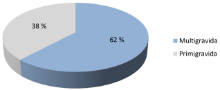
Figure 1. Obstetrics history (Gravidity) of mothers (n=100).
Data were expressed as percentage
Figure 1 shows obstetrics history (Gravidity) of mothers. Most of the women were multigravida (62.0%) and primigravida was seen in (38.0%) of mother.
Table 3. Clinical manifestation of asphyxiated newborn.
Clinical Features | Number of Patients | Percentage (%) |
Respiratory distress | 59 | 59 |
Poor-feeding | 75 | 75 |
Lethargy | 33 | 33 |
Grunting | 48 | 48 |
Weak Muscle tone | 30 | 30 |
Hypothermia | 34 | 34 |
Petechiae | 6 | 6 |
Bleeding diathesis | 5 | 5 |
Convulsion | 34 | 34 |
Values were expressed as frequency and percentage
Table 3 shows clinical manifestation of asphyxiated newborn. Cardinal clinical features were respiratory distress, poor-feeding, lethargy, grunting and petechiae (59.0%, 75.0%, 33.0%, 48.0% and 6.0% respectively). Other clinical features were hypothermia in 34.0% and convulsion 34.0% of neonates.
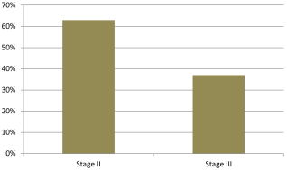
Figure 2. Distribution of HIE stages according to severity of birth asphyxia.
Data were expressed as percentage
Figure 2 shows distribution of HIE stages according to severity of birth asphyxia. Severity of birth asphyxia was graded based on the hypoxic- ischemic encephalopathy (HIE). In this study 63.0% of cases found moderate asphyxia or stage II was 37.0% cases had severe asphyxia or stage III.
Table 4. Assessment of blood glucose level in asphyxiated neonates (n=100).
Blood glucose level | Number of Patients | Percentage (%) |
≤2.5 mmol/l | 26 | 26 |
2.5-6.94 mmol/l | 71 | 71 |
>6.94 mmol/l | 3 | 3 |
Data were expressed as frequency and percentage
Table 4 shows the blood glucose level of asphyxiated neonates. Out of 100 term newborn, 26.0% showed hypoglycaemia, 3.0% showed hyperglycaemia and 71% patients had normal level of blood glucose.
Table 5 shows the association of blood glucose status with severity of birth asphyxia. Abnormal blood glucose status or hypo/hyperglycaemia was positively associated with severity of birth asphyxia. Among the 63 patients withmoderate asphyxia, 14.2% had detected abnormal glucose, whereas in severe asphyxia, 54.0% were detected abnormal blood glucose. Maximum patients of group I had normal blood glucose level. The difference between groups were statistically significant (p<0.05).
Table 5. Association of blood glucose status with severity of birth asphyxia (n=100).
Blood glucose status | Severity of birth asphyxia | P- value |
Moderate Asphyxia (n=63) | Severe Aphyxia (n=37) |
Group 1 | 54 (85.7%) | 17 (45.9%) | <0.0001 |
Group 2 | 9 (14.2%) | 20 (54.0%) | 0.0136 |
Group I: Patient with normal blood glucose level or euglycaemia
Group II: Patients with abnormal blood glucose findings
p-value reached from Chi-square test and <0.05 was statistically significant Data were expressed as frequency and percentage
Table 6. Association of blood glucose findings to time required to control of convulsion (n = 34).
Blood glucose status | Control of Convulsion | Total | P- value |
Within 24 hours (n=19) | ≥24 hours (n=15) |
Group 1 | 15 (78.9%) | 6 (40.0%) | 21 | 0.0112 |
Group 2 | 4 (21.1%) | 9 (60.0%) | 13 | 0.047 |
Group I: Patient with normal blood glucose level or euglycaemia
Group II: Patients with abnormal blood glucose findings
p-value reached from Chi-square test and <0.05 was statistically significant Data were expressed as frequency and percentage
Table 6 shows maximum cases of group-ll (e.g., 60%) were required >= 24 hrs to control convulsion. In this study total 34 patients had developed convulsion and among them 19 cases responds well within 24 hrs after management, and maximum (78.9%) cases were normal blood glucose level. Fifteen cases required ≥24 hrs to control convulsion and maximum (60.0%) cases were abnormal blood glucose level. The difference between groups were statistically significant (p < 0.05).
5. Discussion
Demographic characteristics revealed that, out of 100 patients 58% and 42% belonged to age group >6 hours and <6 hours respectively. The mean age of the neonates was 6.31±0.91 (age range: 0-24) hours. Sex distribution revealed that among 100 patients 60% and 40% were male and female respectively. The male to female ratio was 1.5:1. Among the cases majority of the patient had history of normal vaginal delivery (e.g. 62.0%), LUCS done in 38.0% patients. In this series, maximum numbers of cases (65.0%) were normal birth weight baby, followed by low birth weight in 35.0% patients.
Findings consistent with result of other studies. In a study One hundred newborn were enrolled. There were 52% female and 48% were male neonates
| [13] | Yasmin T, Akther S, Sultana S, & Amin M. Assessment of Cranial Sonographic Findings of Hypoxic Ischemic Brain Injury in Perinatal Asphyxia. Journal of Medicine, 2016; 17(1): 12-16. |
[13]
. Mode of delivery was normal vaginal delivery for 46% neonates and 54% via LUCS for various reasons. In term babies, birth weight between 2.5-3.5 kg was in 40 (71%) cases and between >3.5-4 kg was in 16 (29%) cases. In a retrospective study shows out of 100 neonates 63 were male and 37 were female with 19% babies were in their first day of life, 45% were of two days and 36% of three days. Birth weight of 100 babies was maximum and minimum 3.6 and 2.4 respectively. The mean ± S.D of birth weight of 100 babies were 3.08 ± 0.54
| [14] | Malik AR, Quddusi Al, Fatima N, iqbal I, Javeed AM. Full term babies, correlation of clinical findings of perinatal asphyxia with cranial sonography. Professional Med J 2017; 24(6): 828-833. |
[14]
.
On evaluation of risk factors, present study shows that Out of 100 patients 52%, 30%, 26% and 4% had maternal risk factors like prolonged labour, PROM, hypertensive disorder and mal-presentation respectively. Most of the women were multigravida (62.0%) and primigravidawas seen in (38.0%) of mother.
Risk factors of perinatal asphyxia are antepartum, intrapartum and fetal. Antepartum factors includes advanced maternal age, pre-eclampsia, gestational diabetes, maternal hypertension, anemia, hypertension. Intrapartum risk factors are cephalo- pelvic disproportion (CPD), prolonged labor, premature rupture of membrane, umbilical cord complications like umbilical cord prolapsed. Fetal risk factors include meconium sustained amniotic fluid, abnormal lie or presentation and cord compression or prolapsed
| [14] | Malik AR, Quddusi Al, Fatima N, iqbal I, Javeed AM. Full term babies, correlation of clinical findings of perinatal asphyxia with cranial sonography. Professional Med J 2017; 24(6): 828-833. |
[14]
. Similar findings demonstrated that, perinatal risk factors for asphyxia of newborns revealed that 12% mother had premature rupture membrane, 12% had APH, 8% pre-eclamptic toxemia and hypertension, 6% had Diabetes mellitus and 4% had oligohydramnios during pregnancy
| [13] | Yasmin T, Akther S, Sultana S, & Amin M. Assessment of Cranial Sonographic Findings of Hypoxic Ischemic Brain Injury in Perinatal Asphyxia. Journal of Medicine, 2016; 17(1): 12-16. |
[13]
. Another study reported maternal risk factors present in neonates with abnormal CUS were PIH (53.1%), PROM (25.5%), APH (8.5%) and others (12.7%)
| [15] | Kinikar U, Dhanawade S. Study of cranial ultrasound its correlation with perinatal risk factors and its outcome in preterm neonates admitted to Neonatal intensive care unit. Pediatric Review: International Journal of Pediatric Research. |
[15]
.
Preterm neonates exhibit very few clinical features suggestive of hypoxic-ischemic brain injury. Detection needs meticulous observation of subtle signs and symptoms. In all neonates the problem may be suspected by the following features Impairment or of consciousness or irritability, impairment of muscle tone, tendon reflexes, sucking, Moro response, grasping and oculo cephalic reflex, abnormal pupils, respiration, heart rate; seizures and EEG changes
| [16] | Giri S, Jana T, Tapadar A. Ultrasonographic Evaluation of the Neonatal Brain in Cases of Birth Asphyxia. International Journal of Anatomy, Radiology and Surgery, 2016; 5(1): 58-63. |
[16]
. In this study cardinal clinical features of birth asphyxia were respiratory distress, poor-feeding, lethargy. grunting and petechiae (59.0%, 75.0%, 33.0%, 48.0% and 6.0% respectively). Other clinical features were hypothermia in 34.0% and convulsion 33.0% of neonates. Severity of birth asphyxia was graded based on the hypoxic- ischemic encephalopathy (HIE). Another study demonstrated that, 75% had respiratory distress, 42% had convulsion, 40% had cyanosis, 30% had apnoeic spell and 12% had sepsis
| [13] | Yasmin T, Akther S, Sultana S, & Amin M. Assessment of Cranial Sonographic Findings of Hypoxic Ischemic Brain Injury in Perinatal Asphyxia. Journal of Medicine, 2016; 17(1): 12-16. |
[13]
.
On evaluation of blood sugar status, 29.0% showed abnormal glycaemic status. Among them 26 cases had hypoglycaemia and 3 cases had hyperglycaemia patients had normal level of blood glucose or euglycaemia. In this study abnormal blood glucose status or hypoglycaemia was positively associated with severity of birth asphyxia. Among the 63 patients with moderate asphyxia, 14.2% had detected abnormal glucose, whereas in severe asphyxia, 54.9% were detected abnormal blood glucose. Maximum patients of group I had normal blood glucose level. The difference between groups were statistically significant (p<0.05). In this study fifteen cases required ≥24 hrs to control convulsion and maximum (60.0%) cases were abnormal blood glucose level. The difference between groups were statistically significant (p<0.05).
In a study, total of 1562 live births were recruited. There were 234 (22.5%) newborns with hypoglycemia. Overall, the mean birth weight and gestational age of newborns with hypoglycemia was lower compared to newborns without hypoglycemia (2283.4 g vs. 2788.0 g; 35.7 vs. 37.7 weeks, respectively). Asphyxia, LBW, SGA, LGA, prematurity, pre-eclampsia/eclampsia, meconium aspiration syndrome, RDS, and were significant risk factors for neonatal hypoglycemia
| [7] | Mufidati L, Anggraini A, Wibowo T. Asphyxia as a Risk Factor for Neonatal Hypoglycemia. J Nepal PaediatrSoc 2017; 37(2): 111-116. |
[7]
.
6. Limitations of the Study
This single-center study was limited to patients at Shaheed Suhrawardy Medical College Hospital, which may not reflect the broader national picture. The small sample size, limited investigative facilities, and the tertiary care setting might not represent primary or secondary healthcare centers, potentially skewing results toward more high-risk cases. Additionally, the use of purposive sampling could introduce personal bias. A larger-scale study is needed for more definitive conclusions.
7. Recommendations
As blood glucose is a common, cheap and available diagnostic technique, simultaneously efficient, effective and safe modality, it can be used routinely in primary or secondary level of health care centre as a valuable diagnostic tool for prediction of outcome of hypoxic ischemic brain injury.
8. Conclusion
This study concluded that prevalence of hypoglycaemia was 26%. It was evident from this study is that abnormal blood glucose, e,g, hypoglycaemic birth asphyxiated babies outcome was poor than normoglycaemic babies. Blood glucose assessment can be reliable tools for demonstrating the most frequently occurring forms of cerebral injury in perinatal asphyxia, assessing the evolution of the lesion, and following brain development.
Abbreviations
PNA | Perinatal Asphyxia |
HIE | Hypoxic-Ischemic Encephalopathy |
LBW | Low Birth Weight |
APH | Antepartum Hemorrhage |
Acknowledgments
I would like to express my sincere gratitude for the invaluable support and cooperation provided by the staff, participants, and my co-authors/colleagues who contributed to this study.
Author Contributions
Mukta Thakur: Conceptualization, Data curation, Formal Analysis, Funding acquisition, Investigation, Methodology, Project administration, Resources, Software, Supervision, Validation, Visualization, Writing – original draft, Writing – review & editing
Jakiya Jesmine: Funding acquisition, Investigation, Resources, Software, Visualization
Ajmiri Sultana: Formal Analysis, Project administration, Validation, Writing – original draft, Writing – review & editing
Farjana Afroze Jui: Conceptualization, Methodology, Resources, Software, Visualization, Writing – review & editing
Umme Qulsum Sonia: Funding acquisition, Methodology, Project administration, Software, Supervision, Writing – original draft
Financial Support and Sponsorship
No funding sources.
Ethical Approval
The study was approved by the Institutional Ethics Committee.
Conflicts of Interest
The authors declare no conflicts of interest.
References
| [1] |
Golubnitschaja O, Yeghiazaryan K, Cebioglu M, Morelli M, Marschitz M. Birth asphyxia as the major complication in newborns: moving towards improved individual outcomes by prediction, targeted prevention and tailored medical care. EPМА J. 2011; 2(2): 197-210.
|
| [2] |
MacLennan A. A template for defining a causal relation between acute intrapartum events and cerebral palsy: international consensus statement. BMJ. 1999; 319: 1054-9.
|
| [3] |
Amponsah G, Hagan OCK, E Okai E. Neonatal Hypoglycaemia at Cape Coast Teaching Hospital. J West AfrColl Surg. 2015; 5(2): 100-116.
|
| [4] |
American College of Obstetrics and Gynecology. Task Force on Neonatal Encephalopathy and Cerebral Palsy. American Academy of Pediatrics. Neonatal Encephalopathy and Cerebral Palsy: Defining the Pathogenesis and Pathophysiology. Edited by Washington, DC, American College of Obstetricians and Gynecologists, 2003.
|
| [5] |
Sarnat HB, Sarnat MS. Neonatal encephalopathy following fetal distress. A clinical and electroencephalographic study. Arch Neurol. 1976; 33: 696-705.
|
| [6] |
Leuthner SR, Das UG. Low Apgar scores and the definition of birth asphyxia. PediatrClin North Am. 2004; 51: 737-45.
|
| [7] |
Mufidati L, Anggraini A, Wibowo T. Asphyxia as a Risk Factor for Neonatal Hypoglycemia. J Nepal PaediatrSoc 2017; 37(2): 111-116.
|
| [8] |
Basu P, Som S, Choudhuri, Das H. Contribution of the blood glucose level in perinatal asphyxia. Eur J Pediatr 2009; 168: 833-38.
|
| [9] |
Lopez AD, Mathers CD. Measuring the global burden of disease and epidemiological transitions: 2002-2030. Ann Trop Med Parasitol. 2006; 100: 481-99.
|
| [10] |
Tomashek KM, Crouse CJ, Iyasu S, Johnson CH, Flowers LM. A comparison of morbidity rates attributable to conditions originating in the perinatal period among newborns discharged from United States hospitals, 1989-90 and 1999-2000. Paediatric Perinat Epidemiol. 2006; 20: 24-34.
|
| [11] |
Elamin S, Langhoff-Roos J, Boedker B, Ibrahim SA, Ashmeig AL, Lindmark G. Classification of perinatal death in a developing country. Int J Gynaecol Obstet. 2003: 80: 327-33.
|
| [12] |
Luo ZC, Liu S, Wilkins R, Kramer MS; Fetal and Infant Health Study Group of the Canadian Perinatal Surveillance System. Risks of stillbirth and early neonatal death by day of week. CMAJ. 2004; 170: 337-41.
|
| [13] |
Yasmin T, Akther S, Sultana S, & Amin M. Assessment of Cranial Sonographic Findings of Hypoxic Ischemic Brain Injury in Perinatal Asphyxia. Journal of Medicine, 2016; 17(1): 12-16.
|
| [14] |
Malik AR, Quddusi Al, Fatima N, iqbal I, Javeed AM. Full term babies, correlation of clinical findings of perinatal asphyxia with cranial sonography. Professional Med J 2017; 24(6): 828-833.
|
| [15] |
Kinikar U, Dhanawade S. Study of cranial ultrasound its correlation with perinatal risk factors and its outcome in preterm neonates admitted to Neonatal intensive care unit. Pediatric Review: International Journal of Pediatric Research.
|
| [16] |
Giri S, Jana T, Tapadar A. Ultrasonographic Evaluation of the Neonatal Brain in Cases of Birth Asphyxia. International Journal of Anatomy, Radiology and Surgery, 2016; 5(1): 58-63.
|
Cite This Article
-
APA Style
Thakur, M., Jesmine, J., Sultana, A., Jui, F. A., Sonia, U. Q., et al. (2024). Glycaemic Status Among Neonates in Perinatal Asphyxia with Hypoxic Ischaemic Encephalopathy (Stage II and Stage III) in a Tertiary Level Hospital. American Journal of Pediatrics, 10(4), 179-184. https://doi.org/10.11648/j.ajp.20241004.14
 Copy
|
Copy
|
 Download
ACS Style
Thakur, M.; Jesmine, J.; Sultana, A.; Jui, F. A.; Sonia, U. Q., et al. Glycaemic Status Among Neonates in Perinatal Asphyxia with Hypoxic Ischaemic Encephalopathy (Stage II and Stage III) in a Tertiary Level Hospital. Am. J. Pediatr. 2024, 10(4), 179-184. doi: 10.11648/j.ajp.20241004.14
Copy
|
Download
AMA Style
Thakur M, Jesmine J, Sultana A, Jui FA, Sonia UQ, et al. Glycaemic Status Among Neonates in Perinatal Asphyxia with Hypoxic Ischaemic Encephalopathy (Stage II and Stage III) in a Tertiary Level Hospital. Am J Pediatr. 2024;10(4):179-184. doi: 10.11648/j.ajp.20241004.14
Copy
|
Download
-
@article{10.11648/j.ajp.20241004.14,
  author = {Mukta Thakur and Jakiya Jesmine and Ajmiri Sultana and Farjana Afroze Jui and Umme Qulsum Sonia and Md. Al-Amin Mridha},
  title = {Glycaemic Status Among Neonates in Perinatal Asphyxia with Hypoxic Ischaemic Encephalopathy (Stage II and Stage III) in a Tertiary Level Hospital
},
  journal = {American Journal of Pediatrics},
  volume = {10},
  number = {4},
  pages = {179-184},
  doi = {10.11648/j.ajp.20241004.14},
  url = {https://doi.org/10.11648/j.ajp.20241004.14},
  eprint = {https://article.sciencepublishinggroup.com/pdf/10.11648.j.ajp.20241004.14},
  abstract = {Background: Perinatal asphyxia is major cause of neonatal mortality and morbidity. Hypoxic ischaemic brain injury is the most important consequences of perinatal asphyxia which ultimately results in immediate and delayed form of neuronal death. The aim of this study was to find a relationship between glycaemic status and immediate outcomes of perinatal asphyxia. Methods: This prospective study was carried out in Department of Paediatrics, Shaheed Suhrawardy Medical College Hospital, from 16th April 2019 to 15th October 2019. Total 100 term asphyxiated newborn babies with HIE (Stage II and III) admitted within 24 hours were enrolled according to selection criteria, Blood glucose level and other relevant tests were done in all included patients. Results: The mean age of the neonates was 6.31±0.91 hours. Among the patients, 60% were male and 40% were female. Most cases (65%) had normal birth weight, while 35% were low birth weight. Common clinical features included respiratory distress (59%), poor feeding (75%), lethargy (33%), grunting (48%), and petechiae (6%). Moderate encephalopathy (Stage II) was observed in 63% of cases, and severe asphyxia (Stage III) in 37%. Hypoglycaemia was present in 26% of neonates, hyperglycaemia in 3%, and 71% had normal glucose levels. Hypoglycaemia was significantly associated with severe asphyxia, occurring in 45.9% of severe cases compared to 14.2% of moderate cases (p<0.05). Conclusion: There was significant association between glycaemic abnormalities with severity of perinatal asphyxia and immediate outcome of the asphyxiated newborn.
},
 year = {2024}
}
Copy
|
Download
-
TY  - JOUR
T1  - Glycaemic Status Among Neonates in Perinatal Asphyxia with Hypoxic Ischaemic Encephalopathy (Stage II and Stage III) in a Tertiary Level Hospital

AU  - Mukta Thakur
AU  - Jakiya Jesmine
AU  - Ajmiri Sultana
AU  - Farjana Afroze Jui
AU  - Umme Qulsum Sonia
AU  - Md. Al-Amin Mridha
Y1  - 2024/11/28
PY  - 2024
N1  - https://doi.org/10.11648/j.ajp.20241004.14
DO  - 10.11648/j.ajp.20241004.14
T2  - American Journal of Pediatrics
JF  - American Journal of Pediatrics
JO  - American Journal of Pediatrics
SP  - 179
EP  - 184
PB  - Science Publishing Group
SN  - 2472-0909
UR  - https://doi.org/10.11648/j.ajp.20241004.14
AB  - Background: Perinatal asphyxia is major cause of neonatal mortality and morbidity. Hypoxic ischaemic brain injury is the most important consequences of perinatal asphyxia which ultimately results in immediate and delayed form of neuronal death. The aim of this study was to find a relationship between glycaemic status and immediate outcomes of perinatal asphyxia. Methods: This prospective study was carried out in Department of Paediatrics, Shaheed Suhrawardy Medical College Hospital, from 16th April 2019 to 15th October 2019. Total 100 term asphyxiated newborn babies with HIE (Stage II and III) admitted within 24 hours were enrolled according to selection criteria, Blood glucose level and other relevant tests were done in all included patients. Results: The mean age of the neonates was 6.31±0.91 hours. Among the patients, 60% were male and 40% were female. Most cases (65%) had normal birth weight, while 35% were low birth weight. Common clinical features included respiratory distress (59%), poor feeding (75%), lethargy (33%), grunting (48%), and petechiae (6%). Moderate encephalopathy (Stage II) was observed in 63% of cases, and severe asphyxia (Stage III) in 37%. Hypoglycaemia was present in 26% of neonates, hyperglycaemia in 3%, and 71% had normal glucose levels. Hypoglycaemia was significantly associated with severe asphyxia, occurring in 45.9% of severe cases compared to 14.2% of moderate cases (p<0.05). Conclusion: There was significant association between glycaemic abnormalities with severity of perinatal asphyxia and immediate outcome of the asphyxiated newborn.

VL  - 10
IS  - 4
ER  -
Copy
|
Download