Case Report | | Peer-Reviewed

Dunbar Syndrome: Unusual Cause of Chronic Abdominal Pain in Children

Received: 30 May 2025     Accepted: 18 June 2025     Published: 16 July 2025
Views:       Downloads:
Abstract

Childhood chronic abdominal pain is a diagnostic challenge, with a variety of functional and organic etiologies. Whilst the majority of cases are diagnosed as either functional or dysfunctional, it is imperative that an individual diagnostic assessment is conducted in order to exclude organic causes, thus facilitating a successful management strategy. In instances where alarm-Features are present, it is recommended that investigations be conducted into rare and unusual causes, provided that initial investigations have not yielded a diagnosis. One such rare Entity is Dunbar syndrome, a vascular condition characterized by chronic abdominal pain, which typically manifests during late childhood. This curable cause appears to be more prevalent than what previously hypothesized to be 1.7% in children. We present the case of a 16-year-old female patient has been suffering from severe, unexplained chronic abdominal pain and weight loss for several months. Despite extensive investigations and exploratory laparoscopy, no clear explanation for the patient's symptoms has been found. Our approach was a combination of teamwork, a stepwise approach, and selective investigations. This collaborative effort enabled the successful diagnosis and surgical therapy. We aim to enhance the management of childhood chronic abdominal pain by adapting a cost effective stepwise approach and to raise awareness of Dunbar syndrome.

Published in American Journal of Pediatrics (Volume 11, Issue 3)
DOI 10.11648/j.ajp.20251103.13
Page(s) 127-133
Creative Commons

This is an Open Access article, distributed under the terms of the Creative Commons Attribution 4.0 International License (http://creativecommons.org/licenses/by/4.0/), which permits unrestricted use, distribution and reproduction in any medium or format, provided the original work is properly cited.

Copyright

Copyright © The Author(s), 2025. Published by Science Publishing Group

Keywords

Chronic Abdominal Pain, Organic, Functional, Alarm Criteria, Dunbar Syndrome, Vascular, Decompression

1. Introduction
Chronic abdominal pain is defined as intermittent or persistent pain lasting for at least 2 months. It occurs in 10-19% of children . In general, the causes of abdominal pain can be classified into organic and functional. Functional chronic abdominal pain prevalence is 10.7% of children population . Functional constipation, dyspepsia, irritable bowel syndrome, functional pain not elsewhere specified, and abdominal migraine are functional disorders that may cause chronic abdominal pain in children and have specific diagnostic criteria according to the Rome classification . In principle, all possible organic causes should be excluded first, keeping functional abdominal pain as the diagnosis of exclusion . The variety of organic abdominal and extra-abdominal causes can be classified according to its prevalence as illustrated bellow in “Table 1”:
Table 1. Organic causes of chronic abdominal pain.

Common

Helicobacter Pylori gastritis Carbohydrate malabsorption Celiac disease Constipation Dysmenorrhea Hernia Urinary tract infection

Uncommon

Eosinophilic esophagitis

Inflammatory bowel disease

Food allergy

Nephrolithiasis

Rare

Endometriosis

Familial Mediterranean fever

Chronic hepatitis

Chronic pancreatitis

Lymphoma

Sickel cell anemia.

Vasculitis (HSP, PAN)
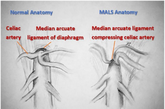
Extremely rare

Lead poisoning Bezoar Angioedema Malrotation Median Arcuate ligament syndrome Superior Mesenteric artery syndrome Ureteropelvic junction stenosis

Celiac artery compression syndrome, or median arcuate ligament syndrome [MALS], or Dunbar syndrome is a rare anomaly. Here, the median arcuate ligament is inferiorly displaced crossing over the base of the celiac artery at the level of L1 vertebral body as shown in” Figure 1”.
Figure 1. Illustration of Dunbar syndrome anatomic abnormality.
It can be seen on abdomen CTA or MRA as a hook-shaped impression of the celiac artery near its origin. The typical regression in the degree of stenosis and improved flow during inspiration as the artery moves away from the pressing ligament can be seen in imaging studies and is considered a characteristic morphological feature in Angiography.
2. Case Presentation
The 16-year-old female patient exhibited a six-month history of chronic abdominal pain and an estimated weight loss of approximately six kilograms. She had no past medical history and was previously in good health. The pain is characterized as episodic, occurring 3 to 4 times per week, and is described as either diffuse or localized in the right lower quadrant, with a duration of hours. In certain cases, the condition can be severe, and may be accompanied by symptoms such as dizziness, fainting, and an inadequate response to analgesics. The aetiology remains unclear, with no obvious trigger identified. The absence of concurrent digestive symptoms, such as diarrhoea or vomiting, as well as urinary or respiratory complaints, is notable. Furthermore, the absence of fever and the absence of a family history suggest that other factors may be involved. It is noteworthy that the patient is sexually inactive and has regular menstrual cycles that are not associated with any discomfort. Upon arrival, the patient was observed and examined. She was not in pain, was alert, and exhibited normal growth and puberty. Her abdomen was soft, with mild diffuse tenderness, predominantly in the right lower quadrant (RLQ). An organ-system review was conducted, which revealed no abnormalities.
3. Previous Investigation and Management on Arrival
Over the course of several months, the patient has undergone extensive medical investigation in a number of medical facilities due to the severity of her pain. The patient's complete blood count, C-reactive protein (CRP) levels, liver and kidney function tests, and urine analysis results were all within normal parameters. Abdominal ultrasound results were also unremarkable. A CT- scan of the patient's abdomen with intravenous and oral contrast was also performed, which revealed mucosal oedema as well as an abnormal distribution of the contrast material in the transverse colon. This was seen to be indicative of a chronic inflammatory disease. However, both upper and lower endoscopies revealed no abnormalities. Furthermore, intestinal biopsies showed nonspecific inflammatory infiltration in the mucosa, without granuloma or clear specific changes.
The patient ultimately was experiencing such severe pain that the surgeon was compelled to perform an exploratory laparoscopy. The procedure revealed normal findings, except for mild pelvic free fluid. Following a comprehensive evaluation, she was referred to a psychiatrist due to severe functional pain that did not fit with any other established diagnostic categories. Her condition demonstrated no response to a combination of analgesics and antidepressants.
4. Stepwise Diagnostic Approach
New laboratory assessment more specific was done by our team:
Hb: 10.6 gr/dl, wbc: 7.6 k. dl, ferritin: 16.ng/ml
CRP (c-reactive protein), KFT (kidney function test), LFT (liver function test), Lipase, urine analysis stool parasitology and cytology all were normal
Fecal calprotectin 110mcg/gr
Celiac disease serology. & H. pylori antigen: negative
ANA (anti nuclear antibodies), Anti smooth muscle antibodies: negative
ASCA (anti saccharomyces cerevisiae antibodies), p ANCA (perinuclear antineutrophil cytoplasmic antibodies): negative
Amyloid-s level: normal, FMF (familial Mediterranean fever) gene mutation: negative
Porphyrin metabolites in urine: negative
New abdominal and pelvis ultrasound: normal
Patient was put on primary treatment for a suspected IBD (inflammatory bowel disease) and arranged for a team discussion meeting.
It was agreed that there is a need to repeat her imaging looking for more specific findings of IBD, newly developed changes, or any vascular causes.
5. Diagnostic Investigation and Intervention
In the absence of magnetic resonance imaging (MRI), a computed tomography (CT) scan angiography (CTA) was performed, which clearly demonstrated a hook-shaped stenosis in the celiac artery trunk. This initially constituted the patient's diagnosis from a radiological point of view, as it remained uncertain whether the finding was associated with the pain or was merely a coincidental detection. A subsequent consultation with a vascular surgeon, followed by diagnostic catheterization, confirmed severe celiac artery stenosis of ≥70%, indicating the diagnosis of Dunbar syndrome.
6. Treatment
The MAL ligament was approached via an upper midline incision. The gastrohepatic ligament was then dissected to reach the hepatic artery, after which the splenic artery and left gastric artery were located. These arteries were traced posteriorly to reach the celiac trunk, which led to the median arcuate ligament. The ligament was found to be inferiorly displaced and severely fibrotic. The artery was observed to be infantile, with a notably small diameter of 4 mm. Electrocautery was subsequently utilized to transect the ligament. The ligament was opened extensively until the celiac artery was entirely free of surrounding fibers.
7. Discussion
The prevalence of chronic abdominal pain in children has been well documented. Approximately 60% of cases are of a functional nature. Nevertheless, it is imperative to preclude organic etiology in each instance and to consider functional pain as the diagnosis of exclusion. The clinical assessment should be directed towards the identification of any potential red flags or pathognomonic findings in the patient's history and clinical examination that could increase the likelihood of an underlying organic etiology as shown in “Table 2”. In order to establish a comprehensive diagnosis, it is imperative that the patient's symptoms of pain are thoroughly and systematically investigated. This should be accompanied by a meticulous abdominal examination, encompassing the urogenital and perianal regions, in order to identify any potential abnormalities.
Table 2. Alarm findings in childhood chronic abdominal pain.

Alarm findings:

In history

Weight loss Vomiting, diarrhea Dysphagia Bloody stool Fever Urinary symptoms Family history of IBD Rash

By physical examination

Faltered growth Oral aphthous Localized pain or tenderness Organomegaly Perianal abnormalities

In the absence of any alarm finding, simple noninvasive investigations might be suggested as shown in “Table 3”.
Table 3. Investigation of chronic abdominal pain without alarms.

Investigation:

Laboratory:

CBC, CRP, ALT, BUN

URINE analysis

Stool parasitology and cytology

Imaging:

Abdominal ultrasound

In instances of chronic abdominal pain accompanied by a red flag or more, a systematic investigation plan should be implemented to first exclude the prevalent treatable organic causes of chronic abdominal pain . We propose a cost-effective stepwise investigation plan as delineated in “Table 4”.
Table 4. Stepwise investigation plan of chronic abdominal pain with alarms.

step

Investigation:

Step 1:

CBC, CRP

LFT, KFT, ELECTROLYTES

RBS

LIPASE

Celiac disease serology (TTG-IgA)

URINE ANALYSIS

STOOL for (parasitology, Occult Blood, cytology H. Pylori Antigen)

Abdominal ultrasound

Simple abdominal x ray

Step2

Fecal calprotectin or lactoferrin

Ferritin

Hemoglobin-s

ANA, anti-smooth muscle antibodies

ANCA, PANCA

Upper gastrointestinal series and follow up

Upper &lower ENDOSCOPY and biopsy

Step3

Amyloid-s

Porphyrin metabolites

Lead blood level

CT-scan abdomen with contrast

CTA scan abdomen

MRA abdomen

Laparoscopy

It is at the clinician's discretion to select the most diagnostic investigation from step 1, based on their clinical findings. However, it is strongly advised that they do not proceed to step 2 or 3 until they have completed the lower step of investigation.
This arrangement is made with the following factors in mind: prevalence, curability, the type of investigation, and the cost-outcome benefit.
Indications for referral to a gastroenterologist :
-suspected IBD
- suspected chronic hepatitis
- no clear cause in the presence of alarm findings
-need for upper or lower endoscopy
Referral to other medical specialties, including surgery, urology, gynecology, and mental health may be arranged as a result of further findings.
In this patient, all preliminary investigative procedures, encompassing a complete blood count (CBC), C-reactive protein (CRP), a kidney function test, a liver function test (LFT), a random blood sugar test, a lipase analysis, an analysis of urine, a stool parasitology examination, an analysis of urine culture, an evaluation of transglutaminase IgA antibodies, a Helicobacter pylori test, and a breath test for the presence of H. pylori, all yielded normal results.
Subsequent investigative procedures were scheduled to the exclusion of less prevalent and uncommon etiologies. The following analyses were conducted: fecal calprotectin, hemoglobin -s, amyloid-s, ANCA, pANCA, and porphyrin metabolites;all were normal.
Both upper and lower endoscopies with biopsies, abdominal CT scans with contrast, and laparoscopies had already been performed, and all of them were normal or showed nonspecific changes.
At this juncture, it is strongly advised that a comprehensive reassessment of the child be undertaken, commencing from the initial stage. This reassessment should entail a meticulous review of the patient's medical history, in addition to a thorough systemic physical examination, with a view to identifying any latent or recent findings. Some investigations might be re- conducted for comparison with previous results, like: CBC, CRP, ALT, lipase, and abdominal US.
A multidisciplinary team meeting was convened (consisting of a pediatrician, a gastroenterologist, a radiologist and a pediatric surgeon) for the purpose of evaluating the case prior to the commencement of further investigations. It was ascertained that, in addition to weight loss, painful crises had escalated, with instances of near syncope and fainting due to pain. Nevertheless, the only atypical results obtained were mild to moderate elevated fecal calprotectin (110 µg/g), mild hypochromic microcytic anemia (10 g/dl), and nonspecific inflammatory changes in the biopsy and imaging study. The patient was prescribed a trial of Mesalzine and Prednisolone, with a subsequent repeat evaluation scheduled for two weeks hence.
The painful crisis remained unresolved, and there was no alteration in fecal calprotectin levels. The predominant finding was mild elevation, attributable to excessive use of analgesics such as ibuprofen. A further selective imaging study was ordered in order to obtain more detailed information about abdominal vascularity, an aspect that had not been addressed in the preceding study, and to evaluate inflammatory changes. A new abdominal ultrasound was performed, yielding normal results. However, given the unavailability of an abdomen MRA or MRI with contrast, a CTA with contrast was conducted instead. a constriction in the form of a hook in the base of the celiac artery was recognized as a significant indicator of the diagnosis as shown in “Figure 2”.
Figure 2. CTA abdomen a hook shaped constriction of celiac trunk.
The incidence of Dunbar syndrome remains uncertain in paediatric populations. As Scholbach et al noted, the prevalence of the condition was 1.7% in children, with 71% of these cases being symptomatic . The clinical spectrum of the condition has been described as ranging from asymptomatic to severe chronic abdominal pain, according to the degree of stenosis and foregut ischemia . In a separate study, 46 patients, all of whom were female and under the age of 21, were successfully treated by surgical release . In addition to abdominal pain, patients may also experience symptoms such as dizziness, postural hypotension and postural tachycardia (POT). The concomitant presence of abdominal pain and POT in a child is a strong indication that the pain is due to the underlying condition and that corrective therapy is required.
Abdominal ultrasound Doppler in the sagittal subcostal view is a reliable method for identifying stenosis of the celiac axis, characterized by high flow velocity. Velocities of 2m/s and above are considered abnormal. A definitive diagnosis generally necessitates the utilization of advanced imaging modalities, such as CTA or MRA, to ensure comprehensive evaluation and precise diagnosis. Conventional ultrasound is inadequate for excluding this diagnosis, as it necessitates the implementation of specific techniques and may fail to discern the celiac trunk origin from the aorta.
Treatment:
The most conventional treatment for Median Arcuate Ligament Syndrome (MALS) involves decompressing the median arcuate ligament (MAL) through an open surgical or laparoscopic approach . Current understanding of MALS pathophysiology suggests that additional neurolysis and extensive excision of the affected celiac plexus should also be performed to address the neurogenic aspects of the syndrome . A retrospective analysis of patient data collected over a decade at the University of North Carolina at Chapel Hill by Kohn et al. indicated that both open and laparoscopic techniques are safe and yield durable mid-range follow-up results . Furthermore, a comprehensive 20-year cohort study published in 2023 by the Vascular Low Frequency Disease Consortium, which included data from 516 MAL release procedures (227 open, 235 laparoscopic, and 54 robotic), found no significant difference in long-term failure rates between open and laparoscopic decompression .
It is crucial to discuss the potential treatment failure rate, which can be as high as 48%, with patients when weighing the risks and benefits of this procedure . Given multiple studies indicating no long-term differences in failure rates, laparoscopic decompression of the celiac artery is rapidly becoming the preferred method for MALS patients. This approach offers several advantages typical of laparoscopic procedures, including reduced postoperative complications such as lower rates of ileus, pain, blood loss, adhesions, and shorter recovery times.
In a direct comparison of open versus laparoscopic treatment for MALS, Jiminez et al. reviewed 400 patients (279 open and 121 laparoscopic) and found that 85% experienced immediate postoperative symptom relief, with only 26 patients later experiencing a recurrence of symptoms . 9.1% of patients treated laparoscopically (11 out of 121) required conversion to laparotomy due to bleeding complications like bleeding and pneumothorax in the laparoscopic group, but no procedure-related fatalities. Common complications in the open group included thrombosed bypass grafts (2%), stroke (1.4%), gastroesophageal reflux disease (1%), pancreatitis (1%), hemothorax (0.3%), and splenic infarctions (0.3%), with no reported deaths .
The choice for vascular reconstruction is typically considered for patients who continue to experience symptoms such as thrill, compression, malformation, or pressure gradients in the celiac artery despite initial external MAL decompression. Long-term compression can lead to hyperplasia in the intimal and adventitial layers of the celiac artery, resulting in significant lumen narrowing . An alternative to open revascularization is laparoscopic MAL release combined with percutaneous transluminal angioplasty (PTA). This approach has shown promising results for patients with persistent stenosis following initial decompression, as evidenced by a limited number of studies .
The significant pain relief observed from celiac plexus block (CPB) in MALS patients supports the notion of celiac ganglion involvement in the condition. Research by Sultan et al. indicates that MAL decompression combined with celiac ganglion sympathectomy (CGS) improves mesenteric blood flow and leads to substantial symptomatic relief. In their study, which had a mean follow-up of 2.8 years, 71% of patients reported freedom from symptoms, even though 50% showed high-velocity flow in the celiac artery on postoperative duplex ultrasound. The rationale for ganglionectomy includes the belief that resection may more effectively prevent the reformation of a compressive band and that ablating the ganglion can alleviate some of the pain associated with MALS .
In this case report, the vascular surgery team elected to commence with a diagnostic catheter angiography, with the objective of determining the optimal course of action for addressing the celiac stenosis, namely whether to implement a stent, perform laparoscopic ligament decompression, or resort to direct open abdomen surgery. The celiac artery was observed to be markedly constricted, necessitating surgical decompression via an open procedure. Surgeons advocated for the preservation of the small infant-sized artery, allowing it to recover naturally following decompression. In the event of persistent symptoms, the insertion of a stent would be considered. fortunately, that the patient no longer experiences episodes of abdominal pain demonstrating a positive outcome during subsequent follow-up visits.
8. Conclusion
The prevalence of chronic abdominal pain in pediatric populations is a subject of considerable interest. A substantial proportion of cases are attributable to functional causes. However, it is imperative to first exclude potential organic etiologies. The persistence of pain accompanied by any alarm sign should never be diagnosed as functional pain, even in the event of normal or disappointing investigation results. Approach of chronic abdominal pain in late childhood girls should consider rare vascular causes like Dunbar syndrome. The characteristic postprandial pain of Dunbar syndrome may be absent in children. Conventional abdominal ultrasound is inadequate to exclude the diagnosis; advanced imaging study as abdominal CTA or MRA is usually needed. The presence of symptoms such as fainting, dizziness, or postural tachycardia in conjunction with MALS serves as a strong indication that it is the underlying cause of the pain, thus necessitating decompression therapy. Stenting is a less invasive procedure; however, the recovery rate following surgical ligament decompression in children is the most favorable.
Abbreviations

MELAS

Median Arcuate Ligament

CTA

Computed Tomography Angiography Scan

MRA

Magnetic Resonance Angiography

CRP

C Reactive Protein

KFT

Kidney Function Test

LFT

Liver Function Test

ASCA

Anti Saccharomyces Cerevisiae Antibodies

pANCA

Perinuclear Antineutrophil Cytoplasmic Antibodies

Conflicts of Interest
The authors declare no conflicts of interest.
References
[1] Chitkara DK, Rawat DJ, Talley NJ. The epidemiology of childhood recurrent abdominal pain in Western countries: a systematic review. Am J Gastroenterol 2005; 100: 1868.
[2] Vermeijden NK, de Silva L, Manathunga S, et al. Epidemiology of Pediatric Functional Abdominal Pain Disorders: A Meta-Analysis. Pediatrics 2025; 155.
[3] American Academy of Pediatrics Subcommittee on Chronic Abdominal Pain, North American Society for Pediatric Gastroenterology Hepatology, and Nutrition. Chronic abdominal pain in children. Pediatrics 2005; 115: e370
[4] Gray L. Chronic abdominal pain in children. Aust Fam Physician 2008; 37: 398.
[5] Wright NJ, Hammond PJ, Curry JI. Chronic abdominal pain in children: help in spotting the organic diagnosis. Arch Dis Child Educ Pract Ed 2013; 98: 32.
[6] Dunbar JD, Molnar W, Beman FF, Marable SA: Compression of the celiac trunk and abdominal angina. Am J Roentgenol Radium Ther Nucl Med. 1965, 95: 731-44.
[7] Scholbach T: Celiac artery compression syndrome in children, adolescents, and young adults: clinical and color duplex sonographic features in a series of 59 cases. J Ultrasound Med. 2006, 25: 299-305.
[8] Moneta GL, Yeager RA, Dalman R, Antonovic R, Hall LD, Porter JM: Duplex ultrasound criteria for diagnosis of splanchnic artery stenosis or occlusion. J Vasc Surg. 1991, 14: 511-20.
[9] Childress K, Gomezsuarez R, Kakavand B: Increased Incidence of Celiac Artery Stenosis in Patients With Postural Orthostatic Tachycardia Syndrome. Poster presentation at NASPGHAN in Washington DC. 2015.
[10] Tracci MC: Median arcuate ligament compression of the mesenteric vasculature. Tech Vasc Interv Radiol. 2015, 18: 43-50.
[11] Kakavand B, Burns R C, Centner A, et al. (March 29, 2024) Median Arcuate Ligament Syndrome in Children: A Single-Center Experience. Cureus 16(3): e57184.
[12] DeCarlo C, Woo K, van Petersen AS, et al. Factors associated with successful median arcuate ligament release in an international, multi-institutional cohort. J Vasc Surg. 2023; 77(2): 567-577.
[13] Kohn GP, Bitar RS, Farber MA, Marston WA, Overby DW, Farrell TM. Treatment options and outcomes for celiac artery compression syndrome. Surg Innov. 2011; 18(4): 338-343.
[14] Jimenez JC, Harlander-Locke M, Dutson EP. Open and laparoscopic treatment of median arcuate ligament syndrome. J Vasc Surg. 2012; 56(3): 869-873.
[15] Do M, Smith TA, Hernan AB, Sterbergh III WC, Abbas AE, Richardson WS. Laparoscopic versus robot-assisted surgery for median arcuate ligament syndrome. J Surg Endosc. 2013; 27: 4060-4066.
[16] Sultan S, Hynes N. Management of median arcuate ligament syndrome (MALS) with decompression and coeliac ganglion sympathectomy (CGS) for chronic mesenteric ischemia (CMI). Procedural, clinical and enduring results with quality-adjusted time spent without symptoms of disease and toxicity of treatment (Q-TWiST). J Vasc Surg. 2010; 51(62S): 6.
Cite This Article
  • APA Style

    Alsaeed, G., Samm, S., khabier, M., Habib, W., Alsaeed, M., et al. (2025). Dunbar Syndrome: Unusual Cause of Chronic Abdominal Pain in Children. American Journal of Pediatrics, 11(3), 127-133. https://doi.org/10.11648/j.ajp.20251103.13

    Copy | Download

    ACS Style

    Alsaeed, G.; Samm, S.; khabier, M.; Habib, W.; Alsaeed, M., et al. Dunbar Syndrome: Unusual Cause of Chronic Abdominal Pain in Children. Am. J. Pediatr. 2025, 11(3), 127-133. doi: 10.11648/j.ajp.20251103.13

    Copy | Download

    AMA Style

    Alsaeed G, Samm S, khabier M, Habib W, Alsaeed M, et al. Dunbar Syndrome: Unusual Cause of Chronic Abdominal Pain in Children. Am J Pediatr. 2025;11(3):127-133. doi: 10.11648/j.ajp.20251103.13

    Copy | Download

  • @article{10.11648/j.ajp.20251103.13,
      author = {Gihad Alsaeed and Subhi Samm and Mohamed khabier and Waiel Habib and Mohamed Alsaeed and Rana Nader Himmat},
      title = {Dunbar Syndrome: Unusual Cause of Chronic Abdominal Pain in Children
    },
      journal = {American Journal of Pediatrics},
      volume = {11},
      number = {3},
      pages = {127-133},
      doi = {10.11648/j.ajp.20251103.13},
      url = {https://doi.org/10.11648/j.ajp.20251103.13},
      eprint = {https://article.sciencepublishinggroup.com/pdf/10.11648.j.ajp.20251103.13},
      abstract = {Childhood chronic abdominal pain is a diagnostic challenge, with a variety of functional and organic etiologies. Whilst the majority of cases are diagnosed as either functional or dysfunctional, it is imperative that an individual diagnostic assessment is conducted in order to exclude organic causes, thus facilitating a successful management strategy. In instances where alarm-Features are present, it is recommended that investigations be conducted into rare and unusual causes, provided that initial investigations have not yielded a diagnosis. One such rare Entity is Dunbar syndrome, a vascular condition characterized by chronic abdominal pain, which typically manifests during late childhood. This curable cause appears to be more prevalent than what previously hypothesized to be 1.7% in children. We present the case of a 16-year-old female patient has been suffering from severe, unexplained chronic abdominal pain and weight loss for several months. Despite extensive investigations and exploratory laparoscopy, no clear explanation for the patient's symptoms has been found. Our approach was a combination of teamwork, a stepwise approach, and selective investigations. This collaborative effort enabled the successful diagnosis and surgical therapy. We aim to enhance the management of childhood chronic abdominal pain by adapting a cost effective stepwise approach and to raise awareness of Dunbar syndrome.},
     year = {2025}
    }
    

    Copy | Download

  • TY  - JOUR
    T1  - Dunbar Syndrome: Unusual Cause of Chronic Abdominal Pain in Children
    
    AU  - Gihad Alsaeed
    AU  - Subhi Samm
    AU  - Mohamed khabier
    AU  - Waiel Habib
    AU  - Mohamed Alsaeed
    AU  - Rana Nader Himmat
    Y1  - 2025/07/16
    PY  - 2025
    N1  - https://doi.org/10.11648/j.ajp.20251103.13
    DO  - 10.11648/j.ajp.20251103.13
    T2  - American Journal of Pediatrics
    JF  - American Journal of Pediatrics
    JO  - American Journal of Pediatrics
    SP  - 127
    EP  - 133
    PB  - Science Publishing Group
    SN  - 2472-0909
    UR  - https://doi.org/10.11648/j.ajp.20251103.13
    AB  - Childhood chronic abdominal pain is a diagnostic challenge, with a variety of functional and organic etiologies. Whilst the majority of cases are diagnosed as either functional or dysfunctional, it is imperative that an individual diagnostic assessment is conducted in order to exclude organic causes, thus facilitating a successful management strategy. In instances where alarm-Features are present, it is recommended that investigations be conducted into rare and unusual causes, provided that initial investigations have not yielded a diagnosis. One such rare Entity is Dunbar syndrome, a vascular condition characterized by chronic abdominal pain, which typically manifests during late childhood. This curable cause appears to be more prevalent than what previously hypothesized to be 1.7% in children. We present the case of a 16-year-old female patient has been suffering from severe, unexplained chronic abdominal pain and weight loss for several months. Despite extensive investigations and exploratory laparoscopy, no clear explanation for the patient's symptoms has been found. Our approach was a combination of teamwork, a stepwise approach, and selective investigations. This collaborative effort enabled the successful diagnosis and surgical therapy. We aim to enhance the management of childhood chronic abdominal pain by adapting a cost effective stepwise approach and to raise awareness of Dunbar syndrome.
    VL  - 11
    IS  - 3
    ER  - 

    Copy | Download

Author Information
  • Syrian Commission for Medical Specialties, Scientific Council of Pediatrics, Ministry of Health, Damascus, Syria. Department of Pediatrics, Dr Sulaiman Alhabib Hospital, Riyadh, Saudi Arabia

  • Department of Pediatrics, Ibn Sina Children Hospital and Training Center, Idlib, Syria

  • Department of Gastroenterology, Islamic Medical Complex, Idlib, Syria

  • Department of Vascular Surgery, Specialized Surgical Hospital, Idlib, Syria

  • Department of Heart and Vascular Surgery, University Hospital of Freiburg-Bad Krozingen, Freiburg, Germany

  • Department of Pediatrics, Dr. Mohammed Alfagieh Hospital, Riyadh, Saudi Arabia