By making it possible to extract intricate and significant biological information from visual imaging, data processing based on microscopy has completely changed contemporary cell biology. Researchers have overcome historical constraints by combining microscopy with sophisticated image processing technologies, opening up new possibilities for comprehending cellular architecture and functions in unprecedented detail. The goal of this study is to present a thorough examination of the methods and new developments in microscopy-driven data analysis, emphasizing both the theoretical underpinnings and real-world applications. The study starts by reviewing many microscopy techniques, including light, fluorescence, confocal, super-resolution, and electron microscopy, emphasizing their unique advantages and functions in contemporary cell biology. After that, it examines crucial picture preprocessing methods that are necessary for data dependability, such as contrast enhancement, background correction, and noise reduction. The segmentation and feature extraction techniques that allow precise cellular component detection and quantification are covered in detail. The article also describes new software and computational tools that facilitate automation and uniformity in the collection, processing, and analysis of images. The importance of quantitative analytic techniques for deciphering biological processes is also highlighted. These techniques include intensity measurement, colocalization, geographical distribution, and statistical analysis. Insights from microscope pictures are further improved by data visualization techniques like 3D rendering and machine learning software. The paper concludes by exploring emerging trends that have the potential to further change the field of microscopy in cell biology, including artificial intelligence, cloud-based platforms, multimodal imaging, and immersive technologies like augmented and virtual reality. To summarize, microscopy-based data processing is crucial to the advancement of cellular biology research, as it provides a wealth of opportunities for discovery through the integration of technology and multidisciplinary innovation.
| Published in | Cell Biology (Volume 13, Issue 1) |
| DOI | 10.11648/j.cb.20251301.11 |
| Page(s) | 1-22 |
| Creative Commons |
This is an Open Access article, distributed under the terms of the Creative Commons Attribution 4.0 International License (http://creativecommons.org/licenses/by/4.0/), which permits unrestricted use, distribution and reproduction in any medium or format, provided the original work is properly cited. |
| Copyright |
Copyright © The Author(s), 2025. Published by Science Publishing Group |
Microscopy, Image Analysis, Feature Extraction, Visualization, Light Microscopy, Electron Microscopy
CLSM | Confocal Laser Scanning Microscopy |
TIRF | Total Internal Reflection Fluorescence Microscopy |
STED | Stimulated Emission Depletion Microscopy |
STORM | Stochastic Optical Reconstruction Microscopy |
PALM | PhotoActivated Localization Microscopy |
FRET | Förster Resonance Energy Transfer |
FRAP | Fluorescence Recovery After Photobleaching |
ROI | Region of Interest |
PSF | Point Spread Function |
FIJI | Fiji Is Just ImageJ |
| [1] | T., A., Sims., Q., Wang. (2022). Microscopy. |
| [2] | Morris JD, Payne CK. Microscopy and Cell Biology: New Methods and New Questions. Annu Rev Phys Chem. 2019 Jun 14; 70: 199-218. |
| [3] | Hooke R. Micrographia: or, some physiological descriptions of minute bodies made by magnifying glasses. 1st ed. London: J. Martyn and J. Allestry; 1665. |
| [4] | Mattiazzi Usaj M, Styles EB, Verster AJ, Friesen H, Boone C, Andrews BJ. High-Content Screening for Quantitative Cell Biology. Trends Cell Biol. 2016 Aug; 26(8): 598-611. |
| [5] | Brian, J., Ford, Hon, FRMS, Hon, FLS. (2023). Discovery with the Light Microscope. |
| [6] | Timothy, J., Hawkins. (2023). Light Microscopy Technologies and the Plant Cytoskeleton. |
| [7] | Anatoly, K., Khitrin., Jonathan, C., Petruccelli., Michael, A., Model. (2017). Bright-Field Microscopy of Transparent Objects: A Ray Tracing Approach.. Microscopy and Microanalysis, |
| [8] | Shuai, Gao., Jianxuan, Xiong., Ali, K., Yetisen., Félix, Salazar-Bloise., Alexander, Koch., Xinghua, Yang., Shengjia, Wang. (2023). Vector Differential Interference Contrast Microscopy Based on a 3-in-1 Phase Mask through a Dynamic Diffractive Optical Element. ACS Photonics, |
| [9] | Catherine, H., Kaschula., Dirk, M., Lang., M., Iqbal, Parker. (2012). Live In-Cell Visualization of Proteins Using Super Resolution Imaging. |
| [10] | Ghiran I. C. (2011). Introduction to fluorescence microscopy. Methods in molecular biology (Clifton, N. J.), 689, 93–136. |
| [11] | Brian, Herman. (1998). 3. Fluorescence Microscopy. Current protocols in cell biology, |
| [12] | Renz M. (2013). Fluorescence microscopy-a historical and technical perspective. Cytometry. Part A: the journal of the International Society for Analytical Cytology, 83(9), 767–779. |
| [13] | Jiayu, Li. (2023). Principles and applications of fluorescent probe imaging technology. |
| [14] | Coling, D., &Kachar, B. (2001). Principles and application of fluorescence microscopy. Current protocols in molecular biology, Chapter 14. |
| [15] | Sanderson, M. J., Smith, I., Parker, I., & Bootman, M. D. (2014). Fluorescence microscopy. Cold Spring Harbor protocols, 2014(10), pdb. top071795. |
| [16] | Zimmer M. (2009). GFP: from jellyfish to the Nobel prize and beyond. Chemical Society reviews, 38(10), 2823–2832. |
| [17] | Elliott A. D. (2020). Confocal Microscopy: Principles and Modern Practices. Current protocols in cytometry, 92(1), e68. |
| [18] | Nwaneshiudu, A., Kuschal, C., Sakamoto, F. H., Anderson, R. R., Schwarzenberger, K., & Young, R. C. (2012). Introduction to confocal microscopy. The Journal of investigative dermatology, 132(12), e3. |
| [19] | Shahriari, N., Grant-Kels, J. M., Rabinovitz, H., Oliviero, M., & Scope, A. (2021). Reflectance confocal microscopy: Principles, basic terminology, clinical indications, limitations, and practical considerations. Journal of the American Academy of Dermatology, 84(1), 1–14. |
| [20] | Chiang, J. C. B., Roy, M., Kim, J., Markoulli, M., & Krishnan, A. V. (2023). In-vivo corneal confocal microscopy: Imaging analysis, biological insights and future directions. Communications biology, 6(1), 652. |
| [21] | Rakhe, Jayamohanan. (2022). Confocal microscopy – Working principle and applications in dermatology. Journal of Skin and Sexually Transmitted Diseases, |
| [22] | Sigal, Y. M., Zhou, R., & Zhuang, X. (2018). Visualizing and discovering cellular structures with super-resolution microscopy. Science (New York, N. Y.), 361(6405), 880–887. |
| [23] | Yang, Z., Samanta, S., Yan, W., Yu, B., & Qu, J. (2021). Super-resolution Microscopy for Biological Imaging. Advances in experimental medicine and biology, 3233, 23–43. |
| [24] | Otomo, K., Hibi, T., Kozawa, Y., & Nemoto, T. (2015). STED microscopy--super-resolution bio-imaging utilizing a stimulated emission depletion. Microscopy (Oxford, England), 64(4), 227–236. |
| [25] | Owen, D. M., Magenau, A., Williamson, D. J., & Gaus, K. (2013). Super-resolution imaging by localization microscopy. Methods in molecular biology (Clifton, N. J.), 950, 81–93. |
| [26] | Amjadian, M., Mostafavi, S. M., Chen, J., Kavehvash, Z., Zhu, J., & Wang, L. (2021). Super-Resolution Photoacoustic Microscopy Using Structured-Illumination. IEEE transactions on medical imaging, 40(9), 2197–2207. |
| [27] | Zhang, Y., Nallathamby, P. D., Vigil, G. D., Khan, A. A., Mason, D. E., Boerckel, J. D., Roeder, R. K., & Howard, S. S. (2018). Super-resolution fluorescence microscopy by stepwise optical saturation. Biomedical optics express, 9(4), 1613–1629. |
| [28] | Volkmann, N., &Hanein, D. (2003). Electron microscopy. Methods of biochemical analysis, 44, 115–133. |
| [29] | Zhao, J., Yu, X., Shentu, X., & Li, D. (2024). The application and development of electron microscopy for three-dimensional reconstruction in life science: a review. Cell and tissue research, 396(1), 1–18. |
| [30] | Treder, K. P., Huang, C., Kim, J. S., & Kirkland, A. I. (2022). Applications of deep learning in electron microscopy. Microscopy (Oxford, England), 71(Supplement_1), i 100–i 115. |
| [31] | Lametschwandtner, A., Lametschwandtner, U., & Weiger, T. (1984). Scanning electron microscopy of vascular corrosion casts--technique and applications. Scanning electron microscopy, (Pt 2), 663–695. |
| [32] | Sorzano, C. O., Jonic, S., Cottevieille, M., Larquet, E., Boisset, N., & Marco, S. (2007). 3D electron microscopy of biological nanomachines: principles and applications. European biophysics journal: EBJ, 36(8), 995–1013. |
| [33] | Linkert, M., Rueden, C. T., Allan, C., Burel, J. M., Moore, W., Patterson, A., Loranger, B., Moore, J., Neves, C., Mac-donald, D., Tarkowska, A., Sticco, C., Hill, E., Rossner, M., Eliceiri, K. W., & Swedlow, J. R. (2010). Metadata matters: access to image data in the real world. The Journal of cell biology, 189(5), 777–782. |
| [34] | Bode, M. (2004). A Few Thoughts About Image File Storage. Microscopy Today, 12(1), 26-29. |
| [35] | Moldovan, G., & Zabel, M. (2020). Quantitative Image Format for Electron Microscopy. Microscopy and Microanalysis, 26(S2), 1176-1178. |
| [36] | Shaw, S. L., & Hinchcliffe, E. H. (2013). 65,000 shades of grey: use of digital image files in light microscopy. Methods in cell biology, 114, 317–336. |
| [37] | Bailey, J. (1994). Converting Microscopy Images Into Other Formats. Microscopy Today, 2(5), 6-7. |
| [38] | Carson, G. S. (1997). Standards pipeline PNG, VRML 97, BIIF, imaging standards. ACM SIGGRAPH Computer Graphics, 31(3), 18-20. |
| [39] | Swedlow, J. R. (2007). The Open Microscopy Environment: A collaborative data modeling and software development project for biological image informatics. In Imaging cellular and molecular biological functions (pp. 71-92). Berlin, Heidelberg: Springer Berlin Heidelberg. |
| [40] | Bourke, P. (1998). Bmp image format. BMP Files. July, 8. |
| [41] | Cornelia, Wetzker. (2023). Example of Fluorescence Lifetime Imaging Microscopy (FLIM) image stack in. ptu format. |
| [42] | Clunie, D. A. (2021). DICOM format and protocol standardization—a core requirement for digital pathology success. Toxicologic Pathology, 49(4), 738-749. |
| [43] | Blackburn, C., Allan, C., Besson, S., Burel, J. M., Carroll, M., Ferguson, R. K., & Swedlow, J. R. (2016, July). The Open Microscopy Environment: open image informatics for the biological sciences. In Software and Cyberinfrastructure for Astronomy IV (Vol. 9913, pp. 823-830). SPIE. |
| [44] | Rigano, A., Ehmsen, S., Ozturk, S. U., Ryan, J., Balashov, A., Hammer, M., &Strambio-De-Castillia, C. (2021). Micro-Meta App: an interactive software tool to facilitate the collection of microscopy metadata based on community-driven specifications. BioRxiv, 2021-05. |
| [45] | Mukaddem, K. T., Beard, E. J., Yildirim, B., & Cole, J. M. (2019). ImageDataExtractor: a tool to extract and quantify data from microscopy images. Journal of chemical information and modeling, 60(5), 2492-2509. |
| [46] | Kunis, S., Hänsch, S., Schmidt, C., Wong, F., Strambio-De-Castillia, C., &Weidtkamp-Peters, S. (2021). MDEmic in a use case for microscopy metadata harmonization: facilitating FAIR principles in practical application with metadata annotation tools. arXiv preprint arXiv: 2103.02942. |
| [47] | Ryan, J., Pengo, T., Rigano, A., Llopis, P. M., Itano, M. S., Cameron, L., & Brown, C. M. (2021). MethodsJ2: a software tool to improve microscopy methods reporting. BioRxiv, 2021-06. |
| [48] | Pepper, J., Senin, A., Jebbia, D., Breen, D., & Greenberg, J. (2022, December). Metadata verification: A workflow for computational archival science. In 2022 IEEE International Conference on Big Data (Big Data) (pp. 2565-2571). IEEE. |
| [49] | Weli, M. M., & Abdullah, O. M. (2024). Digital Image Noise Reduction Based on Proposed Smoothing and Sharpening Filters. The Indonesian Journal of Computer Science, 13(4). |
| [50] | Deng, J., Yan, M., Wang, X., & Bao, J. (2024, June). Image Denoising Algorithm Based on Gaussian-Pepper Noise. In 2024 4th International Conference on Machine Learning and Intelligent Systems Engineering (MLISE) (pp. 16-19). IEEE. |
| [51] | Li, R., della Maggiora, G., Andriasyan, V., Petkidis, A., Yushkevich, A., Kudryashev, M., & Yakimovich, A. (2023). Microscopy image reconstruction with physics-informed denoising diffusion probabilistic model. arXiv preprint arXiv: 2306.02929. |
| [52] | Li, B., Cong, Y., & Mo, H. (2024). Image denoising method integrating ridgelet transform and improved wavelet threshold. PLoS One, 19(9), e0306706. |
| [53] | Taassori, M. (2024). Enhanced Wavelet-Based Medical Image Denoising with Bayesian-Optimized Bilateral Filtering. Sensors, 24(21), 6849. |
| [54] | Trung, T. T., & Ha, S. V. U. (2022, December). Post processing algorithm for background subtraction model based on entropy approximation and style transfer neural network. In 2022 RIVF International Conference on Computing and Communication Technologies (RIVF) (pp. 422-427). IEEE. |
| [55] | Zhao, W., Ai, X., Xiao, X., Xiao, W., Qi, S., Li, J., & Lei, W. (2024). A background correction method for energy-dispersive x-ray fluorescence spectra based on morphological operation. X-Ray Spectrometry, 53(1), 27-37. |
| [56] | SenthilPandi, S., Paulraj, D., & Kumar, N. (2023, November). A Novel Approach for Image Background Elimination. In 2023 International Conference on Research Methodologies in Knowledge Management, Artificial Intelligence and Telecommunication Engineering (RMKMATE) (pp. 1-6). IEEE. |
| [57] | Jiang, C., Chen, T., Lu, C., Wu, Z., Liu, C., Shao, M., & Cao, J. (2023, April). Automatic Inhomogeneous Background Correction for Spatial Target Detection Image Based on Partition Processing. In Photonics (Vol. 10, No. 4, p. 433). MDPI. |
| [58] | Bataineh, B. (2023). Image contrast enhancement for preserving entropy and image visual features. International Journal of Advances in Intelligent Informatics, 9(2). |
| [59] | Manjunath, A., Yatnalli, V., &Bhusare, S. S. (2023). Performance analysis of graph theory-based Contrast limited adaptive histogram equalization for image enhancement. WSEAS Transactions on Systems, 22, 219-230. |
| [60] | Janani, V., & Shanthi, C. (2023, December). Infrared Image Enhancement Using Contrast Limited Adaptive Histogram Equalization and Denoising Convolution Neural Network. In 2023 12th International Conference on System Modeling & Advancement in Research Trends (SMART) (pp. 3-6). IEEE. |
| [61] | Borra, S. R., Tejaswini, N. P., Malathy, V., Kumar, B. M., &Habelalmateen, M. I. (2023, November). Contrast Limited Adaptive Histogram Equalization based Multi-Objective Improved Cat Swarm Optimization for Image Contrast Enhancement. In 2023 International Conference on Integrated Intelligence and Communication Systems (ICIICS) (pp. 1-5). IEEE. |
| [62] | Haj-Hassan, H., Chaddad, A., Tanougast, C., &Harkouss, Y. (2015, April). Comparison of segmentation techniques for histopathological images. In 2015 Fifth International Conference on Digital Information and Communication Technology and its Applications (DICTAP) (pp. 80-85). IEEE. |
| [63] | Obuchowicz, A., Hrebień, M., Nieczkowski, T., & Marciniak, A. (2008). Computational intelligence techniques in image segmentation for cytopathology. Computational intelligence in biomedicine and bioinformatics: current trends and applications, 169-199. |
| [64] | Li, K., & Kanade, T. (2009, July). Nonnegative mixed-norm preconditioning for microscopy image segmentation. In International Conference on Information Processing in Medical Imaging (pp. 362-373). Berlin, Heidelberg: Springer Berlin Heidelberg. |
| [65] | Chiranjeevi, K., Naidu, M. S. R., Mohan, G. K., Manohar, V., Gottapu, S. K., &Indugupalli, A. K. (2024). A Novel Optimization Algorithm for Otsu's Entropy-Based Multi-Level Thresholding for Image Segmentation. Research Reports on Computer Science, 1-24. |
| [66] | Jing, Z., & Tang, B. (2024, August). Improved image segmentation method based on Otsu thresholding and level set techniques. In Journal of Physics: Conference Series (Vol. 2813, No. 1, p. 012017). IOP Publishing. |
| [67] | Fu, J., &Setthawong, R. (2023, December). A Modified Snake Optimizer Algorithm with Otsu-based Method for Satellite Image Segmentation. In Proceedings of the 13th International Conference on Advances in Information Technology (pp. 1-7). |
| [68] | Hadiq, H., Solehatin, S., Djuniharto, D., Muslim, M. A., & Salahudin, S. N. (2023). Comparison of the suitability of the otsu method thresholding and multilevel thresholding for flower image segmentation. Journal of Soft Computing Exploration, 4(4), 242-249. |
| [69] | Rahmawati, A., Yulianti, I., &Nurajizah, S. (2023). Image Segmentation Analysis Using Otsu Thresholding and Mean Denoising for the Identification Coffee Plant Diseases. Jurnal Riset Informatika, 6(1), 7-14. |
| [70] | Gould, S., Gao, T., & Koller, D. (2009). Region-based segmentation and object detection. Advances in neural information processing systems, 22. |
| [71] | Raja, S. K., ABDUL KHADIR, A. S., & Ahamed, S. R. (2009). MOVING TOWARD REGION-BASED IMAGE SEGMENTATION TECHNIQUES: A STUDY. Journal of Theoretical & Applied Information Technology, 5(1). |
| [72] | Farag, A. A. (1992). Edge-based image segmentation. Remote sensing reviews, 6(1), 95-121. |
| [73] | Lourdu, Jennifer, J, R., Joy, Vasantha, Rani, S, P. (2024). Enhanced Edge Detection for Image Segmentation and its Real-Time Implementation. 1-6. |
| [74] | Wang-Su, Jeon., Aram, Kim., Hojin, Jang., Sang-Yong, Rhee. (2024). Real-Time Image Segmentation using Edge Information. Journal of Korea Multimedia Society, 27(6): 675-684. |
| [75] | Shambhu, S., Koundal, D., & Das, P. (2023, April). Edge-based segmentation for accurate detection of malaria parasites in microscopic blood smear images: a novel approach using FCM and MPP algorithms. In 2023 2nd International Conference on Smart Technologies and Systems for Next Generation Computing (ICSTSN) (pp. 1-6). IEEE. |
| [76] | Sahayam, S., & Jayaraman, U. (2024). Integrating Edges into U-Net Models with Explainable Activation Maps for Brain Tumor Segmentation using MR Images. arXiv preprint arXiv: 2401.01303. |
| [77] | Santhoshi, A., &Muthukumaravel, A. (2024, March). Texture and Shape-Based Feature Extraction for Colorectal Tumor Segmentation. In 2024 10th International Conference on Advanced Computing and Communication Systems (ICACCS) (Vol. 1, pp. 315-320). IEEE. |
| [78] | Kumar, S., Pradhan, J., & Pal, A. K. (2021). Adaptive tetrolet based color, texture and shape feature extraction for content based image retrieval application. Multimedia Tools and Applications, 80(19), 29017-29049. |
| [79] | Tsutsumi, M., Saito, N., Koyabu, D., & Furusawa, C. (2022). A method for morphological feature extraction based on variational auto-encoder: an application to mandible shape. bioRxiv, 2022-05. |
| [80] | Li, L., Feng, L., Liu, S. L., Sun, M. X., Wu, J., & Wang, H. B. (2018). Intensity-based co-occurrence local ternary patterns for image retrieval. J. Comput., 29(4), 12-30. |
| [81] | Maheshan, C. M., & Prasanna Kumar, H. (2021). Intensity-Based Feature Extraction of Real-Time Transformer Oil Images. In Advances in VLSI, Signal Processing, Power Electronics, IoT, Communication and Embedded Systems: Select Proceedings of VSPICE 2020 (pp. 379-396). Springer Singapore. |
| [82] | Zhu, D., Semba, S., & Yang, H. (2021). Matching intensity for image visibility graphs: a new method to extract image features. IEEE Access, 9, 12611-12621. |
| [83] | Kurz, D., & Meier, P. (2017). U.S. Patent No. 9,679,384. Washington, DC: U.S. Patent and Trademark Office. |
| [84] | Kusi-Duah, S., Appiah, O., &Appiahene, P. (2022). Performance Evaluation of State-of-the-Art Texture Feature Extraction Techniques on Medical Imagery Tasks. Available at SSRN 4315803. |
| [85] | Mansour, I. R., Miksys, N., Beaulieu, L., Vigneault, É., & Thomson, R. M. (2024). Haralick texture feature analysis for Monte Carlo dose distributions of permanent implant prostate brachytherapy. Brachytherapy. |
| [86] | Mansour, I. R., & Thomson, R. M. (2023). Haralick texture analysis for microdosimetry: characterization of Monte Carlo generated 3D specific energy distributions. Physics in Medicine & Biology, 68(18), 185003. |
| [87] | Le, D. B. T., Narayanan, R., Sadinski, M., Nicholas, K., Nacev, A., Kumar, D., & Venkataraman, S. Application of Haralick Texture Analysis to Differentiate Suspicious Prostate Lesions from Normative Tissue on Low-field MRI. |
| [88] | Shotton, D. M., Rodriguez, A., Guil, N., & Trelles, O. (2000, September). Object tracking and event recognition in biological microscopy videos. In Proceedings 15th International Conference on Pattern Recognition. ICPR-2000 (Vol. 4, pp. 226-229). IEEE. |
| [89] | McAfee, L., Heath, Z., Anderson, W., Hozi, M., Orr, J. W., & Kang, Y. The development of an automated microscope image tracking and analysis system. Biotechnology Progress, e3490. |
| [90] | Augenstreich, J., Poddar, A., Belew, A. T., El-Sayed, N. M., & Briken, V. (2024). da_Tracker: Automated workflow for high throughput single cell and single phagosome tracking in infected cells. bioRxiv. |
| [91] | Tyson, C., Gaire, S., Pegg, I., & Sarkar, A. (2023). Video-Microscopy-Based Automated Trajectory Determination for High-Velocity, Densely Clustered, Indistinguishable Objects Moving in A Directed Force Field. bioRxiv, 2023-10. |
| [92] | Zhao, Z., Wang, J., Horn, M., Ding, Y., He, T., Bai, Z., & Xiao, T. (2023). Object-centric multiple object tracking. In Proceedings of the IEEE/CVF International Conference on Computer Vision (pp. 16601-16611). |
| [93] | Jeong, J. M., Yoon, T. S., & Park, J. B. (2014, September). Kalman filter based multiple objects detection-tracking algorithm robust to occlusion. In 2014 Proceedings of the SICE Annual Conference (SICE) (pp. 941-946). IEEE. |
| [94] | PALE-RAMON, E. L. I. G., Shmaliy, Y. S., Morales-Mendoza, L. J., & González-Lee, M. (2022). Bounding box stabilization for visual object tracking using Kalman and FIR filters. WSEAS Transactions on Signal Processing, 18, 11-20. |
| [95] | Naghshbandi, H., & Damavandi, Y. B. (2022, February). Automated cell tracking using adaptive multi-stage kalman filter in time-laps images. In 2022 International Conference on Machine Vision and Image Processing (MVIP) (pp. 1-8). IEEE. |
| [96] | Yang, Y., Stork, J. A., & Stoyanov, T. (2022). Particle filters in latent space for robust deformable linear object tracking. IEEE Robotics and Automation Letters, 7(4), 12577-12584. |
| [97] | Chen, M. (2021). Cell tracking in time-lapse microscopy image sequences. In Computer Vision for Microscopy Image Analysis (pp. 101-129). Academic Press. |
| [98] | Li, R., Gao, Q., & Rohr, K. (2021, April). Multi-object dynamic memory network for cell tracking in time-lapse microscopy images. In 2021 IEEE 18th International Symposium on Biomedical Imaging (ISBI) (pp. 1029-1032). IEEE. |
| [99] | Zhang, T., & Sun, K. (2021). Deep Semantic edge for cell counting and localization in time-lapse microscopy images. In Pattern Recognition and Computer Vision: 4th Chinese Conference, PRCV 2021, Beijing, China, October 29–November 1, 2021, Proceedings, Part III 4 (pp. 337-349). Springer International Publishing. |
| [100] | Hu, T., Xu, S., Wei, L., Zhang, X., & Wang, X. (2021). CellTracker: an automated toolbox for single-cell segmentation and tracking of time-lapse microscopy images. Bioinformatics, 37(2), 285-287. |
| [101] | Amarteifio, S., Fallesen, T., Pruessner, G., & Sena, G. (2021). A random-sampling approach to track cell divisions in time-lapse fluorescence microscopy. Plant Methods, 17, 1-12. |
| [102] | Thomson, E. (1930). Quantitative microscopic analysis. The Journal of Geology, 38(3), 193-222. |
| [103] | La Ferla, R., Maimone, G., Caruso, G., Azzaro, F., Azzaro, M., Decembrini, F., & Paranhos, R. (2014). Are prokaryotic cell shape and size suitable to ecosystem characterization?. Hydrobiologia, 726, 65-80. |
| [104] | Kriegel, F. L., Köhler, R., Bayat-Sarmadi, J., Bayerl, S., Hauser, A. E., Niesner, R., &Cseresnyes, Z. (2018). Cell shape characterization and classification with discrete Fourier transforms and self-organizing maps. Cytometry Part A, 93(3), 323-333. |
| [105] | Yevick, H. G., & Martin, A. C. (2018). Quantitative analysis of cell shape and the cytoskeleton in developmental biology. Wiley Interdisciplinary Reviews: Developmental Biology, 7(6), e333. |
| [106] | Ryabov, A., Kerimoglu, O., Litchman, E., Olenina, I., Roselli, L., Basset, A., & Blasius, B. (2020). Shape matters: cell geometry determines phytoplankton diversity. bioRxiv, 2020-02. |
| [107] | Culley, S., Caballero, A. C., Burden, J. J., & Uhlmann, V. (2023). Made to measure: an introduction to quantification in microscopy data. arXiv preprint arXiv: 2302.01657. |
| [108] | Jung, S. R., Fujimoto, B. S., & Chiu, D. T. (2017). Quantitative microscopy based on single-molecule fluorescence. Current opinion in chemical biology, 39, 64-73. |
| [109] | Garsha, K. (2008). Quantitative fluorescence microscopy: Considerations and controls. Standardization and Quality Assurance in Fluorescence Measurements II: Bioanalytical and Biomedical Applications, 55-88. |
| [110] | Phillips, K. G., Baker-Groberg, S. M., & McCarty, O. J. (2014). Quantitative optical microscopy: measurement of cellular biophysical features with a standard optical microscope. Journal of visualized experiments: JoVE, (86), 50988. |
| [111] | Palima, D., Villangca, M. J., Bañas, A. R., Kopylov, O., & Glückstad, J. (2015). Quantitative phase in microscopy: back-to-basics measurements. In Focus on Microscopy 2015. |
| [112] | Vega-Lugo, J., da Rocha-Azevedo, B., Dasgupta, A., Touret, N., &Jaqaman, K. (2022). Analysis of conditional colocalization relationships and hierarchies from three-color microscopy images. Biophysical Journal, 121(3), 530a. |
| [113] | McCall, A. D. (2024). Colocalization by cross-correlation, a new method of colocalization suited for super-resolution microscopy. BMC bioinformatics, 25(1), 55. |
| [114] | Seefelder, M., Kochanek, S., & Klein, F. A. (2024). ProteinCoLoc streamlines Bayesian analysis of colocalization in microscopic images. Scientific Reports, 14(1), 13277. |
| [115] | Lopez, S. G., Samwald, S., Jones, S., & Faulkner, C. (2023). On the pixel selection criterion for the calculation of the Pearson's correlation coefficient in fluorescence microscopy. Journal of Microscopy. |
| [116] | Stiekema, M., Gibson, O. N., Veltrop, R. J., Ramaekers, F. C., Broers, J. L., & Zandvoort, M. A. V. (2024). Detailed Colocalization Analysis of A-and B-type Nuclear Lamins: a Workflow Using Super-Resolution STED Microscopy and Deconvolution. bioRxiv, 2024-09. |
| [117] | Farsani, Z. A., & Schmid, V. J. (2021). Co-localization analysis in fluorescence microscopy via maximum entropy copula. The International Journal of Biostatistics, 17(1), 165-175. |
| [118] | Shakhov, A. S., Kovaleva, P. A., Churkina, A. S., Kireev, I. I., & Alieva, I. B. (2022). Colocalization Analysis of Cytoplasmic Actin Isoforms Distribution in Endothelial Cells. Biomedicines, 10(12), 3194. |
| [119] | Summers, H. D., Wills, J. W., & Rees, P. (2022). Spatial statistics is a comprehensive tool for quantifying cell neighbor relationships and biological processes via tissue image analysis. Cell Reports Methods, 2(11). |
| [120] | Parra, E. R. (2021). Methods to determine and analyze the cellular spatial distribution extracted from multiplex immunofluorescence data to understand the tumor microenvironment. Frontiers in Molecular Biosciences, 8, 668340. |
| [121] | Martin, A., Zhang, S., Williamson, A., Tingley, B., Pickus, M., Zurakowski, D., &Grinstaff, M. W. Dispersion indices for universal quantification of fluorescently-labelled subcellular structure spatial distributions. bioRxiv. |
| [122] | Gomariz, A., Portenier, T., Nombela-Arrieta, C., &Goksel, O. (2022). Probabilistic spatial analysis in quantitative microscopy with uncertainty-aware cell detection using deep Bayesian regression. Science Advances, 8(5), eabi8295. |
| [123] | De Santis, I., Zanoni, M., Arienti, C., Bevilacqua, A., & Tesei, A. (2021). Density distribution maps: a novel tool for subcellular distribution analysis and quantitative biomedical imaging. Sensors, 21(3), 1009. |
| [124] | Kermany, D., Ahn, J. Y., Vasquez, M., Zhang, W., Wang, L., Liu, K., Xu, Z., Cho, M. S., Carlos-Alcalde, W., Lee, H., Raghunathan, R., Sheng, J., Hao, X., Zhao, H., Afshar-Kharghan, V., Zhang, X., &; Wong, S. T. C. (2025). Multiscale 3D spatial analysis of the tumor microenvironment using whole-tissue digital histopathology. Cancer Communications. |
| [125] | Johansson, R. (2017). Model-based hypothesis testing in biomedicine: how systems biology can drive the growth of scientific knowledge (Vol. 1877). Linköping University Electronic Press. |
| [126] | Forsgren, E., Cloarec, O., Jonsson, P., Lovell, G., & Trygg, J. (2024). A scalable, data analytics workflow for image-based morphological profiles. Chemometrics and Intelligent Laboratory Systems, 254, 105232. |
| [127] | Wu, Y. L., Tschanz, A., Krupnik, L., & Ries, J. (2020). Quantitative data analysis in single-molecule localization microscopy. Trends in Cell Biology, 30(11), 837-851. |
| [128] | Ryabukha, O. I., &Dronyuk, I. M. (2019). Application of correlation analysis in cytology: Opportunities to study specific activity of follicular thyrocytes. Regulatory Mechanisms in Biosystems, 10(3). |
| [129] | Schnitzbauer, J., Wang, Y., Zhao, S., Bakalar, M., Nuwal, T., Chen, B., & Huang, B. (2018). Correlation analysis framework for localization-based superresolution microscopy. Proceedings of the National Academy of Sciences, 115(13), 3219-3224. |
| [130] | Xu, H., Hu, Y., Zhang, X., Aouizerat, B. E., Yan, C., & Xu, K. (2021). scCorr: A graph-based k-partitioning approach for single-cell gene-gene correlation analysis. bioRxiv, 2021-03. |
| [131] | Binder, B. J., & Simpson, M. J. (2015). Spectral analysis of pair-correlation bandwidth: application to cell biology images. Royal Society Open Science, 2(2), 140494. |
| [132] | Qian, J., Cao, Y., Bi, Y., Wu, H., Liu, Y., Chen, Q., & Zuo, C. (2023). Structured illumination microscopy based on principal component analysis. ELight, 3(1), 4. |
| [133] | Mahmodi, H., Poulton, C. G., Leslie, M. N., Oldham, G., Ong, H. X., Langford, S. J., &Kabakova, I. V. (2024). Principal component analysis in application to Brillouin microscopy data. Journal of Physics: Photonics, 6(2), 025009. |
| [134] | Qian, J., Cao, Y., Bi, Y., Wu, H., Liu, Y., Chen, Q., & Zuo, C. (2023, January). Illumination parameter estimation of structured illumination microscopy based on principal component analysis. In International Conference on Optical and Photonic Engineering (icOPEN 2022) (Vol. 12550, pp. 185-189). SPIE. |
| [135] | Kitao, A. (2022). Principal component analysis and related methods for investigating the dynamics of biological macromolecules. J, 5(2), 298-317. |
| [136] | Yuan, D., & Mancuso, N. (2023). SuSiE PCA: A scalable Bayesian variable selection technique for principal component analysis. Iscience, 26(11). |
| [137] | Nieves, D. J., Pike, J. A., Levet, F., Williamson, D. J., Baragilly, M., Oloketuyi, S., & Owen, D. M. (2023). A framework for evaluating the performance of SMLM cluster analysis algorithms. Nature methods, 20(2), 259-267. |
| [138] | Williamson, D. J., Burn, G. L., Simoncelli, S., Griffié, J., Peters, R., Davis, D. M., & Owen, D. M. (2020). Machine learning for cluster analysis of localization microscopy data. Nature communications, 11(1), 1493. |
| [139] | Griffié, J., Shannon, M., Bromley, C. L., Boelen, L., Burn, G. L., Williamson, D. J., & Rubin-Delanchy, P. (2016). A Bayesian cluster analysis method for single-molecule localization microscopy data. Nature Protocols, 11(12), 2499-2514. |
| [140] | Fishman, E. K., Magid, D., Ney, D. R., Chaney, E. L., Pizer, S. M., Rosenman, J. G., & Robertson, D. D. (1991). Three-dimensional imaging. Radiology, 181(2), 321-337. |
| [141] | Norman, R. X., Chen, Y. C., Recchia, E. E., Loi, J., Rosemarie, Q., Lesko, S. L., & Suzuki, A. (2024). One step 4x and 12x 3D-ExM: robust super-resolution microscopy in cell biology. bioRxiv. |
| [142] | Sun, G., Liu, S., Shi, C., Liu, X., & Guo, Q. (2023). 3DCNAS: A universal method for predicting the location of fluorescent organelles in living cells in three-dimensional space. Experimental Cell Research, 433(2), 113807. |
| [143] | Radulović, S., Sunkara, S., Rachel, R., & Leitinger, G. (2022). Three-dimensional SEM, TEM, and STEM for analysis of large-scale biological systems. Histochemistry and Cell Biology, 158(3), 203-211. |
| [144] | Martišek, D. (2022, December). Mathematical Methods for 3D Reconstruction of Cell Structures. In MENDEL (Vol. 28, No. 2, pp. 83-92). |
| [145] | Georg, M., Fernandez-Cabada, T., Bourguignon, N., Karp, P., Peñaherrera, A. B., Helguera, G., &Mertelsmann, R. (2018). Development of image analysis software for quantification of viable cells in microchips. PloS one, 13(3), e0193605. |
| [146] | Merchant, F., & Castleman, K. (Eds.). (2022). Microscope image processing. Academic press. |
| [147] | Vosatka, K. W., Lavenus, S. B., & Logue, J. S. (2022). A novel Fiji/ImageJ plugin for the rapid analysis of blebbing cells. PloS one, 17(4), e0267740. |
| [148] | Hulsey-Vincent, H., Alvinez, N., Witus, S., Kowalski, J. R., & Dahlberg, C. (2023). A Fiji process for quantifying fluorescent puncta in linear cellular structures. Micropublication Biology, 2023. |
| [149] | Šimunić, I., Jagečić, D., Isaković, J., Dobrivojević Radmilović, M., &Mitrečić, D. (2024). Lusca: FIJI (ImageJ) based tool for automated morphological analysis of cellular and subcellular structures. Scientific Reports, 14(1). |
| [150] | López, A. C., Gómez-Pedrero, J. A., Blanco, A. M., & Sorzano, C. O. S. (2022). Cell-TypeAnalyzer: A flexible Fiji/ImageJ plugin to classify cells according to user-defined criteria. Biological Imaging, 2, e5. |
| [151] | Niazai, S., Rahimzai, A. A., &Atifnigar, H. (2023). Applications of MATLAB in Natural Sciences: A Comprehensive Review. European Journal of Theoretical and Applied Sciences, 1(5), 1006-1015. |
| [152] | Palm, W. J. (2011). Introduction to MATLAB for Engineers. New York: McGraw-Hill. |
| [153] | Hodneland, E., Kögel, T., Frei, D. M., Gerdes, H. H., &Lundervold, A. (2013). CellSegm-a MATLAB toolbox for high-throughput 3D cell segmentation. Source code for biology and medicine, 8, 1-24. |
| [154] | Karmakar, S., Mandal, D., Pratihar, M., Chakraborty, A., Biswas, A., & Talukdar, S. (2023, December). A MATLAB Expedition Into Image Processing. In 2023 7th International Conference on Electronics, Materials Engineering & Nano-Technology (IEMENTech) (pp. 1-6). IEEE. |
| [155] | Sharma, G. (2017). Performance analysis of image processing algorithms using matlab for biomedical applications. Int. J. Eng. Manuf.(IJEM), 7(3), 8-19. |
| [156] | Youan, T., Lingyan, Z., &Gandong, C. (2023, November). Image Processing Method Based on MATLAB in the Application of Belt Tracking with Industrial Robot. In International Conference on Computer Engineering and Networks (pp. 515-526). Singapore: Springer Nature Singapore. |
| [157] | Landau, S., Shor, E., Radisic, M., & Levenberg, S. (2024). Quantitative image analysis of tissue properties: a MATLAB tool for measuring morphology and co-localization in 2D images. bioRxiv, 2024-04. |
| [158] | Singh, P., & Singh, K. (2013). Image encryption and decryption using blowfish algorithm in MATLAB. International Journal of Scientific & Engineering Research, 4(7), 150-154. |
| [159] | Umesh, P. (2012). Image processing in python. CSI Communications, 23(2), 23-24. |
| [160] | Miller, B., & Mick, S. (2020). Data Processing Using Python in DigitalMicrograph. Microscopy and Microanalysis, 26(S2), 1172-1174. |
| [161] | Castaneda, R., Trujillo, C., &Doblas, A. (2022, July). An Open-Source Python library for Digital Holographic Microscopy Imaging. In Computational Optical Sensing and Imaging (pp. JTh2A-1). Optica Publishing Group. |
| [162] | Good, J., & Berriman, G. B. (2019). Image Processing in Python With Montage. arXiv preprint arXiv: 1908.09753. |
| [163] | Janssen, B. J., van der Heide, A., Roeven, H. G., & Thomassen, J. A. M. (2018). U.S. Patent No. 10,014,158. Washington, DC: U.S. Patent and Trademark Office. |
| [164] | Saxton, W. O. (2013). Computer techniques for image processing in electron microscopy (Vol. 10). Academic Press. |
| [165] | Morgado, L., Gómez-de-Mariscal, E., Heil, H. S., & Henriques, R. (2024). The rise of data-driven microscopy powered by machine learning. Journal of Microscopy. |
| [166] | Cunha, I., Latron, E., Bauer, S., Sage, D., &Griffié, J. (2024). Machine learning in microscopy–insights, opportunities and challenges. Journal of Cell Science, 137(20). |
| [167] | Chechekhina, E., Voloshin, N., Kulebyakin, K., & Tyurin-Kuzmin, P. (2024). Code-Free Machine Learning Solutions for Microscopy Image Processing: Deep Learning. Tissue Engineering Part A. |
| [168] | Day, A. L., Wahl, C. B., Gupta, V., Dos Reis, R., Liao, W. K., Mirkin, C. A., & Agrawal, A. (2024). Machine Learning-Enabled Image Classification for Automated Electron Microscopy. Microscopy and Microanalysis, ozae042. |
| [169] | Altarawneh, M. O. K. H. L. E. D., Al-qaisi, A., & SALAMAH, J. B. (2019). Evaluation of cloud computing platform for image processing algorithms. J. Eng. Sci. Technol, 14, 2345-2358. |
| [170] | Mathivanan, B., Nandhashree, K. R., Radhakrishnan, C., Ananthi, S., Suganthi, P., & Asokan, R. (2022, November). Deployment of Scalable Cloud Based Image Processing Application Using Fuzzy Coupled Learning Technique. In 2022 1st International Conference on Computational Science and Technology (ICCST) (pp. 1-5). IEEE. |
| [171] | Walia, S., & Kumar, K. (2019). Digital image forgery detection: a systematic scrutiny. Australian Journal of Forensic Sciences, 51(5), 488-526. |
| [172] | Pandey, N. K., & Diwakar, M. (2020, March). A review on cloud based image processing services. In 2020 7th International Conference on Computing for Sustainable Global Development (INDIACom) (pp. 108-112). IEEE. |
APA Style
Sarkar, A., Khatun, R., Sengupta, S., Bhattacharya, M. (2025). Microscopy-based Data Processing in Cell Biology. Cell Biology, 13(1), 1-22. https://doi.org/10.11648/j.cb.20251301.11
ACS Style
Sarkar, A.; Khatun, R.; Sengupta, S.; Bhattacharya, M. Microscopy-based Data Processing in Cell Biology. Cell Biol. 2025, 13(1), 1-22. doi: 10.11648/j.cb.20251301.11
@article{10.11648/j.cb.20251301.11,
  author = {Agnidipta Sarkar and Rojina Khatun and Sudeshna Sengupta and Malavika Bhattacharya},
  title = {Microscopy-based Data Processing in Cell Biology
},
  journal = {Cell Biology},
  volume = {13},
  number = {1},
  pages = {1-22},
  doi = {10.11648/j.cb.20251301.11},
  url = {https://doi.org/10.11648/j.cb.20251301.11},
  eprint = {https://article.sciencepublishinggroup.com/pdf/10.11648.j.cb.20251301.11},
  abstract = {By making it possible to extract intricate and significant biological information from visual imaging, data processing based on microscopy has completely changed contemporary cell biology. Researchers have overcome historical constraints by combining microscopy with sophisticated image processing technologies, opening up new possibilities for comprehending cellular architecture and functions in unprecedented detail. The goal of this study is to present a thorough examination of the methods and new developments in microscopy-driven data analysis, emphasizing both the theoretical underpinnings and real-world applications. The study starts by reviewing many microscopy techniques, including light, fluorescence, confocal, super-resolution, and electron microscopy, emphasizing their unique advantages and functions in contemporary cell biology. After that, it examines crucial picture preprocessing methods that are necessary for data dependability, such as contrast enhancement, background correction, and noise reduction. The segmentation and feature extraction techniques that allow precise cellular component detection and quantification are covered in detail. The article also describes new software and computational tools that facilitate automation and uniformity in the collection, processing, and analysis of images. The importance of quantitative analytic techniques for deciphering biological processes is also highlighted. These techniques include intensity measurement, colocalization, geographical distribution, and statistical analysis. Insights from microscope pictures are further improved by data visualization techniques like 3D rendering and machine learning software. The paper concludes by exploring emerging trends that have the potential to further change the field of microscopy in cell biology, including artificial intelligence, cloud-based platforms, multimodal imaging, and immersive technologies like augmented and virtual reality. To summarize, microscopy-based data processing is crucial to the advancement of cellular biology research, as it provides a wealth of opportunities for discovery through the integration of technology and multidisciplinary innovation.
},
 year = {2025}
}
TY  - JOUR
T1  - Microscopy-based Data Processing in Cell Biology

AU  - Agnidipta Sarkar
AU  - Rojina Khatun
AU  - Sudeshna Sengupta
AU  - Malavika Bhattacharya
Y1  - 2025/06/23
PY  - 2025
N1  - https://doi.org/10.11648/j.cb.20251301.11
DO  - 10.11648/j.cb.20251301.11
T2  - Cell Biology
JF  - Cell Biology
JO  - Cell Biology
SP  - 1
EP  - 22
PB  - Science Publishing Group
SN  - 2330-0183
UR  - https://doi.org/10.11648/j.cb.20251301.11
AB  - By making it possible to extract intricate and significant biological information from visual imaging, data processing based on microscopy has completely changed contemporary cell biology. Researchers have overcome historical constraints by combining microscopy with sophisticated image processing technologies, opening up new possibilities for comprehending cellular architecture and functions in unprecedented detail. The goal of this study is to present a thorough examination of the methods and new developments in microscopy-driven data analysis, emphasizing both the theoretical underpinnings and real-world applications. The study starts by reviewing many microscopy techniques, including light, fluorescence, confocal, super-resolution, and electron microscopy, emphasizing their unique advantages and functions in contemporary cell biology. After that, it examines crucial picture preprocessing methods that are necessary for data dependability, such as contrast enhancement, background correction, and noise reduction. The segmentation and feature extraction techniques that allow precise cellular component detection and quantification are covered in detail. The article also describes new software and computational tools that facilitate automation and uniformity in the collection, processing, and analysis of images. The importance of quantitative analytic techniques for deciphering biological processes is also highlighted. These techniques include intensity measurement, colocalization, geographical distribution, and statistical analysis. Insights from microscope pictures are further improved by data visualization techniques like 3D rendering and machine learning software. The paper concludes by exploring emerging trends that have the potential to further change the field of microscopy in cell biology, including artificial intelligence, cloud-based platforms, multimodal imaging, and immersive technologies like augmented and virtual reality. To summarize, microscopy-based data processing is crucial to the advancement of cellular biology research, as it provides a wealth of opportunities for discovery through the integration of technology and multidisciplinary innovation.

VL  - 13
IS  - 1
ER  -
Department of Biotechnology, Techno India University, Kolkata, India
Department of Biotechnology, Techno India University, Kolkata, India
Department of Biotechnology, Techno India University, Kolkata, India
Department of Biotechnology, Techno India University, Kolkata, India
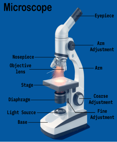
Figure 1. Light Microscope.
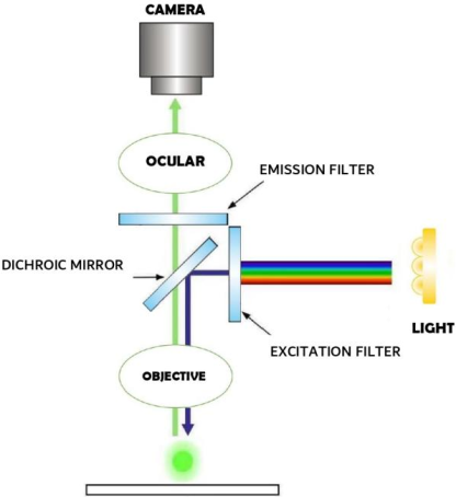
Figure 2. Functioning of a Fluorescence Microscope.
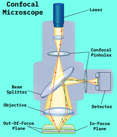
Figure 3. Functioning of the Confocal Microscope.
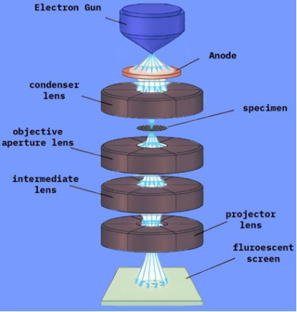
Figure 4. Transmission electron microscopy.
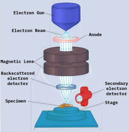
Figure 5. Scanning electron microscopy.
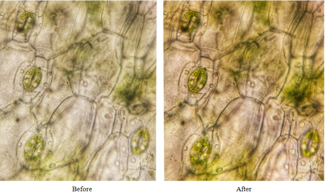
Figure 6. Before and after an image of stomata during image preprocessing.
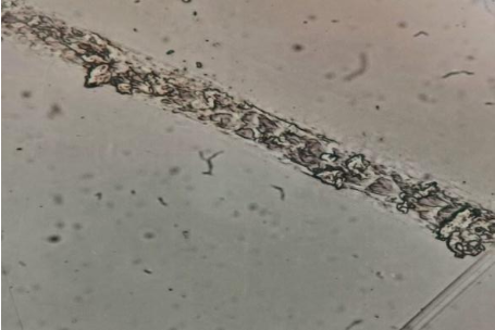
Figure 7. Cell shape and size help identify these cells as zebra fish gill cells.
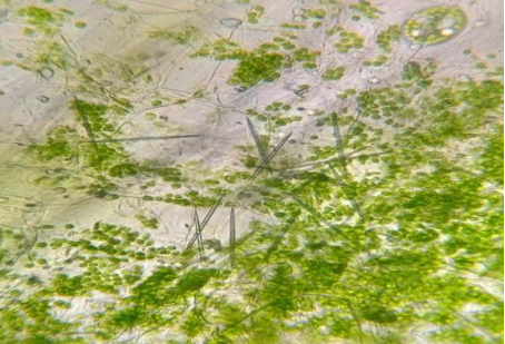
Figure 8. Cell shape and size help identify these cells as chlorophyl
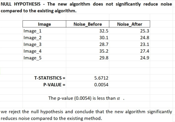
Figure 9. Shows a sample hypothetical testing on images before and after noise reduction done by 2 different algorithms.
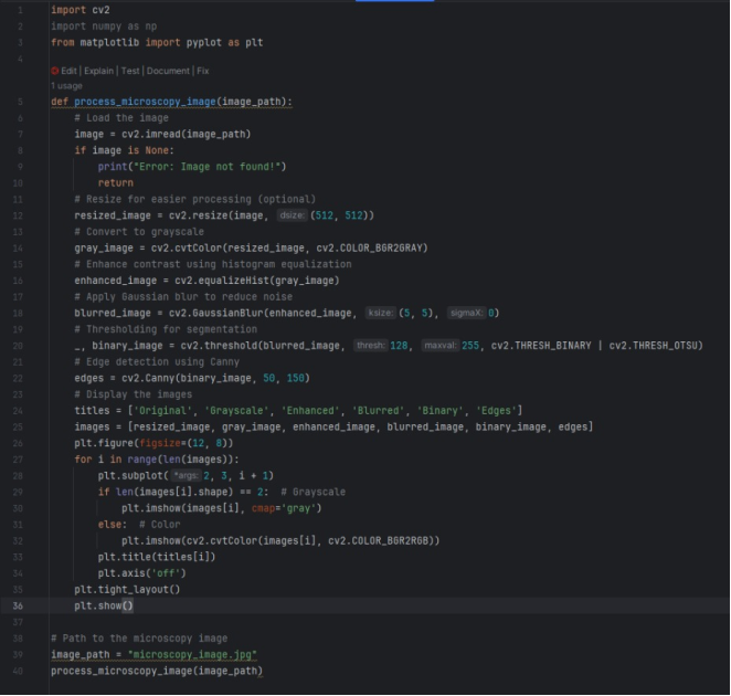
Figure 10. A Python application that uses the well-known OpenCV and NumPy modules to prepare a picture for microscopy. The application may segment regions of interest, apply denoising filters, and improve contrast [142].

Figure 11. Cloud-based systems have fundamentally altered image processing in microscopy by providing scalable, efficient, and reasonably priced solutions. For instance, Microsoft Azure (L) and Google Collaboratory R [158].
Information

