Research Article | | Peer-Reviewed

A Prospective Clinical Study on Transgingival Lag Screws for Management of Dentoalveolar Fractures

Received: 10 March 2025     Accepted: 25 March 2025     Published: 14 April 2025
Views:       Downloads:
Abstract

Dentoalveolar fracture (DAF) is a common injury which comprises of alveolar fractures which include 2-8% of facial fractures. Traditional methods of management of these fractures may not be feasible all the time. The aim of this study is to evaluate the efficiency of transgingival lag screws (TGLS) in management of dentoalveolar fractures. 20 patients with dentoalveolar fractures, either isolated or concomitant with other facial fractures were included in the study. All patients were treated using transgingival lagscrews under local or general anaesthesia and were evaluated for stability, reduction, bone loss, periodontal health and functional rehabilitation with a follow up period of 3 months. All the subjects healed without any complications. The screws were retrieved by the end of 3rd month. A descriptive statistical analysis was done for the assessed parameters. Good fracture stability was noted in 90% of the cases in immediate post operative period. Anatomical reduction assessed radiographically was ideal. Sound periodontal health was maintained effortlessly, inversely reflecting on minimal bone loss. Rehabilitative period was minimal with preinjury average bite force establishment by the end of 3 months. The study concluded that TGLS is an effective alternative to achieve reduction and stabilization in management of dentoalveolar fractures without jeopardizing the blood supply.

Published in International Journal of Clinical Oral and Maxillofacial Surgery (Volume 11, Issue 1)
DOI 10.11648/j.ijcoms.20251101.12
Page(s) 14-20
Creative Commons

This is an Open Access article, distributed under the terms of the Creative Commons Attribution 4.0 International License (http://creativecommons.org/licenses/by/4.0/), which permits unrestricted use, distribution and reproduction in any medium or format, provided the original work is properly cited.

Copyright

Copyright © The Author(s), 2025. Published by Science Publishing Group

Keywords

Dentoalveolar Fractures, Facial Fractures, Lag Screw, Transgingival

1. Introduction
Dentoalveolar fracture can be termed as a fracture of the bone surrounding the tooth, supporting soft tissues and the alveolar bone housing the teeth without any extension to the basal bones of the maxilla or mandible . 2–8% of all cranio-facial injuries comprise of fracture of the alveolar process .
One of the cornerstones to effective maxillofacial trauma management is ensuring adequate occlusal function, which requires prompt treatment of dentoalveolar injuries as well as any facial fractures. The blood supply to the broken pieces may be compromised by the fracture line and soft tissue damage, necessitating repair and repositioning. More often than not, the fractured segments are immobilized by wire-composite splint or maxillo-mandibular fixation (MMF) using arch bars or bone screws .
Lag screw fixation can be helpful in situations where typical fixation of the fractured component is not feasible due to the absence of the teeth required for splinting. Lag screw osteosynthesis is a technique that offers the benefits of reliable and stable internal fixation. Lag screws have long been a vital tool for surgeons, having been chiefly used by Brons et al. and subsequently made popular by Niederdellmann et al . The primary goal of our study is to assess the outcomes of employing lag screw osteosynthesis to manage dentoalveolar process fractures with closed reduction. The aim of the study is to evaluate the efficiency of transgingival lag screws for dentoalveolar fractures of maxilla and mandible. The objectives include assessment of fracture stability, anatomical reduction, bone resorption of alveolar process, wound infection and periodontal health, root damage and rehabilitative period.
2. Materials and Methodology
A clinical study was conducted in the Department of Facio-maxillary Surgery, Sanjay Gandhi Institute of Trauma and Orthopaedics, Rajiv Gandhi University, India for a period of 10 months between Feb 2024 to Dec 2024 which included the patients diagnosed with dentoalveolar fracture satisfying the inclusion criteria. All the participants were explained about the study and informed consent was taken before participation. The study protocol was approved by the Ethical Committee as per university regulations.
All the patients above 18 years with dentoalveolar fracture with or without other concomitant fractures were included in the study. Exclusion criteria was primary dentition, communited fractures and medically compromised patients. Diagnosis was made based on clinical and radiographical examination. 3D CT face was done to gauge and study the fracture pattern pre-operatively. The factors which were assessed in the study were fracture stability, anatomical reduction, bone resorption, occlusion and periodontal health.
2.1. Technique
Under standard aseptic conditions, patient was prepared either under general or local anaesthesia. The fractured fragments were manually reduced and repositioned. A pilot drill (1.5 mm drill bit) was used to drill a hole through the gingiva from the fractured segment perpendicular to the stable bone. Next, a slightly larger diameter drill bit (2 mm drill bit) was cautiously used to drill through the same hole of the fracture cortex alone. Following this, stainless-steel lag screws (S. K. Surgicals) of 2 x 10 mm, 2 x 12 mm or 2 x 14 mm (used according to the prerequisite) were driven into the bone bicortically through the gingiva in a compression manner. Minimum of 2 screws are generally recommended for stability. Maximum number of screws which we used in our study were 4 for adequate stability. Patient was advised 5 days of antibiotic and analgesic cover. Strict oral hygiene maintenance was instructed. Regular follow up was done. The lag screws were retained for 3 months and then retrieved under local anaesthesia.
2.2. Statistical Analysis
Statistical Package for social Sciences (SPSS) 20.0 software was used to perform statistical analysis. Chi-square test and Fischer exact test were used to assess the parameters/variables at different intervals. Probability value (p <0.05) was considered statistically b significant.
3. Results
A total of 20 subjects who sustained dentoalveolar fracture were included in the study and all were assessed for each parameter. Good fracture stability was accomplished within 1 week of screw placement in 18 (90%) of the cases (Table 1). Good anatomical reduction was noted in all the cases (20) which were assessed radiographically. Only 3 (15%) cases showed mild bone resorption after the first month of post-op period which resolved by the end of third month. Only 2 cases showed signs of infection at week 1 (Table 2). Infection was seen in cases where the site was contaminated with debris due to the trauma resulting in an contaminated wound at the fracture site and in patients with poor oral hygiene. These tribulations were managed with regular irrigation with antiseptic solution (5% Povidine Iodine+0.9% Normal Saline) and stringent oral hygiene maintenance.
Not more than 3(15%) cases showed root damage caused due to the procedure. Isolated DAFs were managed under local anaesthesia. DAF associated with concurrent facial fractures were managed under general anaesthesia along with fixation of other fractures. Sensory feedback from the patient when screw was close to the root couldn’t be elicited when placement was under general anaesthesia, unlike when done under local anaesthesia. When root damage was suspected, these patients were kept under observation and pulp vitality was assessed accordingly at frequent intervals. If the outcome was a non-vital tooth, root canal treatment was advised for the same. Pre-operative assessment with periapical radiographs or CBCT would give us a better picture of the available bone dimensions to plans for screw placement avoiding root damage.
Bite force device was used to review the post-operative bite force established by the patient by umpiring against their post-injury state. An average range of anterior bite force was considered acceptable. Rehabilitative period was established by evaluating the bite force by the end of 3 months where all the cases yielded good restoration of bite force to an average preinjury state.
4. Discussion
Dentoalveolar fractures represent a trivial proportion of facial injuries. The patient's age, gender, and causation all have a significant impact on the frequency and type of these injuries . Teeth associated with alveolar process fractures are characterized by mobility of the alveolar process where numerous teeth will typically move as a unit when mobility is checked . The underlying mechanism of dentoalveolar injury correlates to direct trauma to the teeth or secondary to a blow to the chin resulting in the mandibular dentition being forced into the maxillary dentition. The maxillary central incisors are the teeth in the permanent dentition that are most frequently affected .
Alveolar fractures associated with dental injuries result in functional, aesthetic and psychological impediment. Conventional methods for the management of DAF include various wiring technique, figure of eight wiring, placement of arch bar, acid etch composite wire and modified cap splint. High alveolar process fractures are also managed with plates and screws . Rahpyema et al suggested maintenance of apical force either with the use of MMF or with Intermaxillary fixation (IMF) screws or circum-mandibular or suspension wiring for these fractures for better reduction and stabilization. Bone screws stabilization of dentoalveolar fracture have also been advocated .
Wiring techniques are often cumbersome and requires more intra-operative time for placement. In addition, twisted and cut ends of the wires and arch bar are sharp and it may cause trauma to buccal mucosa and may penetrate the gingiva . Furthermore, conventional wiring methods are not practical in some situations where trauma causes tooth loss involving the fracture segment. Although the first line of treatment for these fractures is closed reduction, open reduction and fixation are taken into consideration for dentoalveolar fractures that cannot be treated by closed reduction. The vascularity of tissues gives dentoalveolar injuries their unique ability to recover. Traditional plate osteosynthesis necessitates stripping of the periosteum to expose the alveolar fracture. This manoeuvre of raising a flap for exposure along with any soft tissue injuries will jeopardise the blood supply of the broken fragments requiring repositioning and fixation . A novel method to counter the pitfalls of these technique came with the introduction of transgingival screw as an alternate to wiring, splinting and open reduction and fixation.
Brons and Boering introduced the lag screw technique in maxillofacial surgery in 1970 in the management of mandibular fractures. Less implant material/hardware, less cost, a simpler method, and less surgical exposure are the benefits of employing a single screw . Lag screw osteosynthesis is a process in which two bone fragments under pressure with the help of screws inserted in lag fashion led to a stable union . The fundamental concept of a lag screw is to compress two fractured fragments together. The screw is threaded into the opposite cortex and slides through a hole in the near cortex. Compression is aided by creating a countersink in the outer cortex of the screw's head. In the absence of a gliding hole, the fragments remain fixed in place but do not experience any compression. Tightening the screw compresses the fractured pieces by advancing the screw head against the adjacent cortex. Lag screws have been versatile implants which are used for management of various facial bone fractures such as mandibular symphysis, condyle, angle, and in few midface regions .
The goal of modern treatment for fractures of the alveolar process is to restore the original occlusion while ensuring optimal function along with natural aesthetics . Transgingival lag screws are a recent technique where a miniature version of lag screws are used for dentoalveolar process fracture segment stabilization. Despite other facial fractures, lag screws can be utilised to treat dentoalveolar process fractures and maxillofacial skeleton fractures without compromising other treatment options for the same condition .
Niederdellmann et al. characterized the concept of lag screw osteosynthesis as the stable amalgamation of bone fragments through the application of screws under pressure that are subjected to tension . The long-term stability of screw fixation is influenced by several factors, including the quantity of screws utilized, the technique of screw insertion, the placement of screws across both cortices, and the grip strength of the screws, which is determined by the thickness of the cortical bone. The reliability of this type of osteosynthesis depends entirely on the compression between the fragments . Our study found good fracture stability which was established from immediate post-operative period assessed clinically. By the end of 3 months absolute stability was seen reflecting a good union. All the fractures of alveolar process concomitant with or without other facial fracture were reduced anatomically with the help of lag screws. A significant improvement was observed in the radiographs taken after the surgery.
In their study, Zachariades et al. found that the infection rate ranged from 1% for fractures treated during the first week to 4% for those treated within the second week or later. Poor local conditions were another aspect that contributed to the infection . Coburn et al. reported a few hurdles with standard lag screws, including bone sequestra development, iatrogenic injury to the roots of teeth that results in loss, and screw fracture after insertion . There are various drawbacks associated with conventional techniques of using arch bars, prefabricated arch bar splint or wiring for management such as prolonged duration of surgery, high risk of needle stick injury and injury to surrounding soft tissues. Arch bars can pose challenges in hygiene maintenance and can thus affect gingival health. Poor oral conditions lead to compromising periodontal conditions. Bone degradation is driven by the immune and inflammatory reactions of the host in response to microbial threats. The rotund nature of these materials can cause more plaque comparatively, leading to periodontally compromised gingiva which in turn may cause marginal alveolar bone resorption. With the use of transgingival lag screws, this hindrance can be negated with the ease of maintenance that comes along with it.
A poor drill choice could result in a larger hole, which could cause screws to loosen and engage incorrectly, decreasing stability . Conversely, complication of screw loosening was not noted in our study. Most encountered complication during screw placement is iatrogenic damage of roots . Usually when we drill, we experience a bur drop into the medullary bone after having perforated the buccal cortex, before palatal/lingual cortex. The likelihood that the bur is partially or completely in a tooth root should be taken into consideration if this change in resistance is not felt. Corelating to this, our study witnessed 3(15%) of the cases enduring root damage caused iatrogenically. Pre-operative assessment with elementary intra-oral periapical radiographs would help us in better understanding of the interdental bone availability for the screw placement. Judicious placement of the screw is important to avoid perforation of mucosa after bicortical placement. Utilization of short screws and the direction of drilling would be of value to avoid root perforation and damage to vital structures. However, in our study, we have not encountered any damage to the nasopalatine arteries, mental foramen or maxillary sinus. Rehabilitative period was calculated by evaluating the biteforce which was re-established to the average force by the end of 3 months.
Since flap elevation was not required, the introduction of transgingival lag screws to stabilize the fractured pieces did not impair the blood supply to the alveolar process. The hardware used is minimal. Once tightened, the resulting compression bridges the distance between fragments facilitating bone healing. The flapless technique also reduces the operation time. The removal of the transgingival implants used was near simple and uneventful. In few cases where gingival overgrowth was seen over the head of the screw, a small stab incision was placed to expose the screw head and then retrieved under local anaesthesia.
A smaller sample size considered in our study could come across as a limitation. Further research is required to substantiate the applicability of this transgingival lag screw as a standard practice for fractures of the dentoalveolar process.
5. Conclusion
Transgingival lag screw osteosynthesis is a simple and rapid flapless procedure that can be utilized as a successful substitute for closed reduction of dentoalveolar fractures. It is constructive when there are numerous missing dentition with extensive injuries involving the overlying soft tissue. Any injuries to the overlying soft tissue may influence the blood supply to the underlying fracture fragment. This minimally invasive technique serves as a boon resulting in a successful osteosynthesis without stripping the fracture segment of its blood supply. Our results prove that TGLS is a feasible alternative compared to other conventional techniques in management of dentoalveolar fractures.
Abbreviations

DAF

Dentoalveolar Fracture

TGLS

Transgingival Lag Screw

MMF

Maxillo-Mandibular Fixation

IMF

Intermaxillary Fixation

Author Contributions
Abhinandan Patel: Conceptualization, Validation
Girish Gowda: Methodology, Project administration
Sirisha Sampangi Pushpa: Conceptualization, Data curation, Wirting – original draft
Preethi Bhat: Supervision, Visualization, Writing- review and editing
Siri Shetty: Data curation, Writing- review and editing
Funding
This work is not supported by any external funding.
Data Availability Statement
Not applicable.
Conflicts of Interest
The authors declare no conflicts of interest.
Appendix
Table 1. Fracture Stability.

Absent n (%)

Present n (%)

P value

Pre operative

20 (100)

-

1 week

2 (10)

18 (90)

0.001

1 month

-

20 (100)

0.001

3 months

-

20 (100)

0.001

Table 2. Infection and Periodontal health.

Absent n (%)

Present n (%)

P value

Pre operative

19 (95)

1 (5)

1 week

18 (90)

2 (10)

1.00

1 month

20 (100)

-

0.50

3 months

20 (100)

-

0.50

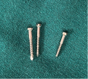
Figure 1. Transgingival lag screws of different sizes.
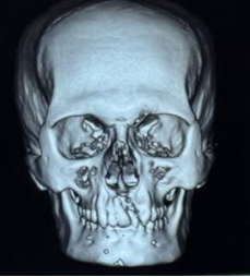
Figure 2. Pre-operative radiograph.
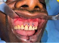
Figure 3. Intra-operative photograph after TGLS placement.
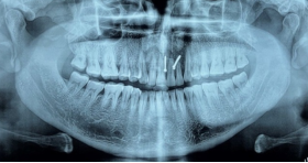
Figure 4. Post-operative Orthopantomogram.
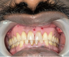
Figure 5. Post-operative photograph after TGLS removal with re-established occlusion.
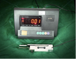
Figure 6. Bite force device.
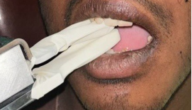
Figure 7. Bite force assessment.
References
[1] Nyárády Z, Orsi E, Nagy K, Olasz L, Nyárády J. Transgingival lag-screw osteosynthesis of alveolar process fracture. International journal of oral and maxillofacial surgery. 2010 Aug 1; 39(8): 779-82.
[2] Rahpeyma A, Khajehahmadi S. Apical Force Application in the Management of Dentoalveolar Fractures: Indications, Procedure and Limitations. Journal of Dental Materials and Techniques. 2015 Sep 1; 4(3): 115-6.
[3] MacLeod SP, Rudd TC. Update on the management of dentoalveolar trauma. Current opinion in otolaryngology & head and neck surgery. 2012 Aug 1; 20(4): 318-24.
[4] Sharma A, Rastogi S, Shukla M, Choudhury R, Tripathi S, Iqbal J. Use of transgingival lag screw osteosynthesis in the management of alveolar process fracture. Craniomaxillofacial Trauma & Reconstruction. 2019 Mar; 12(1): 27-33
[5] Glendor UL. Aetiology and risk factors related to traumatic dental injuries–a review of the literature. Dental traumatology. 2009 Feb; 25(1): 19-31.
[6] Oikarinen K, Kassila D. Causes and types of traumatic tooth injuries treated in a public dental health clinic. Dental Traumatology. 1987 Aug; 3(4): 172-7.
[7] RamosJorge ML, Peres MA, Traebert J, Ghisi CZ, De Paiva SM, Pordeus IA, Marques LS. Incidence of dental trauma among adolescents: a prospective cohort study. Dental Traumatology. 2008 Apr; 24(2): 159-63.
[8] Purohit J, Kumar GJ, Datta A, Maiti S, Hinge A, Kapoor S, Tiwari RV. Recent advances & historical management in dentoalveolar fracture-A review. Journal of Advanced Medical and Dental Sciences Research. 2020; 8(1): 129-36.
[9] Kasatwar JNA, Surana S, Rajanikanth K, Kala A, Dakshinkar P. Management of dento-alveolar fractures with trans-gingival lag screws: a novel technique. International Journal of Medical and Oral Research. 2017; 2(2): 1–6.
[10] Gibbons AJ. Bone screw stabilization of a dentoalveolar fracture. Journal of oral and maxillofacial surgery. 2007 Jul 1; 65(7): 1439.
[11] Qureshi AA, Reddy UK, Warad NM, Badal S, Jamadar AA, Qurishi N. Intermaxillary fixation screws versus Erich arch bars in mandibular fractures: A comparative study and review of literature. Annals of maxillofacial surgery. 2016 Jan 1; 6(1): 25-30.
[12] Li Z, Hu TQ, Li ZB. Open reduction by vestibular approach in the treatment of segmental alveolar fracture. Dental Traumatology. 2012 Dec; 28(6): 470-3.
[13] Brons R, Boering G. Fractures of the mandibular body treated by stable internal fixation: a preliminary report. Journal of oral surgery (American Dental Association: 1965). 1970 Jun; 28(6): 407-15.
[14] Elsayed SA. Cortical lag screw fixation for the management of mandibular injuries. Journal of the Korean Association of Oral and Maxillofacial Surgeons. 2020 Dec 31; 46(6): 393-402.
[15] Niederdellmann H, UHLIG G. Lag-screw osteosynthesis: a new procedure for treating fractures of the mandibular angle.
[16] Chotkowski G, Eggleston TI, Buchbinder D. Lag screw fixation of a nonstable zygomatic complex fracture: case report. Journal of oral and maxillofacial surgery. 1997 Feb 1; 55(2): 183-5.
[17] Rao E, Naveen S, Rao RC, Kollabathula K, Srirambhatla M, Gandham S. Principle of lag-screw fixation in mandibular trauma. Journal of International Society of Preventive and Community Dentistry. 2019 May 1; 9(3): 282-9.
[18] Zachariades N, Mezitis M, Papademetriou I. Use of lag screws for the management of mandibular trauma. Oral Surgery, Oral Medicine, Oral Pathology, Oral Radiology, and Endodontology. 1996 Feb 1; 81(2): 164-7.
[19] Coburn DG, Kennedy DW, Hodder SC. Complications with intermaxillary fixation screws in the management of fractured mandibles. British Journal of Oral and Maxillofacial Surgery. 2002 Jun 1; 40(3): 241-3.
[20] Nandini GD, Balakrishna R, Rao J. Self tapping screws v/s Erich arch bar for inter maxillary fixation: a comparative clinical study in the treatment of mandibular fractures. Journal of maxillofacial and oral surgery. 2011 Jun; 10: 127-31.
[21] Jain A, Taneja S, Rai A. What is a better modality of maxillomandibular fixation: bone-supported arch bars or Erich arch bars? A systematic review and meta-analysis. British Journal of Oral and Maxillofacial Surgery. 2021 Oct 1; 59(8): 858-66.
[22] Burman S, Lal B, Alagarsamy R, Kumar J, Ankush A, Rai AJ, Yunus M. Evidence-based management of isolated dentoalveolar fractures: a systematic review. Journal of the Korean Association of Oral and Maxillofacial Surgeons. 2024 Jun 1; 50(3): 123-33.
[23] Majumdar A, Brook IM. Iatrogenic injury caused by intermaxillary fixation screws. The British Journal of Oral & Maxillofacial Surgery. 2002 Feb 1; 40(1): 84-5.
Cite This Article
  • APA Style

    Patel, A., Gowda, G., Pushpa, S. S., Bhat, P., Shetty, S. (2025). A Prospective Clinical Study on Transgingival Lag Screws for Management of Dentoalveolar Fractures. International Journal of Clinical Oral and Maxillofacial Surgery, 11(1), 14-20. https://doi.org/10.11648/j.ijcoms.20251101.12

    Copy | Download

    ACS Style

    Patel, A.; Gowda, G.; Pushpa, S. S.; Bhat, P.; Shetty, S. A Prospective Clinical Study on Transgingival Lag Screws for Management of Dentoalveolar Fractures. Int. J. Clin. Oral Maxillofac. Surg. 2025, 11(1), 14-20. doi: 10.11648/j.ijcoms.20251101.12

    Copy | Download

    AMA Style

    Patel A, Gowda G, Pushpa SS, Bhat P, Shetty S. A Prospective Clinical Study on Transgingival Lag Screws for Management of Dentoalveolar Fractures. Int J Clin Oral Maxillofac Surg. 2025;11(1):14-20. doi: 10.11648/j.ijcoms.20251101.12

    Copy | Download

  • @article{10.11648/j.ijcoms.20251101.12,
      author = {Abhinandan Patel and Girish Gowda and Sirisha Sampangi Pushpa and Preethi Bhat and Siri Shetty},
      title = {A Prospective Clinical Study on Transgingival Lag Screws for Management of Dentoalveolar Fractures
    },
      journal = {International Journal of Clinical Oral and Maxillofacial Surgery},
      volume = {11},
      number = {1},
      pages = {14-20},
      doi = {10.11648/j.ijcoms.20251101.12},
      url = {https://doi.org/10.11648/j.ijcoms.20251101.12},
      eprint = {https://article.sciencepublishinggroup.com/pdf/10.11648.j.ijcoms.20251101.12},
      abstract = {Dentoalveolar fracture (DAF) is a common injury which comprises of alveolar fractures which include 2-8% of facial fractures. Traditional methods of management of these fractures may not be feasible all the time. The aim of this study is to evaluate the efficiency of transgingival lag screws (TGLS) in management of dentoalveolar fractures. 20 patients with dentoalveolar fractures, either isolated or concomitant with other facial fractures were included in the study. All patients were treated using transgingival lagscrews under local or general anaesthesia and were evaluated for stability, reduction, bone loss, periodontal health and functional rehabilitation with a follow up period of 3 months. All the subjects healed without any complications. The screws were retrieved by the end of 3rd month. A descriptive statistical analysis was done for the assessed parameters. Good fracture stability was noted in 90% of the cases in immediate post operative period. Anatomical reduction assessed radiographically was ideal. Sound periodontal health was maintained effortlessly, inversely reflecting on minimal bone loss. Rehabilitative period was minimal with preinjury average bite force establishment by the end of 3 months. The study concluded that TGLS is an effective alternative to achieve reduction and stabilization in management of dentoalveolar fractures without jeopardizing the blood supply.
    },
     year = {2025}
    }
    

    Copy | Download

  • TY  - JOUR
    T1  - A Prospective Clinical Study on Transgingival Lag Screws for Management of Dentoalveolar Fractures
    
    AU  - Abhinandan Patel
    AU  - Girish Gowda
    AU  - Sirisha Sampangi Pushpa
    AU  - Preethi Bhat
    AU  - Siri Shetty
    Y1  - 2025/04/14
    PY  - 2025
    N1  - https://doi.org/10.11648/j.ijcoms.20251101.12
    DO  - 10.11648/j.ijcoms.20251101.12
    T2  - International Journal of Clinical Oral and Maxillofacial Surgery
    JF  - International Journal of Clinical Oral and Maxillofacial Surgery
    JO  - International Journal of Clinical Oral and Maxillofacial Surgery
    SP  - 14
    EP  - 20
    PB  - Science Publishing Group
    SN  - 2472-1344
    UR  - https://doi.org/10.11648/j.ijcoms.20251101.12
    AB  - Dentoalveolar fracture (DAF) is a common injury which comprises of alveolar fractures which include 2-8% of facial fractures. Traditional methods of management of these fractures may not be feasible all the time. The aim of this study is to evaluate the efficiency of transgingival lag screws (TGLS) in management of dentoalveolar fractures. 20 patients with dentoalveolar fractures, either isolated or concomitant with other facial fractures were included in the study. All patients were treated using transgingival lagscrews under local or general anaesthesia and were evaluated for stability, reduction, bone loss, periodontal health and functional rehabilitation with a follow up period of 3 months. All the subjects healed without any complications. The screws were retrieved by the end of 3rd month. A descriptive statistical analysis was done for the assessed parameters. Good fracture stability was noted in 90% of the cases in immediate post operative period. Anatomical reduction assessed radiographically was ideal. Sound periodontal health was maintained effortlessly, inversely reflecting on minimal bone loss. Rehabilitative period was minimal with preinjury average bite force establishment by the end of 3 months. The study concluded that TGLS is an effective alternative to achieve reduction and stabilization in management of dentoalveolar fractures without jeopardizing the blood supply.
    
    VL  - 11
    IS  - 1
    ER  - 

    Copy | Download

Author Information
  • Department of Facio-Maxillary Surgery, Sanjay Gandhi Institue of Trauma and Orthopaedics, Bengaluru, India

  • Department of Facio-Maxillary Surgery, Sanjay Gandhi Institue of Trauma and Orthopaedics, Bengaluru, India

  • Department of Facio-Maxillary Surgery, Sanjay Gandhi Institue of Trauma and Orthopaedics, Bengaluru, India

    Biography: Sirisha Sampangi Pushpa, a Fellow in the Department of Facio-Maxillary Surgery currently pursuing fellowship in Maxillo-facial trauma at the Sanjay Gandhi Institute of Trauma and Ortho-paedics. Completed her Master in Oral and Maxillofacial Surgery in the year 2023. She is well-versed in managing complex trauma cases. Her primary focus and passion lies in emergency care, working diligently to provide timely and effective management for patients with facial injuries. In addition to trauma, her extended interests extend to minor surgical procedures and maxillofacial cleft surgeries.

    Research Fields: Minor surgeries, Oral pathology, Maxillofacial trauma, Craniofacial reconstruction, Implantology, Regenerative medicine, Anaesthesia and Pain management

  • Department of Facio-Maxillary Surgery, Sanjay Gandhi Institue of Trauma and Orthopaedics, Bengaluru, India

  • Department of Facio-Maxillary Surgery, Sanjay Gandhi Institue of Trauma and Orthopaedics, Bengaluru, India