Abstract
Background: Facial nerve dysfunction is divided into hypofunctional type (facial paralysis) and hyperfunctional type (hemifacial spasm and blepharospasm type of Major syndrome). Treatment of hemifacial spasm often requires surgical craniotomy and microvascular decompression, while severe Major syndrome requires expensive deep brain electrostimulation. Facial nerve block or radiofrequency ablation performed by the pain department is safer, more effective, convenient and economical to treat facial nerve dysfunction. Objective: To formulate an expert consensus on facial nerve block to better promote the homogeneous treatment technology of facial nerve block and radiofrequency ablation. Methods: Chinese pain experts in facial nerve block and radiofrequency treatment were organized to systematically review the anatomy and physiological functions of the facial nerve, sort out the clinical manifestations of facial nerve dysfunction, and reach expert consensus on the indications, contraindications, operating procedures, and prevention and treatment of related complications of facial nerve block and radiofrequency treatment based on the relevant literature and expert experience of facial nerve block and radiofrequency treatment. Conclusion: The consensus of Chinese pain experts on facial nerve block can provide a relatively authoritative and systematic guidance document for nerve block and radiofrequency treatment of facial nerve dysfunction diseases, so as to facilitate the better promotion and application of this technology.
|
Published in
|
International Journal of Pain Research (Volume 2, Issue 3)
|
|
DOI
|
10.11648/j.ijpr.20260203.18
|
|
Page(s)
|
162-172 |
|
Creative Commons
|

This is an Open Access article, distributed under the terms of the Creative Commons Attribution 4.0 International License (http://creativecommons.org/licenses/by/4.0/), which permits unrestricted use, distribution and reproduction in any medium or format, provided the original work is properly cited.
|
|
Copyright
|
Copyright © The Author(s), 2026. Published by Science Publishing Group
|
Keywords
Facial Nerve, Anatomy, Nerve Conduction Block, Computer-Tomography Guided, Grading Criteria for Facial Paralysis, Grading Criteria for Hemifacial Spasm
1. Introduction
The facial nerve is the VII pair of cranial nerves, which mainly govern the facial expression muscle movement and the anterior 2/3 of the tongue taste. facial nerve dysfunction often manifests as expression muscle paralysis or spasm. Facial nerve block is a common method for treating facial nerve dysfunction diseases. In order to promote the standardized development of facial nerve block technology in China, Chinese Journal of Painology organized domestic experts with rich experience in the clinical application of this technology to jointly formulate the Chinese expert consensus on facial nerve block technology for clinicians' reference.
2. Facial Nerve Anatomy and Function
The facial nerve, also known as the intermediate facial nerve
| [1] | Burmeister HP, Hause F, Baltzer PA, et al. Improvement of visualization of the intermediofacial nerve in the temporal bone using 3T magnetic resonance imaging: part 1: the facial nerve [J]. J Comput Assist Tomogr, 2009, 33 (5): 782-788.
https://doi.org/10.1097/RCT.0b013e31818da6bb |
[1]
, strictly speaking, refers to the special visceral motor nerves that originate from the facial nerve nucleus and innervate the facial muscles such as the expression muscles in the second gill arch. The intermediate nerve is the other motor and sensory fibers that make up the facial nerve. Therefore, the facial nerve refers to a mixed nerve composed of four components: special and general visceral efferent fibers, special visceral afferent fibers, and general somatic afferent fibers
| [2] | Standring S: Gray's Anatomy E‑Book: The anatomical basis of clinical practice (ed 41). London, UK, Elsevier Health Sciences, 2015. |
[2]
.
2.1. Special Visceral Motor Fiber
It is composed of the axons of the facial nerve nucleus and is the most important part of the facial nerve. The axon emitted from the facial nerve nucleus exits the brain in the Pons - cerebellar triangle, exits the cranial cavity through the inner ear portal together with the acoustic nerve through the prepontine cistern, and after exiting the cranial cavity, it travels with the intermediate nerve and the VIII pair of cranial nerves, and is wrapped in a common dura sheath, and enters the bony duct (Facial nerve canal) in the mastoid process through the inner ear portal. The remaining fibers exit the skull from the styloid mastoid foramen, first divide into the posterior auricular nerve branch, stylohyoid muscle and digastric branch
| [3] | Kikuta S, Iwanaga J, Watanabe K, et al. Anatomical study of the posterior auricular branch of the facial nerve: application to parotid surgery [J]. J Craniofac Surg. 2020, 31 (1): 300-302. https://doi.org/10.1097/SCS.0000000000005883 |
[3]
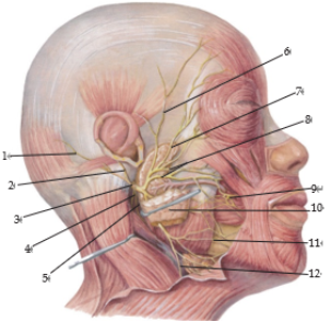
, and then enter the parotid gland to divide into the temporal branch, zygomatic branch, buccal branch, mandibular marginal branch and cervical branch, innervating stylohyoid muscle, digastric posterior abdomen, expression muscle and platysma muscle
| [4] | Kikuta S, Iwanaga J, Watanabe K, et al. The feasibility of using the posterior auricular branch of the facial nerve as a donor for facial nerve reanimation procedures: a cadaveric study [J]. J Oral Maxillofac Surg, 2019, 77 (7): 1470. e1-1470. e8. https://doi.org/10.1016/j.joms.2019.02.043 |
| [5] | Kwak HH, Park HD, Youn KH, et al. Branching patterns of the facial nerve and its communication with the auriculotemporal nerve [J]. Surg Radiol Anat, 2004, 26 (6), 494-500. https://doi.org/10.1007/s00276‑004‑0259‑6 |
| [6] | Mitsukawa N, Moriyama H, Shiozawa K, et al. Study on distribution of terminal branches of the facial nerve in mimetic muscles (Orbicularis Oculi Muscle and Orbicularis Oris Muscle) [J]. Ann Plast Surg, 2014, 72 (1): 71-74.
https://doi.org/10.1097/SAP.0b013e318284eca0 |
| [7] | Moreau S, Bourdon N, Salame E, et al. Facial nerve: vascular-related anatomy at the stylomastoid foramen [J]. Ann Otol Rhinol Laryngol. 2000, 109(9): 849-852.
https://doi.org/10.1177/000348940010900912 |
[4-7]
(see
Figure 1).
Figure 1. Anatomical distribution of facial nerve and branches on the face.
Note: After the facial nerve composed of special visceral motor fibers dries out of the styloid and mastoid foramen, it first divides into the posterior auricular nerve branch, styloid hyoid muscle and digastric muscle branch, and then enters the parotid gland to divide into the temporal branch, zygomatic branch, buccal branch, mandibular marginal branch and cervical branch, which innervates the styloid hyoid muscle, digastric muscle posterior abdomen, expression muscle and platysma muscle; In the figure, 1 is the occipital branch of the facial nerve, 2 is the auricular branch of the facial nerve, 3 is the posterior auricular nerve, 4 is the digastric branch, 5 is the styloid hyoid muscle branch, 6 is the temporal branch of the facial nerve, 7 is the zygomatic branch of the facial nerve, 8 is the internal plexus of the parotid gland, 9 is the buccal branch of the facial nerve, 10 is the parotid gland, 11 is the mandibular marginal branch of the facial nerve, and 12 is the cervical branch of the facial nerve.
2.2. General Visceral Motor Fiber
The axons of the salivary gland nucleus and lacrimal gland nucleus cells belong to the parasympathetic pre ganglionic fibers. The fibers originating from the lacrimal gland nucleus travel forward at the knee ganglion, leave the facial nerve trunk, and form the petrosal nerve. After replacement in the pterygopalatine ganglion, they innervate the lacrimal gland, nasal palatine mucosal gland, and blood vessels; The fibers originating from the superior salivary gland pass through the knee ganglion and branch out into the chorda tympani branch in the middle of the facial nerve canal
. They leave the facial nerve trunk with the tympanic cord, pass through the tympanic cavity, and split out of the skull through the rock drum. They then join the lingual nerve and alternate in the submandibular ganglion before being distributed to the sublingual gland and submandibular gland.
2.3. Sensory Fiber of Cranial Nerve of the VII Pair
Most are located within the knee ganglion of the facial canal. Among all sensory fibers, special visceral sensory fibers are the most, conducting taste impulses in the anterior 2/3 of the tongue and the soft palate. Generally, the number of somatosensory fibers is very small, and they mainly transmit pain, warmth and tactile impulses in the external auditory canal, around the external auricular portal and the skin behind the auricle.
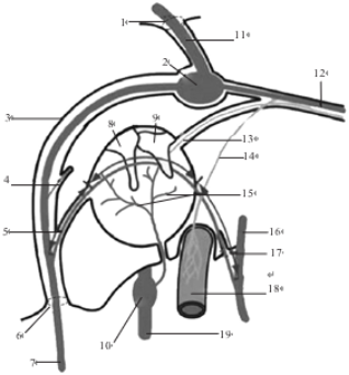
It can be seen that after the VII pair of cranial nerves exits the cranial cavity, they divide into the great petrous nerve, the cord tympanic branch and the stapes muscle branch in the facial nerve canal (
Figure 2), and after exiting the stylomastoid foramen, they divide into the posterior auricular nerve branch, the cervical branch, the temporal branch, the zygomatic branch, the buccal branch and the mandibular marginal branch, which mainly govern the facial expression muscle movement, the secretion of head and face glands and the anterior 2/3 taste of the tongue. The facial nerve runs in the skeletal facial nerve canal from entering the inner ear gate to exiting the stylomastoid foramen, so it is difficult to perform nerve block operation in this section; However, the facial nerve enters the parotid gland about 16 mm after exiting the stylomastoid foramen
| [9] | Salame K, Ouaknike GER, Arensburg B, et al. Microsurgical anatomy of the facial nerve trunk [J]. Clin Anat, 2002, 15 (2): 93-99. https://doi.org/10.1002/ca.1102 |
[9]
and is plexiform and scattered into multiple branches
(
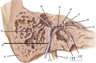
Figure 1). Each branch has a large variability and lacks clear bone markers, so it is very difficult to perform highly selective branch block; However, the stylomastoid foramen has clear bone anatomical landmarks, and the position of the facial nerve trunk here is relatively fixed, so the stylomastoid foramen is the best target choice for facial nerve block
(
Figure 3).
Figure 2. Schematic diagram of facial nerve running and branching anatomy in facial nerve canal.
Note: The facial nerve enters the facial nerve canal in the mastoid process from the inner ear portal. After the geniculate ganglion replaces neurons, special visceral motor fibers emit stapedial muscle branches to innervate the stapedial muscle. Generally, visceral motor fibers emit petrous nerve and tympanic cord branches. The former innervates the lacrimal gland, nasopalatine mucosal glands and internal carotid artery after substitution in the pterygopalatine ganglion. The latter crosses the tympanum and bursts out of the skull from the rock tympanum, joins the lingual nerve, and innervates the sublingual gland and the submandibular gland; In the figure, 1 is the entrance of the internal auditory tract, 2 is the geniculate ganglion, 3 is the facial nerve canal, 4 is the stapes branch, 5 is the cord tympanic branch, 6 is the stylomastoid foramen, 7 is the motor branch of the facial nerve, 8 is the malleus, 9 is the incus, 10 is the inferior glossopharyngeal ganglion (cranial nerve IX), 11 is the facial nerve (cranial nerve VII), 12 is the petrous great nerve branch, 13 is the petrous small nerve branch, 14 is the petrous deep nerve branch, 15 is the tympanic plexus, 16 is the lingual nerve (cranial nerve V), 17 is the petrous bulge, 18 is the internal carotid artery, and 19 is the glossopharyngeal nerve (cranial nerve IX)).
Figure 3. Anatomical view of facial nerve canal and facial nerve outlet-stylomastoid foramen.
Note: The styloid mastoid foramen is located between the root of the styloid process and the mastoid process. It is a trumpet-shaped bony foramen with a narrow top and a wide bottom. It is the outlet of the facial nerve trunk in the facial nerve canal and the best puncture target of facial nerve block. In the figure, 1 is the mastoid chamber, 2 is the facial nerve, 3 is the mastoid process, 4 is the facial nerve canal, 5 is the stapes muscle, 6 is the incus, 7 is the malleus, 8 is the knee ganglion, 9 is the petrous nerve, 10 is the tympanic cord, 11 is the promontory, 12 is the styloid process, and 13 is the stylomastoid foramen.
3. Clinical Manifestations of Facial Nerve Dysfunction
3.1. Facial Nerve Central Paralysis
The facial nerve is mainly a special visceral motor nerve, which mainly innervates the expression muscle. Therefore, the main clinical manifestation of facial nerve dysfunction is expression muscle dysfunction, that is, expression muscle paralysis or spasm. Because the expression muscles above the eye cleft are controlled by the bilateral cerebral cortex, the expression muscles below the eye cleft are controlled by the contralateral cerebral cortex. Thus, unilateral central facial nerve injury (such as cerebrovascular accident or tumor invasion of facial nerve center) is mainly manifested as expression muscle paralysis below the contralateral eye cleft: that is, paralysis of the contralateral orbicularis oculi muscle and orbicularis oris muscle. Patients often become shallow in the contralateral nasolabial fold when they are still, the corner of the mouth droops, and the corner of the mouth tilts to the central lesion side when teeth are shown; However, the expression muscles above the eye cleft still have the contralateral healthy facial nerve innervation, so closing the eyes, raising the eyebrows and frowning are normal, and the frontal lines are also equal to the contralateral depth, which is called facial nerve central paralysis (supranuclear paralysis).
3.2. Peripheral Paralysis of Facial Nerve
Unilateral peripheral facial nerve injury, such as mastoid fracture, iatrogenic injury during craniotomy micro-vascular decompression (MVD), facial nerve root of the anterior pontic cistern, radiofrequency injury of stylomastoid foramen, facial nerve trunk, Parotid gland surgery injures facial nerve branches, or inflammation, such as non-specific inflammation of the facial nerve in the facial canal (Bell palsy) and geniculate ganglion zoster virus infection (Ramsay Hunt syndrome): the former is called facial nerve palsy, which is a facial nerve palsy caused by non-specific inflammation and edema of the facial nerve in the facial canal, which leads to facial nerve compression or local blood circulation disorder, and is common after cold or upper respiratory tract infection
; The latter is caused by herpes zoster virus infection of the geniculate ganglion, also known as geniculate gangliitis. In addition to the former, there are dull sensation or severe pain in the auricle and external auditory canal, herpes appears on the external auditory canal and tympanic membrane, and may also be accompanied by hearing and balance impairment
| [14] | Kim SH, Jung J, Jung SY, et al. Comparative prognosis in patients with Ramsay-Hunt syndrome and Bell's palsy [J]. Eur Arch Otorhinolaryngol, 2019, 276 (4): 1011‑1016.
https://doi.org/10.1007/s00405‑019‑05300‑3 |
[14]
. All expression muscles above and below the eye cleft on this side are affected, which is manifested by the disappearance of frontal wrinkles on this side, enlargement of the eye cleft or even inability to close the eyes, flat nasolabial folds, oblique corners of the mouth, sagging corners of the mouth and more obvious facial skew when smiling or exposing teeth. You can't do movements such as frowning, closing your eyes, puffing and pouting on this side. When puffing your cheeks and whistling, the lips on the affected side can't close and air leaks. When eating, food residues often stay in the tooth-cheek space on the diseased side, and saliva often drips down from this side, and the tear points turn out with the lower eyelid, so that the tears can't be drained normally and tears overflow, which is called facial nerve peripheral paralysis (subnuclear paralysis).
In order to assess the degree of facial paralysis, our expert group proposed the Chinese grading standard for the degree of facial paralysis with reference to the 6-grade standard proposed by House-Brackmann
and other facial nerve function grades
| [16] | Lee HY, Park MS, Byun JY, et al. Agreement between the facial nerve grading system 2.0 and the house-Brackmann grading system in patients with Bell Palsy [J]. Clin Exp Otorhinolaryngol, 2013, 6 (3): 135-139.
https://doi.org/10.3342/ceo.2013.6.3.135 |
[16]
, as shown in Annex 1.
3.3. Hemifacial Spasm
Facial nerve disorders are not limited to decreased muscle strength. When the facial nerve is over-excited, it will manifest as expression muscle spasm. Common in hemifacial spasm (HFS)
and segmental cranio-cervical dystonia. HFS is a facial movement disorder controlled by the facial nerve, which is more common in adults. At the time of attack, the facial muscles on one side show irregular, involuntary and painless clonus, which can be induced or aggravated by emotional agitation, mental tension and voluntary facial movement, and can be appropriately relieved when relaxed. It often starts around the eyes, and then gradually progresses down to the cheeks, around the mouth and even the neck. According to the etiology, HFS is divided into primary (caused by vascular compression of the facial nerve root) and secondary hemifacial spasm (caused by the ponto-cerebellar angle tumor occupying space and compressing the facial nerve root of the anterior pontic cistern); Segmental craniocervical dystonia, also known as Meige syndrome (MS)
, occurs at about 60 years old, and the incidence rate is higher in women than in men. It usually involves involuntary twitching of bilateral eyelids and/or perioral muscles, which is manifested as frequent bilateral blinking, difficulty opening eyes, and/or grinding teeth, closed teeth, and twisting of the lips and jaw. As the disease progresses, this involuntary twitch gradually spreads throughout the face and even the neck muscles. At present, the cause of MS is still unclear, and it is generally believed that it may be mainly related to the transmitter changes or ion channel abnormalities in the Cortical striatum globus-pallidus-thalamus-cortical pathway. According to the different cranial nerves involved, MS is divided into Blepharospasm type (involuntary twitching of the eyelids only, mainly involving the facial nerve), tension disorder of the oral and mandibular muscles (involuntary spasmodic movements of the oral and mandibular muscles, involving the trigeminal nerve), and tension disorder of the eyelids combined with the oral and mandibular muscles (this is a typical type of MS, manifested as involuntary twitching of the eyelids combined with the oral and mandibular muscles, often with strange expressions and simultaneous involvement of the facial nerve and trigeminal nerve), and other types can be combined with the corresponding dystonia caused by accessory nerve and hypoglossal nerve involvement on the basis of the above types.
In order to evaluate the degree of hemifacial spasm, Cohen proposed a five-grade standard
| [19] | Tambasco N, Simoni S, Sacchini E, et al. Validation of the hemifacial spasm grading scale: a clinical tool for hemifacial spasm [J]. Neurol Sci, 2019, 40 (9): 1887-1892.
https://doi.org/10.1007/s10072‑019‑03921‑4 |
[19]
, and this expert group proposed a Chinese grading standard for the degree of hemifacial spasm according to the situation of Chinese people, see Annex 2.
From the manifestations of HFS and MS, the former is a peripheral disorder of facial nerve (facial nerve root compression by accompanying blood vessels, demyelination and abnormal discharge, unilateral expression muscle spasm), while the latter is a central disorder of facial nerve (abnormal excitation of nucleus, bilateral expression muscle involvement).
4. Conservative Therapy and Nerve Block Therapy for Facial Nerve Dysfunction
Common facial nerve disorders are divided into neurological inhibitory disorders and hyperactive disorders. The former is manifested as facial paralysis, common facial neuritis (Bell palsy), herpes zoster virus infection (Ramsay Hunt syndrome), mastoid fracture or surgical iatrogenic injury to the peripheral facial nerve, etc. The latter manifests as expression muscle spasm, which is common in HFS or MS.
For facial nerve disorders, in addition to the treatment of causes (anti-inflammatory for Bell palsy, anti-viral for Ramsay Hunt syndrome, surgical anastomosis for facial nerve dissection injury, craniotomy and microvascular decompression for HFS to relieve vascular compression on nerves, and anti-epileptic drugs for MS to inhibit the production of ectopic electrical excitation)
| [20] | Li Peipei, Han Yuefeng. Advances in the treatment of Bell's facial paralysis [J]. International Journal of Otolaryngology Head and Neck Surgery, 2016, 40 (3): 142‑147.
https://doi.org/10.3760.cma.j.issn.1673‑4106.2016.03.004 |
| [21] | Menchetti I, McAllister K, Walker D, et al. Surgical interventions for the early management of Bell's palsy [J]. Cochrane Database Syst Rev. 2021, 1 (1): CD007468.
https://doi.org/10.1002/14651858.CD007468.pub4 |
| [22] | Chinese Medical Association Neurology Branch, Chinese Medical Association Neurology Branch, Chinese Medical Association Neurology Branch, Chinese Medical Association Neurology Branch, Chinese Medical Association Neurology Branch Electromyography and Clinical Neuroelectrophysiology Group. Chinese guidelines for diagnosis and treatment of idiopathic facial nerve palsy [J]. Chinese Journal of Neurology, 2016, (2): 84‑86.
https://doi.org/10.3760/cma.j.issn.1006‑7876.2016.02.002 |
[20-22]
.
Facial nerve block is an effective treatment: for patients with neurological inhibition, facial nerve block can directly inject anti-inflammatory drugs such as glucocorticoids and neurotrophic drugs around the facial nerve trunk to eliminate inflammation and promote nerve function repair; For patients with facial nerve hyperfunction, facial nerve block can directly inhibit the excessive excitation of facial nerve and quickly relieve expression muscle spasm
.
Narrowly defined nerve block refers to the use of chemical drugs injected around nerves to regulate or block their conduction function, while broadly defined nerve block also includes the use of physical methods such as radiofrequency to block the conduction function of nerves. Especially for patients with facial nerve hyperfunction, it is difficult to control the extent and extent of block when using long-acting destructive nerve block drugs (absolute ethanol, phenolic glycerol, doxorubicin, methylene blue), and it must be accomplished by more controllable radiofrequency ablation block technology
| [25] | Huang Bing, Lin Huidan, Du Xindan, et al. Therapeutic effect of CT-guided radiofrequency thermocoagulation by percutaneous puncture of the stylomastoid foramen in the treatment of primary hemifacial spasm [J]. Chinese Journal of Neuromedicine, 2019, 18 (9): 933‑938.
https://doi.org/10.3760.cma.j.issn.1671‑8925.2019.09.010 |
| [26] | Hori T, Fukushima T, Terao H, et al. facial nerve coagulation in the management of facial spasm [J]. J Neurosurg, 1981, 54 (5), 655-658. https://doi.org/10.3171/jns.1981.54.5.0655 |
[25, 26]
to prevent severe iatrogenic facial paralysis.
4.1. Indications and Contraindications for Facial Nerve Block
Facial nerve block is suitable for all patients except for facial nerve disconnection injury. Patients with primary facial nerve disease, but those with a clear cause need to be treated at the same time. For patients with facial nerve function inhibition, narrowly defined nerve block technology or pulsed radio frequency should be used to adjust or promote facial nerve function recovery, while for patients with facial nerve hyperfunction, broadly defined nerve block treatment technology represented by standard continuous radio frequency ablation should be used to achieve long-term therapeutic effect
| [27] | Huang B, Yao M, Chen QL, et al. Awake CT-guided percutaneous stylomastoid foramen puncture and radiofrequency ablation of facial nerve for treatment of hemifacial spasm [J]. J Neurosurg, 2021, 16:1-7.
https://doi.org/10.3171/2020.10.JNS203209 |
[27]
. Patients with severe local or systemic infection at the site to be punctured and severe coagulation dysfunction should be listed as contraindications for facial nerve block.
4.2. Operation Technique of Facial Nerve Block by Puncture of Stylomastoid Foramen
4.2.1. Preoperative Preparation
The diagnosis and differential diagnosis were confirmed again, and the indications for facial nerve treatment were confirmed and the contraindications were excluded. Inform patients about the advantages and disadvantages of facial nerve block and alternatives, and sign informed consent. Prepare nerve block medication, emergency medication, radio frequency instrument, monitor, emergency endotracheal intubation kit and simple respirator. And intravenous indwelling catheter preparation infusion channel before treatment.
4.2.2. Stylomastoid Foramen Puncture and Facial Nerve Block Operation
The distance between stylomastoid foramen and the skin at the earlobe is only 3~5 cm, and the average depth 3.62 cm
| [27] | Huang B, Yao M, Chen QL, et al. Awake CT-guided percutaneous stylomastoid foramen puncture and radiofrequency ablation of facial nerve for treatment of hemifacial spasm [J]. J Neurosurg, 2021, 16:1-7.
https://doi.org/10.3171/2020.10.JNS203209 |
[27]
, and the puncture can be performed by bare hands or under ultrasound guidance. Because it is adjacent to the jugular foramen and the internal carotid artery in the inner and posterior part, in order to improve the puncture accuracy, reduce puncture complications, and achieve the purpose of precision medicine, this consensus recommends a more accurate and definite CT-guided stylomastoid foramen puncture facial nerve block treatment. The operation method is as follows:
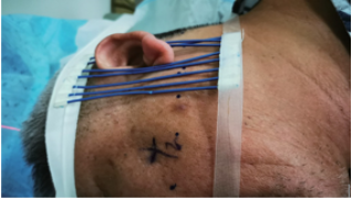
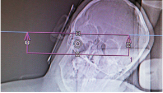
The patient was instructed to lie on his side on the CT table, and place a CT positioning grid in front and behind the earlobe (
Figure 4), monitor blood pressure, electrocardiogram, pulse oximetry, and administer oxygen through a nasal cannula. The CT localization image of the skull was taken in the paranasal sinus mode
, and then the mastoid area was scanned in CT axial position with a layer thickness of 3 mm (
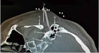
Figure 5). The layer containing the stylomastoid foramen was selected as the puncture layer and the puncture path was designed on this layer: the CT self-contained measurement tool software was used to straighten the line from the stylomastoid foramen (puncture target) on the affected side forward and upward to avoid the bone barrier of the temporal drum. The intersection point between this line and the skin in front of the affected mastoid is the puncture point of the anterior mastoid approach. The puncture depth (distance from the target point to the puncture point) and the puncture angle (angle between the puncture route and the sagittal plane) were measured with the CT built-in software tool ruler (
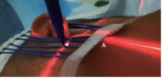
Figure 6). Find the puncture plane with CT positioning line and mark the puncture point with reference to the positioning grid (
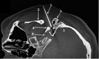
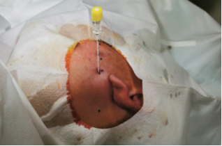
Figure 7). Perform local anesthesia on the puncture point after routine sterilization and laying sterile hole towel. Then, use a No. 7 ordinary puncture needle with a length of 5~10 cm or a radiofrequency puncture needle with a bare end of 5 mm to puncture to the target point according to the designed puncture path under CT guidance (
Figures 8 and 9), and confirm by three-dimensional reconstruction observation of CT scan (
Figure 10) that the puncture needle tip is in the stylomastoid foramen.
Figure 4. Positioning image taken by CT with positioning grid placed in lateral recumbent position.
Note: The self-made positioning bars of the interventional catheter are placed longitudinally before and after the earlobe to determine the puncture point in combination with the development of the positioning bars on the skin on the CT scan image.
Figure 5. CT scan frame for axial scan of mastoid region.
Figure 6. Puncture Path Design.
Note: In the figure, 1 is the puncture depth of 4.18 cm, 2 is the puncture angle (angle with the sagittal plane of 13°), 3 is the mandible, 4 is the mastoid process, 5 is the root of the styloid process, 6 is the stylomastoid foramen, and 7 is the jugular foramen. In the figure, the multiple white dots close to the skin are developed by positioning grates, so as to mark the skin puncture points by reference.
Figure 7. Find the puncture plane and mark the puncture point with the CT positioning red line.
Note: The point indicated by arrow A in the figure is the puncture point. This positioning red line is the CT scan plane position (SP-650.5) shown in
Figure 4, which is located at the level of the lower edge of the ear lobe. The puncture point is between the 3rd and 4th shadow points of the positioning bars on the skin shown in
Figure 4.
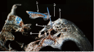
Figure 8. Successful puncture to the stylomastoid foramen on the affected side according to the puncture path.
Note: In the figure, 1 is the puncture needle, 2 is the mandible, 3 is the mastoid process, 4 is the root of the styloid process, 5 is the stylomastoid foramen, and 6 is the jugular foramen.
Figure 9. Appearance when puncture is successful.
Note: In the figure, the puncture point is between two adjacent marking points on the lower edge of the earlobe, and the remaining marking points indicate the puncture level.
Figure 10. CT three-dimensional reconstruction after successful puncture.
Note: the root of the styloid process. 1 is the mandible, 2 is the puncture needle, 3 is the mastoid process, 4 is the styloid process, 5 is the styloid-mastoid orifice, and 6 is the jugular foramen.
At this time, if the facial nerve function inhibition type disease is treated, narrowly defined facial nerve block is performed, nerve block drugs are injected, and glucocorticoids are added betamethasone sodium phosphate 0.5 ml to 1.0 ml, 2% lidocaine 1 ml,
| [22] | Chinese Medical Association Neurology Branch, Chinese Medical Association Neurology Branch, Chinese Medical Association Neurology Branch, Chinese Medical Association Neurology Branch, Chinese Medical Association Neurology Branch Electromyography and Clinical Neuroelectrophysiology Group. Chinese guidelines for diagnosis and treatment of idiopathic facial nerve palsy [J]. Chinese Journal of Neurology, 2016, (2): 84‑86.
https://doi.org/10.3760/cma.j.issn.1006‑7876.2016.02.002 |
[22]
Vitamin B 1 ml mixture total 3 ml. If facial nerve pulse radiofrequency is performed to regulate and promote facial nerve function recovery, it can be treated with a temperature of 42℃, voltage of 30-40 V, frequency of 2 Hz, pulse width of 20 ms, radiofrequency time of 300 s, or high voltage long-term pulse radiofrequency, temperature of 42 ℃, pulse width of 20 ms, voltage of 40-80 V, radiofrequency time of 900 s
. After pulsed radio frequency, narrowly defined facial nerve drug blockade can still be used after pulse radiofrequency.
If the treatment of facial nerve hyperfunction disease requires standard continuous radiofrequency ablation block of the facial nerve to destroy part of the conduction function of the facial nerve to achieve the therapeutic effect, then the motor function of the facial nerve is tested by low-frequency (2 Hz) current stimulation of the radiofrequency instrument first: when the current stimulation below 0.8 mA can induce rhythmic twitching of the facial expression muscle on the affected side (the same frequency as the electrical stimulation, video 1), the test result is considered positive; Otherwise, the radiofrequency needle tip position was adjusted to re-stimulate the test until a positive result appeared before standard continuous radiofrequency ablation could be performed
. In order to prevent excessive damage to facial nerve conduction function caused by standard continuous radio frequency, a continuous standard radio frequency mode with stepwise temperature rise from low to high should be adopted: the initial temperature parameter is set to 60℃, the time parameter is set to 30 s, the patient is instructed to bulge his cheeks and close his eyes tightly after the radio frequency starts, and the patient's facial expression muscle function is closely observed during the radio frequency process. Once the affected side bulges his cheeks and leaks, and his eyes are not tight, the radio frequency treatment is immediately terminated; If the patient still has bulging gills without air leakage and the affected eyes can still be closed tightly after 30 seconds of radiofrequency under this temperature parameter, the temperature parameter is increased by 5℃ and the time remains unchanged, that is, 65℃ and 30 seconds continuous radiofrequency is performed. If the gills are still bulging without air leakage, the temperature parameter is increased by 5℃, and the standard continuous radiofrequency ablation is performed at 70℃ and 30 seconds, until the patient's bulging gills have mild air leakage and the eyes are not tightly closed, and the radiofrequency treatment is terminated; If the therapeutic effect cannot be achieved after 30 seconds when the temperature is adjusted up to 90 ℃, the needle tip is adjusted again or the puncture path is changed before testing and radiofrequency treatment is performed
| [25] | Huang Bing, Lin Huidan, Du Xindan, et al. Therapeutic effect of CT-guided radiofrequency thermocoagulation by percutaneous puncture of the stylomastoid foramen in the treatment of primary hemifacial spasm [J]. Chinese Journal of Neuromedicine, 2019, 18 (9): 933‑938.
https://doi.org/10.3760.cma.j.issn.1671‑8925.2019.09.010 |
| [27] | Huang B, Yao M, Chen QL, et al. Awake CT-guided percutaneous stylomastoid foramen puncture and radiofrequency ablation of facial nerve for treatment of hemifacial spasm [J]. J Neurosurg, 2021, 16:1-7.
https://doi.org/10.3171/2020.10.JNS203209 |
| [30] | Huang Bing, Du Xindan, Huang Hao, et al. CT-guided radiofrequency puncture of stylomastoid foramen in the treatment of hemifacial spasm [J]. Chinese Journal of Pain Science, 2020, 16 (2): 121‑126.
https://doi.org/10.3760/cma.j.issn.2096‑8019.2020.02.005 |
[25, 27, 30]
.
During radiofrequency therapy, blood pressure, heart rate, blood oxygen saturation and other vital signs should be closely monitored, and when heart rate and blood pressure fluctuate greatly, medication should be administered in time
. Fentanyl citrate 1 μ g/kg analgesia can be administered intravenously during radiofrequency ablation
| [32] | Chen Dongliang, Zhong Junfeng, Huang Bing, et al. Process and monitoring management of extracranial radiofrequency thermocoagulation comfort therapy for trigeminal neuralgia [J]. Chinese Journal of General Practice, 2021, 19 (1): 141‑145. https://doi.org/10.16766/j.cnki.issn.1674‑4152.001751 |
[32]
to improve therapeutic comfort.
4.3. Precautions for Facial Nerve Block
4.3.1. Facial Nerve Block Can Be Treated Repeatedly, But Glucocorticoids Should Not Be Used Repeatedly
Although glucocorticoids are potent in eliminating inflammation, they also have numerous adverse reactions. Medication contraindications should be excluded before use. For patients with peptic ulcer, diabetes, and mental illness, it should be prohibited or reduced as appropriate. Even if there is no medication contraindication, it should not be used repeatedly in a short time to prevent complications related to glucocorticoids.
4.3.2. Try to Avoid the Use of Chemically Damaging Drugs for Facial Nerve Block
In facial nerve block, due to the uncontrollable distribution of the injected drug
| [33] | Luo Yongxiang, Huang Bing, Yao Ming, et al. Complications and preventive measures of diseases associated with chemodestructive lumbar sympathetic nerve block therapy [J]. Chinese Journal of Neurosurgery, 2017, 33 (6): 592-596.
https://doi.org/10.3760/cma.j.issn.1001-2346.2017.06.012 |
[33]
, In order to prevent the involvement of its adjacent vagus nerve, glossopharyngeal nerve and accessory nerves, it is not advisable to use chemically destructive drugs. Even for hyperactive disorders of facial nerve (HFS and MS), more controllable radio frequency techniques should be used to avoid facial paralysis of uncontrollable degree.
4.3.3. A Blunt Puncture Needle Should Be Used When Facial Nerve Block During facial nerve block, the use of sharp puncture needles should be avoided to prevent uncontrollable puncture and cutting injuries to the facial nerve. It is recommended to use a blunt puncture needle. When the needle tip is close to the stylomastoid foramen, the needle should be gently inserted. Because even standard continuous radiofrequency ablation for hyperactive disorders of facial nerve should be controlled partial damage, part of the facial nerve motor function needs to be preserved to ensure that the expression muscle can complete voluntary movement. Uncontrollable damage of facial nerve by puncture needle may cause serious facial paralysis complications.
The standard continuous radiofrequency ablation should start at 60℃, and the temperature should rise step by step. The standard continuous radiofrequency ablation should start at 60 ℃ and gradually increase the temperature. If it reaches 3 points of the Chinese standard for facial paralysis (mild facial paralysis), radiofrequency ablatoin should be stopped immediately, and the patient should be informed of any complications of facial paralysis before surgery
| [25] | Huang Bing, Lin Huidan, Du Xindan, et al. Therapeutic effect of CT-guided radiofrequency thermocoagulation by percutaneous puncture of the stylomastoid foramen in the treatment of primary hemifacial spasm [J]. Chinese Journal of Neuromedicine, 2019, 18 (9): 933‑938.
https://doi.org/10.3760.cma.j.issn.1671‑8925.2019.09.010 |
| [27] | Huang B, Yao M, Chen QL, et al. Awake CT-guided percutaneous stylomastoid foramen puncture and radiofrequency ablation of facial nerve for treatment of hemifacial spasm [J]. J Neurosurg, 2021, 16:1-7.
https://doi.org/10.3171/2020.10.JNS203209 |
| [30] | Huang Bing, Du Xindan, Huang Hao, et al. CT-guided radiofrequency puncture of stylomastoid foramen in the treatment of hemifacial spasm [J]. Chinese Journal of Pain Science, 2020, 16 (2): 121‑126.
https://doi.org/10.3760/cma.j.issn.2096‑8019.2020.02.005 |
[25, 27, 30]
.
The standard radiofrequency treatment of HFS needs to achieve partial radiofrequency damage to the facial nerve, in order to achieve the effect of eliminating hemifacial spasm and not causing severe facial paralysis. Therefore, the selection of radiofrequency parameters is crucial. The temperature parameters should rise stepwise from low to high according to the motion effect of low-frequency current test. Once the cheeks are bulged and the eyes are not tight, the radiofrequency treatment should be terminated immediately to prevent the irreversible serious facial paralysis complications. Moreover, the advantages and disadvantages of radiofrequency block must be explained in detail to patients and their families before surgery, especially mild facial paralysis must occur after radiofrequency ablation, and full communication with patients must be made before treatment. Inform the patient that it takes 1-3 months after treatment for facial paralysis to recover on its own. If the patient cannot accept transient facial paralysis complication, standard radiofrequency ablation therapy should not be performed for him. Alternative treatment options such as botulinum toxin injection
| [34] | Gutierrez SAS, Yu JRT, Yalung PM, et al. Real-world experience with botulinum toxin A for the treatment of hemifacial spasm: a study of 1138 injections [J]. Clin Neurol Neurosurg, 2021, 205: 106632. https://doi.org/10.1016/j.clineuro.2021.106632 |
[34]
or craniotomy microvascular decompression
can be recommended for the patient
| [36] | Cranial Nerve Disease Diagnosis and Treatment Center of Shanghai Jiaotong University. Chinese expert consensus on the diagnosis and treatment of hemifacial spasm [J]. Chinese Journal of Minimally Invasive Neurosurgery, 2014 (11): 528‑532. https://doi.org/10.11850/j.issn.1009‑122X.2014.11.018 |
[36]
.
Abbreviations
HFS | Hemi-Facial Spasm |
MS | Meige Syndrome |
MVD | Micro-Vascular Decompression |
CT | Computer Tomography |
Author Contributions
Bing Huang: Conceptualization, Project administration, Writing – original draft, Writing – review & editing
Xue Li: Data curation, Funding acquisition, Investigation, Methodology
Hua Bo Liu: Data curation, Funding acquisition, Investigation, Methodology
Xin Dan Du: Data curation, Funding acquisition, Investigation, Methodology
Ke Ma: Resources, Visualization
Ling Jie Xia: Resources, Visualization
Sen Ming Zhao: Resources, Visualization
Xian Zhao: Resources, Visualization
Hao Huang: Resources, Visualization
Ming Yao: Supervision, Writing – review & editing
Peng Yao: Resources, Visualization
Baolin Duan: Resources, Visualization
Bifa Fan: Supervision, Writing – review & editing
Daying Zhang: Supervision, Writing – review & editing
Jianguo Guo: Data curation, Funding acquisition, Investigation, Methodology
Funding
Zhejiang Social Public Welfare Research and Development Project (LGF20H090021); General Project of Hangzhou Agriculture and Social Development in 2020 (20201203B173); Key Disciplines Jointly Established by Provinces and Cities in Zhejiang Province‑Pain Medicine (2019‑ss‑ttyx); Zhejiang Health Science and Technology Plan Project in 2022 (2022ZH012).
Conflicts of Interest
The authors declare no conflicts of interest.
Appendix
Appendix I
Chinese Grading Standard for Facial Paralysis (The Chinese facial palsy grading scale).
Close your eyes forcefully: 1, 2 and 3 points for exposing eyelashes, cornea and sclera, respectively;
Facial stillness: symmetry, mild asymmetry, and significant asymmetry were 1, 2, and 3 points respectively;
Mouth puffing: slight air leakage, obvious air leakage, severe air leakage, and exposed teeth are respectively.
1, 2, 3, 4 points;
The total score is 10 points, with 1~3 points, 4~6 points and 7~10 points corresponding to light, moderate and severe.
Appendix II
Chinese classification criteria for hemifacial spasm (The Chinese hemifacial spasm grading scale).
Spasm site: involving eyelid, lower face and neck are 1, 2 and 3 points respectively; Amplitude of spasm: mild tremor, obvious tics and tonic spasm were 1, 2 and 3 points respectively;
Frequency of spasticity: Tactile-induced spasticity, spontaneous spasticity less than 10% time, less than30% time and more than 30% time are 1, 2, 3 and 4 points respectively;
The total score is 10 points, with 1~3 points, 4~6 points and 7~10 points corresponding to light, moderate and severe.
References
| [1] |
Burmeister HP, Hause F, Baltzer PA, et al. Improvement of visualization of the intermediofacial nerve in the temporal bone using 3T magnetic resonance imaging: part 1: the facial nerve [J]. J Comput Assist Tomogr, 2009, 33 (5): 782-788.
https://doi.org/10.1097/RCT.0b013e31818da6bb
|
| [2] |
Standring S: Gray's Anatomy E‑Book: The anatomical basis of clinical practice (ed 41). London, UK, Elsevier Health Sciences, 2015.
|
| [3] |
Kikuta S, Iwanaga J, Watanabe K, et al. Anatomical study of the posterior auricular branch of the facial nerve: application to parotid surgery [J]. J Craniofac Surg. 2020, 31 (1): 300-302.
https://doi.org/10.1097/SCS.0000000000005883
|
| [4] |
Kikuta S, Iwanaga J, Watanabe K, et al. The feasibility of using the posterior auricular branch of the facial nerve as a donor for facial nerve reanimation procedures: a cadaveric study [J]. J Oral Maxillofac Surg, 2019, 77 (7): 1470. e1-1470. e8.
https://doi.org/10.1016/j.joms.2019.02.043
|
| [5] |
Kwak HH, Park HD, Youn KH, et al. Branching patterns of the facial nerve and its communication with the auriculotemporal nerve [J]. Surg Radiol Anat, 2004, 26 (6), 494-500.
https://doi.org/10.1007/s00276‑004‑0259‑6
|
| [6] |
Mitsukawa N, Moriyama H, Shiozawa K, et al. Study on distribution of terminal branches of the facial nerve in mimetic muscles (Orbicularis Oculi Muscle and Orbicularis Oris Muscle) [J]. Ann Plast Surg, 2014, 72 (1): 71-74.
https://doi.org/10.1097/SAP.0b013e318284eca0
|
| [7] |
Moreau S, Bourdon N, Salame E, et al. Facial nerve: vascular-related anatomy at the stylomastoid foramen [J]. Ann Otol Rhinol Laryngol. 2000, 109(9): 849-852.
https://doi.org/10.1177/000348940010900912
|
| [8] |
Miloro M, Redlinger S, Pennington DM, et al. In situ location of the temporal branch of the facial nerve [J]. J Oral Maxillofac Surg, 2007, 65 (12): 2466-2469.
https://doi.org/10.1016/j.joms.2007.04.013
|
| [9] |
Salame K, Ouaknike GER, Arensburg B, et al. Microsurgical anatomy of the facial nerve trunk [J]. Clin Anat, 2002, 15 (2): 93-99.
https://doi.org/10.1002/ca.1102
|
| [10] |
Liu Pan, Bai Shuke, Peng Liwei, et al. Anatomical study of the relationship between facial nerve branches and mouth angle plane [J]. Chinese Journal of Stomatology, 2017, 52 (10): 637‑640.
https://doi.org/10.3760/cma.j.issn.1002‑0098.2017.10.011
|
| [11] |
Sharma N, Varshney R. Morphometry of stylomastoid foramen and its clinical application in facial nerve block [J]. Saudi J Anaesth, 2015, 9 (1): 60-63.
https://doi.org/10.4103/1658‑354x.146314
|
| [12] |
Zhang W, Xu L, Luo T, et al. The etiology of Bell's palsy: A review [J]. Neurol. 2020, 267(7): 1896‑1905.
https://doi.org/10.1007/s00415‑019‑09282‑4
|
| [13] |
Agostini F, Mangone M, Santilli V, et al. Idiopathic facial palsy: umbrella review of systematic reviews and meta-analyses [J]. J Biol Regul Homeost Agents, 2020, 34 (4): 1245‑1255.
https://doi.org/10.23812/20‑339‑A
|
| [14] |
Kim SH, Jung J, Jung SY, et al. Comparative prognosis in patients with Ramsay-Hunt syndrome and Bell's palsy [J]. Eur Arch Otorhinolaryngol, 2019, 276 (4): 1011‑1016.
https://doi.org/10.1007/s00405‑019‑05300‑3
|
| [15] |
House JW, Brackmann DE. Facial nerve grading system [J]. Otolaryngol Head and Neck Surg, 1985, 93 (2): 146-147.
https://doi.org/10.1177/019459988509300202
|
| [16] |
Lee HY, Park MS, Byun JY, et al. Agreement between the facial nerve grading system 2.0 and the house-Brackmann grading system in patients with Bell Palsy [J]. Clin Exp Otorhinolaryngol, 2013, 6 (3): 135-139.
https://doi.org/10.3342/ceo.2013.6.3.135
|
| [17] |
Chaudhry N, Srivastava A, Joshi L. Hemifacial spasm: the past, present and future [J]. J Neurol Sci, 2015, 356 (1-2): 27-31.
https://doi.org/10.1016/j.jns.2015.06.032
|
| [18] |
Pandey S, Sharma S. Meige's syndrome: history, epidemiology, clinical features, pathogenesis and treatment [J]. J Neurol Sci, 2017, 372: 162‑170.
https://doi.org/10.1016/j.jns.2016.11.053
|
| [19] |
Tambasco N, Simoni S, Sacchini E, et al. Validation of the hemifacial spasm grading scale: a clinical tool for hemifacial spasm [J]. Neurol Sci, 2019, 40 (9): 1887-1892.
https://doi.org/10.1007/s10072‑019‑03921‑4
|
| [20] |
Li Peipei, Han Yuefeng. Advances in the treatment of Bell's facial paralysis [J]. International Journal of Otolaryngology Head and Neck Surgery, 2016, 40 (3): 142‑147.
https://doi.org/10.3760.cma.j.issn.1673‑4106.2016.03.004
|
| [21] |
Menchetti I, McAllister K, Walker D, et al. Surgical interventions for the early management of Bell's palsy [J]. Cochrane Database Syst Rev. 2021, 1 (1): CD007468.
https://doi.org/10.1002/14651858.CD007468.pub4
|
| [22] |
Chinese Medical Association Neurology Branch, Chinese Medical Association Neurology Branch, Chinese Medical Association Neurology Branch, Chinese Medical Association Neurology Branch, Chinese Medical Association Neurology Branch Electromyography and Clinical Neuroelectrophysiology Group. Chinese guidelines for diagnosis and treatment of idiopathic facial nerve palsy [J]. Chinese Journal of Neurology, 2016, (2): 84‑86.
https://doi.org/10.3760/cma.j.issn.1006‑7876.2016.02.002
|
| [23] |
Wen Xuebin, Yang Congxian, Xing Qianqian, et al. Effect of facial acupoint injection combined with nerve block in the treatment of hemifacial spasm [J]. Chinese Journal of Anesthesiology, 2018, 38 (1): 44-47.
https://doi.org/10.3760/cma.j.issn.0254?1416.2018.01.006
|
| [24] |
Zhang Limei, Wang Weizhi, Yan Xiaobo. Treatment of hemifacial spasm with local injection of botulinum toxin A and facial trunk block [J]. Chinese Journal of Neurology, 2001, 34 (2): 73.
https://doi.org/10.3760/j.issn:1006‑7876.2001.02.015
|
| [25] |
Huang Bing, Lin Huidan, Du Xindan, et al. Therapeutic effect of CT-guided radiofrequency thermocoagulation by percutaneous puncture of the stylomastoid foramen in the treatment of primary hemifacial spasm [J]. Chinese Journal of Neuromedicine, 2019, 18 (9): 933‑938.
https://doi.org/10.3760.cma.j.issn.1671‑8925.2019.09.010
|
| [26] |
Hori T, Fukushima T, Terao H, et al. facial nerve coagulation in the management of facial spasm [J]. J Neurosurg, 1981, 54 (5), 655-658.
https://doi.org/10.3171/jns.1981.54.5.0655
|
| [27] |
Huang B, Yao M, Chen QL, et al. Awake CT-guided percutaneous stylomastoid foramen puncture and radiofrequency ablation of facial nerve for treatment of hemifacial spasm [J]. J Neurosurg, 2021, 16:1-7.
https://doi.org/10.3171/2020.10.JNS203209
|
| [28] |
Xing Wei, Huang Bing, Yao Ming. CT-guided radiofrequency therapy of extracranial non-semilunar ganglia Line exposure and protection [J]. Chinese Journal of Pain Science, 2020, 16 (3): 43-50.
https://doi.org/10.3760/cma.j.issn.2096‑8019.2020.03.005
|
| [29] |
Li Limei, Liu Xiaodong, Zhang Zhili. Therapeutic effect of high voltage long-duration pulsed radiofrequency surgery for primary hemifacial spasm [J]. Chinese Journal of Pain Science, 2021, 17 (6): 614‑620.
https://doi.org/10.3760/cma.j.cn101658‑20200410‑00062
|
| [30] |
Huang Bing, Du Xindan, Huang Hao, et al. CT-guided radiofrequency puncture of stylomastoid foramen in the treatment of hemifacial spasm [J]. Chinese Journal of Pain Science, 2020, 16 (2): 121‑126.
https://doi.org/10.3760/cma.j.issn.2096‑8019.2020.02.005
|
| [31] |
Ren Xiaomei, Shen Xiuhua, Huang Bing, et al. Nursing care of extracranial non-semilunar radiofrequency thermocoagulation for primary trigeminal neuralgia [J]. Chinese Journal of General Practice, 2017, 15 (11): 1987-1989.
https://doi.org/10.16766/j.cnki.issn.1674‑4152.2017.11.049
|
| [32] |
Chen Dongliang, Zhong Junfeng, Huang Bing, et al. Process and monitoring management of extracranial radiofrequency thermocoagulation comfort therapy for trigeminal neuralgia [J]. Chinese Journal of General Practice, 2021, 19 (1): 141‑145.
https://doi.org/10.16766/j.cnki.issn.1674‑4152.001751
|
| [33] |
Luo Yongxiang, Huang Bing, Yao Ming, et al. Complications and preventive measures of diseases associated with chemodestructive lumbar sympathetic nerve block therapy [J]. Chinese Journal of Neurosurgery, 2017, 33 (6): 592-596.
https://doi.org/10.3760/cma.j.issn.1001-2346.2017.06.012
|
| [34] |
Gutierrez SAS, Yu JRT, Yalung PM, et al. Real-world experience with botulinum toxin A for the treatment of hemifacial spasm: a study of 1138 injections [J]. Clin Neurol Neurosurg, 2021, 205: 106632.
https://doi.org/10.1016/j.clineuro.2021.106632
|
| [35] |
Ghali M, Srinivasan VM, Viswanathan A. Microvascular decompression for hemifacial spasm [J]. Int Ophthalmol Cli, 2018, 58 (1): 111‑121.
https://doi.org/10.1097/IIO.0000000000000209
|
| [36] |
Cranial Nerve Disease Diagnosis and Treatment Center of Shanghai Jiaotong University. Chinese expert consensus on the diagnosis and treatment of hemifacial spasm [J]. Chinese Journal of Minimally Invasive Neurosurgery, 2014 (11): 528‑532.
https://doi.org/10.11850/j.issn.1009‑122X.2014.11.018
|
Cite This Article
-
APA Style
Huang, B., Li, X., Liu, H. B., Du, X. D., Ma, K., et al. (2026). Application of Facial Nerve Block Technology in Facial Nerve Dysfunction Diseases Chinese Pain Expert Consensus. International Journal of Pain Research, 2(3), 162-172. https://doi.org/10.11648/j.ijpr.20260203.18
 Copy
|
Copy
|
 Download
ACS Style
Huang, B.; Li, X.; Liu, H. B.; Du, X. D.; Ma, K., et al. Application of Facial Nerve Block Technology in Facial Nerve Dysfunction Diseases Chinese Pain Expert Consensus. . 2026, 2(3), 162-172. doi: 10.11648/j.ijpr.20260203.18
Copy
|
Download
AMA Style
Huang B, Li X, Liu HB, Du XD, Ma K, et al. Application of Facial Nerve Block Technology in Facial Nerve Dysfunction Diseases Chinese Pain Expert Consensus. . 2026;2(3):162-172. doi: 10.11648/j.ijpr.20260203.18
Copy
|
Download
-
@article{10.11648/j.ijpr.20260203.18,
  author = {Bing Huang and Xue Li and Hua Bo Liu and Xin Dan Du and Ke Ma and Ling Jie Xia and Sen Ming Zhao and Xian Zhao and Hao Huang and Ming Yao and Peng Yao and Baolin Duan and Bifa Fan and Daying Zhang and Jianguo Guo},
  title = {Application of Facial Nerve Block Technology in Facial Nerve Dysfunction Diseases Chinese Pain Expert Consensus},
  journal = {International Journal of Pain Research},
  volume = {2},
  number = {3},
  pages = {162-172},
  doi = {10.11648/j.ijpr.20260203.18},
  url = {https://doi.org/10.11648/j.ijpr.20260203.18},
  eprint = {https://article.sciencepublishinggroup.com/pdf/10.11648.j.ijpr.20260203.18},
  abstract = {Background: Facial nerve dysfunction is divided into hypofunctional type (facial paralysis) and hyperfunctional type (hemifacial spasm and blepharospasm type of Major syndrome). Treatment of hemifacial spasm often requires surgical craniotomy and microvascular decompression, while severe Major syndrome requires expensive deep brain electrostimulation. Facial nerve block or radiofrequency ablation performed by the pain department is safer, more effective, convenient and economical to treat facial nerve dysfunction. Objective: To formulate an expert consensus on facial nerve block to better promote the homogeneous treatment technology of facial nerve block and radiofrequency ablation. Methods: Chinese pain experts in facial nerve block and radiofrequency treatment were organized to systematically review the anatomy and physiological functions of the facial nerve, sort out the clinical manifestations of facial nerve dysfunction, and reach expert consensus on the indications, contraindications, operating procedures, and prevention and treatment of related complications of facial nerve block and radiofrequency treatment based on the relevant literature and expert experience of facial nerve block and radiofrequency treatment. Conclusion: The consensus of Chinese pain experts on facial nerve block can provide a relatively authoritative and systematic guidance document for nerve block and radiofrequency treatment of facial nerve dysfunction diseases, so as to facilitate the better promotion and application of this technology.},
 year = {2026}
}
Copy
|
Download
-
TY  - JOUR
T1  - Application of Facial Nerve Block Technology in Facial Nerve Dysfunction Diseases Chinese Pain Expert Consensus
AU  - Bing Huang
AU  - Xue Li
AU  - Hua Bo Liu
AU  - Xin Dan Du
AU  - Ke Ma
AU  - Ling Jie Xia
AU  - Sen Ming Zhao
AU  - Xian Zhao
AU  - Hao Huang
AU  - Ming Yao
AU  - Peng Yao
AU  - Baolin Duan
AU  - Bifa Fan
AU  - Daying Zhang
AU  - Jianguo Guo
Y1  - 2026/09/09
PY  - 2026
N1  - https://doi.org/10.11648/j.ijpr.20260203.18
DO  - 10.11648/j.ijpr.20260203.18
T2  - International Journal of Pain Research
JF  - International Journal of Pain Research
JO  - International Journal of Pain Research
SP  - 162
EP  - 172
PB  - Science Publishing Group
SN  - 3070-1562
UR  - https://doi.org/10.11648/j.ijpr.20260203.18
AB  - Background: Facial nerve dysfunction is divided into hypofunctional type (facial paralysis) and hyperfunctional type (hemifacial spasm and blepharospasm type of Major syndrome). Treatment of hemifacial spasm often requires surgical craniotomy and microvascular decompression, while severe Major syndrome requires expensive deep brain electrostimulation. Facial nerve block or radiofrequency ablation performed by the pain department is safer, more effective, convenient and economical to treat facial nerve dysfunction. Objective: To formulate an expert consensus on facial nerve block to better promote the homogeneous treatment technology of facial nerve block and radiofrequency ablation. Methods: Chinese pain experts in facial nerve block and radiofrequency treatment were organized to systematically review the anatomy and physiological functions of the facial nerve, sort out the clinical manifestations of facial nerve dysfunction, and reach expert consensus on the indications, contraindications, operating procedures, and prevention and treatment of related complications of facial nerve block and radiofrequency treatment based on the relevant literature and expert experience of facial nerve block and radiofrequency treatment. Conclusion: The consensus of Chinese pain experts on facial nerve block can provide a relatively authoritative and systematic guidance document for nerve block and radiofrequency treatment of facial nerve dysfunction diseases, so as to facilitate the better promotion and application of this technology.
VL  - 2
IS  - 3
ER  -
Copy
|
Download





 Share This Article
 Twitter
 Linked In
 Facebook